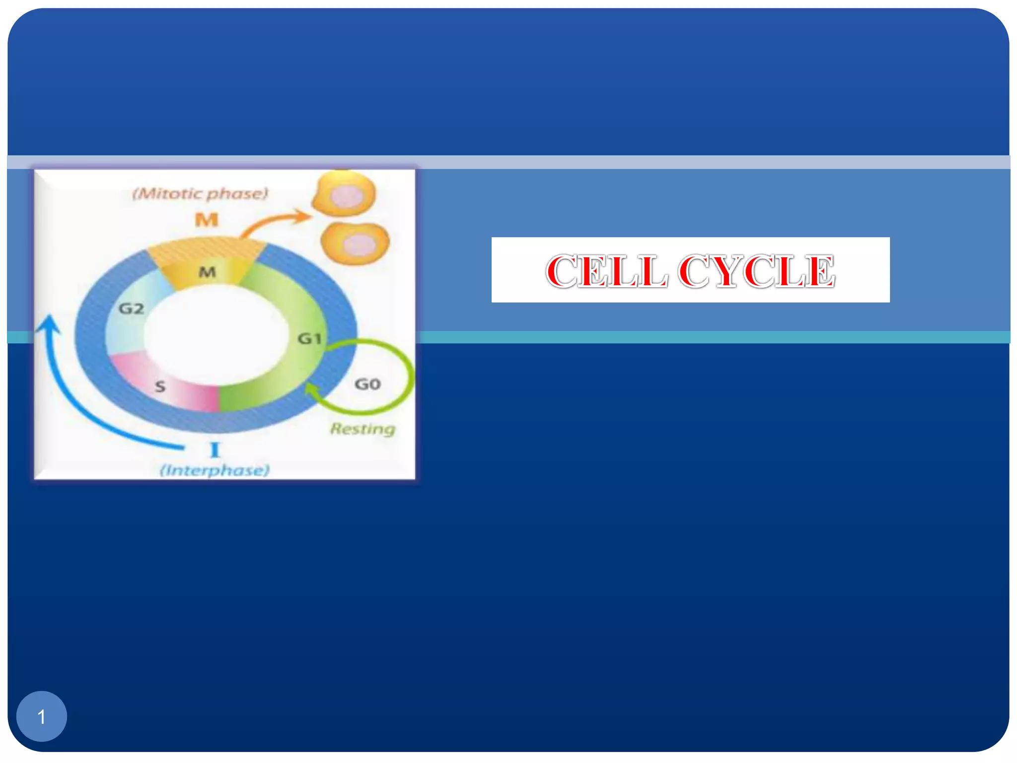
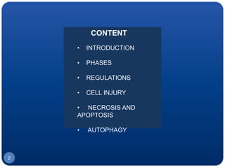
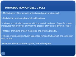
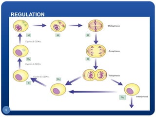
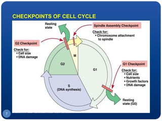
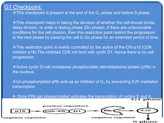
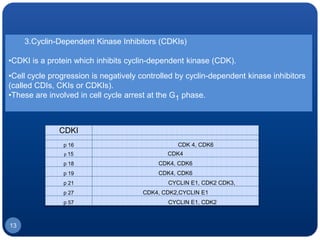
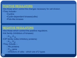
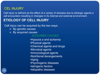
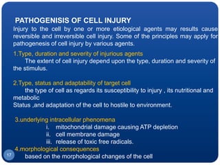
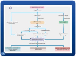
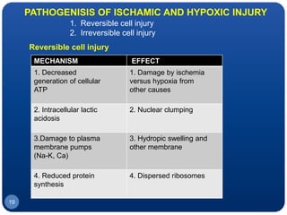
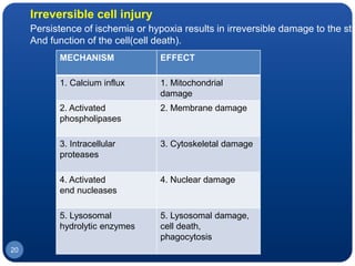
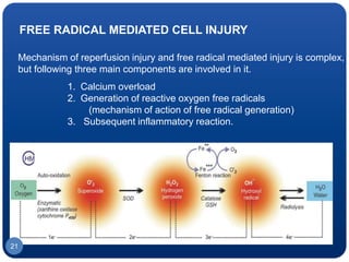
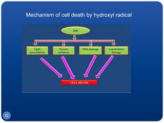
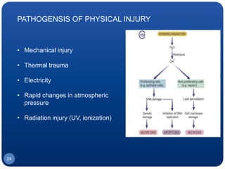
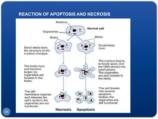
1
CONTENT
• INTRODUCTION
• PHASES
• REGULATIONS
• CELL INJURY
• NECROSIS AND
APOPTOSIS
• AUTOPHAGY
2
3
INTRODUCTION OF CELL CYCLE
Multiplication of the somatic (mitosis) and germ (meiosis)cell.
Cells is the most complex of all cell functions.
 Mitosis is controlled by genes which encode for release of specific protein
molecules that promotes or inhibit the process of mitosis at different steps.
mitosis promoting protein molecules are cyclin A,B and E.
These cyclins activate Cyclin Depended Kinase(CDK).which are conjuction
with cyclins.
After the mitosis complete cyclins,CDK will degrade.
4
PHASES OF CELL
CYCLE
•G1 (pre - mitotic gap)
•S phase
•G2 (pre – mitotic phase)
•M phase
I. Prophase
II. Metaphase
III. Anaphase
IV. Telophase
• G0 phase
5
REGULATION
6
7
CHECKPOINTS OF CELL CYCLE
8
G1 Checkpoint:
This checkpoint is present at the end of the G1 phase and before S phase.
This checkpoint helps in taking the decision of whether the cell should divide,
delay division, or enter a resting phase (Go phase). If there are unfavourable
conditions for the cell division, then this restriction point restrict the progression
to the next phase by passing the cell to Go phase for an extended period of time.
This restriction point is mainly controlled by the action of the CKI-p16 (CDK
inhibitor p16).The inhibited CDK not bind with cyclin D1, hence there is no cell
progression.
Active cyclin D-cdk complexes phosphorylate retinoblastoma protein (pRb) in
the nucleus.
Un-phosphorylated pRb acts as an inhibitor of G1 by preventing E2F-mediated
transcription.
 Once PRb get phosporylated activates the transcription of cyclins E and A,
Which then intract withCDK2 to allow for G1-S phase transition.
 This brings the cell to the end of the first checkpoint (unphosphorylated Rb
inhibits the E2F).
9
G2 Checkpoint
This restriction point is located at the end of the G2 phase. This checks the number
of factors which
are essential for the cell division.
Maturation-promoting factor or mitosis promoting factor or M-phase promoting
factor-(MPF) is a protein composed of cyclin-B and CDK-1. This protein promotes the
G2 phase into
the entrance of M-phase. MPF is activated at the end of G2 by a phosphatase (Chk)
which removes an inhibitory phosphate group added earlier.
The main functions of MPF in this restriction point are: a. Triggers the formation of
mitotic spindle.
b. Promotes chromosome condensation.
c. Causes nuclear envelop breakdown.
If there are any damages are noticed in this restriction point, then the phasphatase
not activate the
MPF, resulting in the arrest of cell cycle in G phase till the repair of the damaged
10
M-Checkpoint:
This occurs at metaphase.
Anaphase-promoting complex (APC) regulates this checkpoint.This is also
called spindle checkpoint.
This checks whether all chromosomes are properly attached to the spindle or
not.This also governs the alignment of the chromosomes and integrity of the
spindles. If there are mistakes then it delays the cell in entering into anaphase
from metaphase.
11
CELL CYCLE REGULATORS
The cell cycle is regulated by cycles.
1.Cyclins
2.cyclin-dependent kinases (CDKs)
3.cyclin-dependent kinase inhibitors (CDKIs).
1. CYLCLINS:
Their concentration varies during the cell cycle. Cyclins are the family of proteins
which regulates the cell cycle.
There are several types of cyclins that are active in different parts of the cell cycle
and causes phosphorylation of CDK.
There are also several “orphan” cyclins for which no CDK partner has
beenidentified.
For example, cyclin F is an orphan cyclin that is essential for G2/M transition.
There are two main groups of cyclins.
12
2.Cyclin-Dependent kinases (CDKs)
•These are a family of protein kinases that regulates the cell cycle.
•They are present in all known eukaryotic cells.
•Inactive on their own but becomes active when attached to cyclins.
13
3.Cyclin-Dependent Kinase Inhibitors (CDKIs)
•CDKI is a protein which inhibits cyclin-dependent kinase (CDK).
•Cell cycle progression is negatively controlled by cyclin-dependent kinase inhibitors
(called CDIs, CKIs or CDKIs).
•These are involved in cell cycle arrest at the G1 phase.
CDKI
p 16 CDK 4, CDK6
p 15 CDK4
p 18 CDK4, CDK6
p 19 CDK4, CDK6
p 21 CYCLIN E1, CDK2 CDK3,
p 27 CDK4, CDK2,CYCLIN E1
p 57 CYCLIN E1, CDK2
14
POSITIVE REGULATORS
•Are those which control the changes necessary for cell division.
•They include:-
•Cyclins
•Cyclin-dependent kinases(cdks)
•Polo-like kinases
NEGATIVE REGULATORS
•Are those which control the positive regulators.
•Ink family (Inhibitors of kinases)
P19 ,P15
•CIP family (cdks inhibitory proteins)
P21, P57
•They include :-
Rb proteins
P53 gene
Inhibitors of cdks - which are of 2 types
15
ETIOLOGY OF CELL INJURY
Cell injury can be acquired by the two ways
 By genetic causes
 By acquired causes
ACQUIRED CAUSES
•Hypoxia a and ischemia
•Physical agents
•Chemical agents and drugs
•Microbial agents
•Immunological agents
•Nutritional derangements
•Aging
•Psychogenic diseases
•Iatrogenic factors
•Idiopathic diseases16
Cell injury is defined as the effect of a variety of stresses due to etiologic agents a
cell encounters resulting in changes in its internal and external environment.
CEL INJURY
PATHOGENISIS OF CELL INJURY
Injury to the cell by one or more etiological agents may results cause
reversible and irreversible cell injury. Some of the principles may apply for
pathogenesis of cell injury by various agents.
1.Type, duration and severity of injurious agents
The extent of cell injury depend upon the type, duration and severity of
the stimulus.
2.Type, status and adaptability of target cell
the type of cell as regards its susceptibility to injury , its nutritional and
metabolic
Status ,and adaptation of the cell to hostile to environment.
3.underlying intracellular phenomena
i. mitochondrial damage causing ATP depletion
ii. cell membrane damage
iii. release of toxic free radicals.
4.morphological consequences
based on the morphological changes of the cell17
18
PATHOGENISIS OF ISCHAMIC AND HYPOXIC INJURY
1. Reversible cell injury
2. Irreversible cell injury
Reversible cell injury
MECHANISM EFFECT
1. Decreased
generation of cellular
ATP
1. Damage by ischemia
versus hypoxia from
other causes
2. Intracellular lactic
acidosis
2. Nuclear clumping
3.Damage to plasma
membrane pumps
(Na-K, Ca)
3. Hydropic swelling and
other membrane
4. Reduced protein
synthesis
4. Dispersed ribosomes
19
Irreversible cell injury
Persistence of ischemia or hypoxia results in irreversible damage to the stru
And function of the cell(cell death).
MECHANISM EFFECT
1. Calcium influx 1. Mitochondrial
damage
2. Activated
phospholipases
2. Membrane damage
3. Intracellular
proteases
3. Cytoskeletal damage
4. Activated
end nucleases
4. Nuclear damage
5. Lysosomal
hydrolytic enzymes
5. Lysosomal damage,
cell death,
phagocytosis
20
FREE RADICAL MEDIATED CELL INJURY
Mechanism of reperfusion injury and free radical mediated injury is complex,
but following three main components are involved in it.
1. Calcium overload
2. Generation of reactive oxygen free radicals
(mechanism of action of free radical generation)
3. Subsequent inflammatory reaction.
21
Mechanism of cell death by hydroxyl radical
22
Pathogenesis of chemical cell injury
Chemically induce cell injury by one of the following by direct
cytotoxicity and chemical into reactive metabolites.
Direct cytotoxic effects:
e.g. chemotherapeutic agents in cancer, toxic heavy metals (cyanide,
mercury, lead, iron)
Conversion to reactive toxic metabolites:
e.g. CCl4, acetaminophen, bromobenzene
23
PATHOGENSIS OF PHYSICAL INJURY
• Mechanical injury
• Thermal trauma
• Electricity
• Rapid changes in atmospheric
pressure
• Radiation injury (UV, ionization)
24
REACTION OF APOPTOSIS AND NECROSIS
25
APOPTOSIS AND NECROSIS
NECROSIS
1. Necrosis is defined as a localised area of death of tissue followed
later by degradation of tissue by hydrolytic enzymes liberated from
dead cells; it is invariably accompanied by inflammatory reaction.
2. Necrosis can be caused by various agents such as hypoxia,
chemical and physical agents, microbial agents, immunological
injury, etc.
3. Based on etiology and morphologic appearance, there are 5 types
of necrosis:
1. COAGULATIVE NECROSIS
2. LIQUEFACTION (COLLIQUATIVE) NECROSIS
3. CASEOUS NECROSIS
4. FAT NECROSIS
5. FIBRINOID NECROSIS
26
APOPTOSIS
1. Apoptosis is a form of ‘coordinated and internally programmed
cell death’ having significance in a variety of physiologic and
pathologic conditions (apoptosis=falling off or dropping off , as
that of leaves or petals).
2. Apoptosis is not accompanied by any inflammation and collateral
tissue damage.
MOLECULAR MECHANISMS OF APOPTOSIS
Several physiologic and pathologic processes activate
apoptosis in a variety of ways. However, in general the
following molecular events sum up the sequence involved in
apoptosis
27
1. Initiators of apoptosis
i) Withdrawal of normal cell survival signals
ii) Agents of cell injury
2. Initial steps in apoptosis
i) Intrinsic (mitochondrial) pathway
ii) Extrinsic (cell death receptor initiated)
pathway
3. Final phase of apoptosis
4. Phagocytosis
MOLECULAR MECHANISMS OF
APOPTOSIS
28
INTRINSIC AND EXTRINSIC PATHWAY
29
MECHANISM OF CELL DEATH IN OXIDATIVE STRESS MODEL
1. Hydrogen peroxide induced apoptosis
2. Reactive nitrogen species and apoptosis
3. Toll like receptor pathways and apoptosis
4. Lipid metabolites and apoptosis
5. Photodynamic therapy
6. Ionizing radiation
7. Cigarette smoke
MECHANISM OF CELL DEATH IN OXIDATIVE LUNG INJURY AND
ISCHEMIA/REPERFUSION INJURY
1. Hyperoxia
2. LPS induced lung injury
3. Ischemia/perfusion(I/R)
30
AUTOPHAGY
The term ‘autophagy’, derived from the Greek meaning ‘eating of self.’
It is one type of programmed cell death (PCD) distinct from the apoptosis a
This is regulated pathway for internal organelle or protein degradation.
Finally the delivered content become degrade and recycled.
31
TYPES OF AUTOPHAGY
•Macro-autophagy
• Micro-autophagy
•Chaperone-mediated autophagy(CMA)
32
1.MACRO AUTOPHAGY
Macro-autophagy delivers cytoplasmic cargo to the lysosome
through the intermediary of a double membrane-bound
vesicle, referred to as an autophagosome, that fuses with the
lysosome to form an autolysosome.
33
2.MICRO AUTOPHAGY
In micro-autophagy, by contrast, cytosolic components are
directly taken up by the lysosome itself through invagination
of the lysosomal membrane.
Both macro-and micro-autophagy are able to engulf large
structures through both selective and non-selective
mechanisms.
In chaperone-mediated autophagy (CMA), targeted proteins
are translocated across the lysosomal membrane in a
complex with chaperone proteins (such as Hsc-70) that are
recognized by the lysosomal membrane receptor lysosomal-
associated membrane protein 2A (LAMP-2A), resulting in their
unfolding and degradation
3.CHAPERONE-MEDIATED AUTOPHAGY
34
BASIC AUTOPHAGY MACHINERY
35
(a)control of phagophore formation by Beclin-1/VPS34 at the ER
and other membranes in response to stress signalling
pathways;
(b)Atg5–Atg12 conjugation, interaction with Atg16L and
multimerization at the phagophore;
(c) LC3 processing and insertion into the extending phagophore
membrane;
(d) capture of random or selective targets for degradation,
completion of the autophagosome accompanied by recycling
of some LC3-II/ATG8 by ATG4, followed by;
(e)Autophagy is induced by hypoxia and low cytosolic ATP levels
that feed through REDD1 and AMP-kinase to inhibit mTOR
activity through reduced Rheb GTPase activity. Conversely,
autophagy is inhibited by increased growth factor signalling
through the insulin receptor and its adaptor, IRS1, as well as
other growth factor receptors that activate the Class I group of
36
REFERENCES
1.Text book of pathology by Harsh Mohan 6th,7th edition.
2. HM/CH-1/L-4,cell injury
3. Danielle Glick1,2, Sandra Barth1, and Kay
F.Macleod1,2,*Autophagy: cellular and molecular mechanisms,
NIH Public Access Author Manuscript,2010.
37

Cell cycle and regulation

  • 1.
  • 2.
    CONTENT • INTRODUCTION • PHASES •REGULATIONS • CELL INJURY • NECROSIS AND APOPTOSIS • AUTOPHAGY 2
  • 3.
  • 4.
    INTRODUCTION OF CELLCYCLE Multiplication of the somatic (mitosis) and germ (meiosis)cell. Cells is the most complex of all cell functions.  Mitosis is controlled by genes which encode for release of specific protein molecules that promotes or inhibit the process of mitosis at different steps. mitosis promoting protein molecules are cyclin A,B and E. These cyclins activate Cyclin Depended Kinase(CDK).which are conjuction with cyclins. After the mitosis complete cyclins,CDK will degrade. 4
  • 5.
    PHASES OF CELL CYCLE •G1(pre - mitotic gap) •S phase •G2 (pre – mitotic phase) •M phase I. Prophase II. Metaphase III. Anaphase IV. Telophase • G0 phase 5
  • 6.
  • 7.
  • 8.
    8 G1 Checkpoint: This checkpointis present at the end of the G1 phase and before S phase. This checkpoint helps in taking the decision of whether the cell should divide, delay division, or enter a resting phase (Go phase). If there are unfavourable conditions for the cell division, then this restriction point restrict the progression to the next phase by passing the cell to Go phase for an extended period of time. This restriction point is mainly controlled by the action of the CKI-p16 (CDK inhibitor p16).The inhibited CDK not bind with cyclin D1, hence there is no cell progression. Active cyclin D-cdk complexes phosphorylate retinoblastoma protein (pRb) in the nucleus. Un-phosphorylated pRb acts as an inhibitor of G1 by preventing E2F-mediated transcription.  Once PRb get phosporylated activates the transcription of cyclins E and A, Which then intract withCDK2 to allow for G1-S phase transition.  This brings the cell to the end of the first checkpoint (unphosphorylated Rb inhibits the E2F).
  • 9.
    9 G2 Checkpoint This restrictionpoint is located at the end of the G2 phase. This checks the number of factors which are essential for the cell division. Maturation-promoting factor or mitosis promoting factor or M-phase promoting factor-(MPF) is a protein composed of cyclin-B and CDK-1. This protein promotes the G2 phase into the entrance of M-phase. MPF is activated at the end of G2 by a phosphatase (Chk) which removes an inhibitory phosphate group added earlier. The main functions of MPF in this restriction point are: a. Triggers the formation of mitotic spindle. b. Promotes chromosome condensation. c. Causes nuclear envelop breakdown. If there are any damages are noticed in this restriction point, then the phasphatase not activate the MPF, resulting in the arrest of cell cycle in G phase till the repair of the damaged
  • 10.
    10 M-Checkpoint: This occurs atmetaphase. Anaphase-promoting complex (APC) regulates this checkpoint.This is also called spindle checkpoint. This checks whether all chromosomes are properly attached to the spindle or not.This also governs the alignment of the chromosomes and integrity of the spindles. If there are mistakes then it delays the cell in entering into anaphase from metaphase.
  • 11.
    11 CELL CYCLE REGULATORS Thecell cycle is regulated by cycles. 1.Cyclins 2.cyclin-dependent kinases (CDKs) 3.cyclin-dependent kinase inhibitors (CDKIs). 1. CYLCLINS: Their concentration varies during the cell cycle. Cyclins are the family of proteins which regulates the cell cycle. There are several types of cyclins that are active in different parts of the cell cycle and causes phosphorylation of CDK. There are also several “orphan” cyclins for which no CDK partner has beenidentified. For example, cyclin F is an orphan cyclin that is essential for G2/M transition. There are two main groups of cyclins.
  • 12.
    12 2.Cyclin-Dependent kinases (CDKs) •Theseare a family of protein kinases that regulates the cell cycle. •They are present in all known eukaryotic cells. •Inactive on their own but becomes active when attached to cyclins.
  • 13.
    13 3.Cyclin-Dependent Kinase Inhibitors(CDKIs) •CDKI is a protein which inhibits cyclin-dependent kinase (CDK). •Cell cycle progression is negatively controlled by cyclin-dependent kinase inhibitors (called CDIs, CKIs or CDKIs). •These are involved in cell cycle arrest at the G1 phase. CDKI p 16 CDK 4, CDK6 p 15 CDK4 p 18 CDK4, CDK6 p 19 CDK4, CDK6 p 21 CYCLIN E1, CDK2 CDK3, p 27 CDK4, CDK2,CYCLIN E1 p 57 CYCLIN E1, CDK2
  • 14.
    14 POSITIVE REGULATORS •Are thosewhich control the changes necessary for cell division. •They include:- •Cyclins •Cyclin-dependent kinases(cdks) •Polo-like kinases NEGATIVE REGULATORS •Are those which control the positive regulators. •Ink family (Inhibitors of kinases) P19 ,P15 •CIP family (cdks inhibitory proteins) P21, P57 •They include :- Rb proteins P53 gene Inhibitors of cdks - which are of 2 types
  • 15.
  • 16.
    ETIOLOGY OF CELLINJURY Cell injury can be acquired by the two ways  By genetic causes  By acquired causes ACQUIRED CAUSES •Hypoxia a and ischemia •Physical agents •Chemical agents and drugs •Microbial agents •Immunological agents •Nutritional derangements •Aging •Psychogenic diseases •Iatrogenic factors •Idiopathic diseases16 Cell injury is defined as the effect of a variety of stresses due to etiologic agents a cell encounters resulting in changes in its internal and external environment. CEL INJURY
  • 17.
    PATHOGENISIS OF CELLINJURY Injury to the cell by one or more etiological agents may results cause reversible and irreversible cell injury. Some of the principles may apply for pathogenesis of cell injury by various agents. 1.Type, duration and severity of injurious agents The extent of cell injury depend upon the type, duration and severity of the stimulus. 2.Type, status and adaptability of target cell the type of cell as regards its susceptibility to injury , its nutritional and metabolic Status ,and adaptation of the cell to hostile to environment. 3.underlying intracellular phenomena i. mitochondrial damage causing ATP depletion ii. cell membrane damage iii. release of toxic free radicals. 4.morphological consequences based on the morphological changes of the cell17
  • 18.
  • 19.
    PATHOGENISIS OF ISCHAMICAND HYPOXIC INJURY 1. Reversible cell injury 2. Irreversible cell injury Reversible cell injury MECHANISM EFFECT 1. Decreased generation of cellular ATP 1. Damage by ischemia versus hypoxia from other causes 2. Intracellular lactic acidosis 2. Nuclear clumping 3.Damage to plasma membrane pumps (Na-K, Ca) 3. Hydropic swelling and other membrane 4. Reduced protein synthesis 4. Dispersed ribosomes 19
  • 20.
    Irreversible cell injury Persistenceof ischemia or hypoxia results in irreversible damage to the stru And function of the cell(cell death). MECHANISM EFFECT 1. Calcium influx 1. Mitochondrial damage 2. Activated phospholipases 2. Membrane damage 3. Intracellular proteases 3. Cytoskeletal damage 4. Activated end nucleases 4. Nuclear damage 5. Lysosomal hydrolytic enzymes 5. Lysosomal damage, cell death, phagocytosis 20
  • 21.
    FREE RADICAL MEDIATEDCELL INJURY Mechanism of reperfusion injury and free radical mediated injury is complex, but following three main components are involved in it. 1. Calcium overload 2. Generation of reactive oxygen free radicals (mechanism of action of free radical generation) 3. Subsequent inflammatory reaction. 21
  • 22.
    Mechanism of celldeath by hydroxyl radical 22
  • 23.
    Pathogenesis of chemicalcell injury Chemically induce cell injury by one of the following by direct cytotoxicity and chemical into reactive metabolites. Direct cytotoxic effects: e.g. chemotherapeutic agents in cancer, toxic heavy metals (cyanide, mercury, lead, iron) Conversion to reactive toxic metabolites: e.g. CCl4, acetaminophen, bromobenzene 23
  • 24.
    PATHOGENSIS OF PHYSICALINJURY • Mechanical injury • Thermal trauma • Electricity • Rapid changes in atmospheric pressure • Radiation injury (UV, ionization) 24
  • 25.
    REACTION OF APOPTOSISAND NECROSIS 25
  • 26.
    APOPTOSIS AND NECROSIS NECROSIS 1.Necrosis is defined as a localised area of death of tissue followed later by degradation of tissue by hydrolytic enzymes liberated from dead cells; it is invariably accompanied by inflammatory reaction. 2. Necrosis can be caused by various agents such as hypoxia, chemical and physical agents, microbial agents, immunological injury, etc. 3. Based on etiology and morphologic appearance, there are 5 types of necrosis: 1. COAGULATIVE NECROSIS 2. LIQUEFACTION (COLLIQUATIVE) NECROSIS 3. CASEOUS NECROSIS 4. FAT NECROSIS 5. FIBRINOID NECROSIS 26
  • 27.
    APOPTOSIS 1. Apoptosis isa form of ‘coordinated and internally programmed cell death’ having significance in a variety of physiologic and pathologic conditions (apoptosis=falling off or dropping off , as that of leaves or petals). 2. Apoptosis is not accompanied by any inflammation and collateral tissue damage. MOLECULAR MECHANISMS OF APOPTOSIS Several physiologic and pathologic processes activate apoptosis in a variety of ways. However, in general the following molecular events sum up the sequence involved in apoptosis 27
  • 28.
    1. Initiators ofapoptosis i) Withdrawal of normal cell survival signals ii) Agents of cell injury 2. Initial steps in apoptosis i) Intrinsic (mitochondrial) pathway ii) Extrinsic (cell death receptor initiated) pathway 3. Final phase of apoptosis 4. Phagocytosis MOLECULAR MECHANISMS OF APOPTOSIS 28
  • 29.
  • 30.
    MECHANISM OF CELLDEATH IN OXIDATIVE STRESS MODEL 1. Hydrogen peroxide induced apoptosis 2. Reactive nitrogen species and apoptosis 3. Toll like receptor pathways and apoptosis 4. Lipid metabolites and apoptosis 5. Photodynamic therapy 6. Ionizing radiation 7. Cigarette smoke MECHANISM OF CELL DEATH IN OXIDATIVE LUNG INJURY AND ISCHEMIA/REPERFUSION INJURY 1. Hyperoxia 2. LPS induced lung injury 3. Ischemia/perfusion(I/R) 30
  • 31.
    AUTOPHAGY The term ‘autophagy’,derived from the Greek meaning ‘eating of self.’ It is one type of programmed cell death (PCD) distinct from the apoptosis a This is regulated pathway for internal organelle or protein degradation. Finally the delivered content become degrade and recycled. 31 TYPES OF AUTOPHAGY •Macro-autophagy • Micro-autophagy •Chaperone-mediated autophagy(CMA)
  • 32.
    32 1.MACRO AUTOPHAGY Macro-autophagy deliverscytoplasmic cargo to the lysosome through the intermediary of a double membrane-bound vesicle, referred to as an autophagosome, that fuses with the lysosome to form an autolysosome.
  • 33.
    33 2.MICRO AUTOPHAGY In micro-autophagy,by contrast, cytosolic components are directly taken up by the lysosome itself through invagination of the lysosomal membrane. Both macro-and micro-autophagy are able to engulf large structures through both selective and non-selective mechanisms. In chaperone-mediated autophagy (CMA), targeted proteins are translocated across the lysosomal membrane in a complex with chaperone proteins (such as Hsc-70) that are recognized by the lysosomal membrane receptor lysosomal- associated membrane protein 2A (LAMP-2A), resulting in their unfolding and degradation 3.CHAPERONE-MEDIATED AUTOPHAGY
  • 34.
  • 35.
    35 (a)control of phagophoreformation by Beclin-1/VPS34 at the ER and other membranes in response to stress signalling pathways; (b)Atg5–Atg12 conjugation, interaction with Atg16L and multimerization at the phagophore; (c) LC3 processing and insertion into the extending phagophore membrane; (d) capture of random or selective targets for degradation, completion of the autophagosome accompanied by recycling of some LC3-II/ATG8 by ATG4, followed by; (e)Autophagy is induced by hypoxia and low cytosolic ATP levels that feed through REDD1 and AMP-kinase to inhibit mTOR activity through reduced Rheb GTPase activity. Conversely, autophagy is inhibited by increased growth factor signalling through the insulin receptor and its adaptor, IRS1, as well as other growth factor receptors that activate the Class I group of
  • 36.
    36 REFERENCES 1.Text book ofpathology by Harsh Mohan 6th,7th edition. 2. HM/CH-1/L-4,cell injury 3. Danielle Glick1,2, Sandra Barth1, and Kay F.Macleod1,2,*Autophagy: cellular and molecular mechanisms, NIH Public Access Author Manuscript,2010.
  • 37.