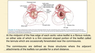
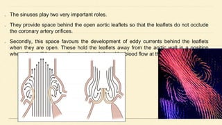
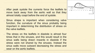
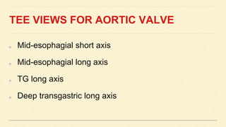
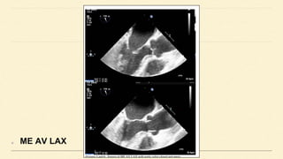
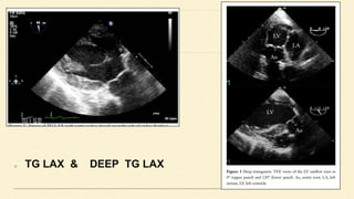
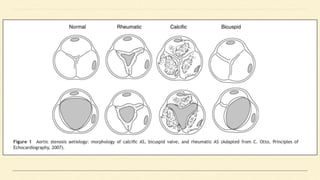
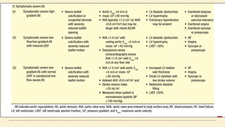
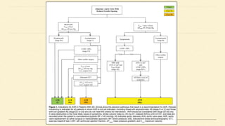
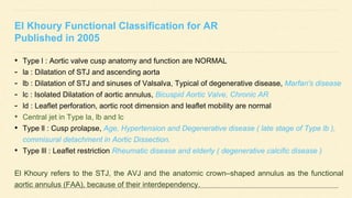
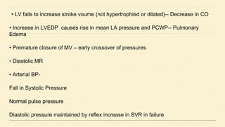
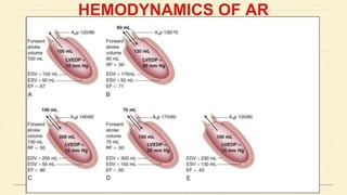
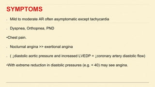
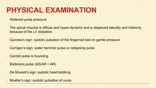
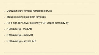
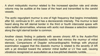
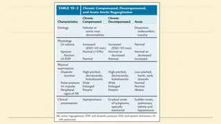
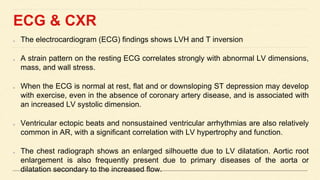
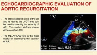
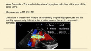
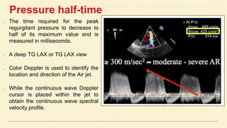
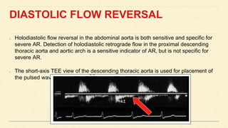
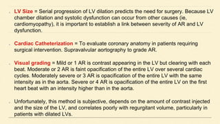
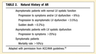
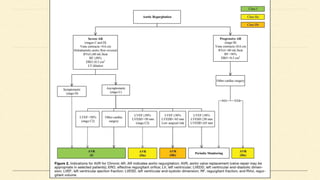
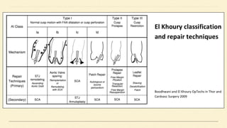
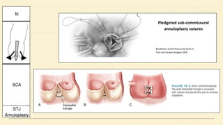
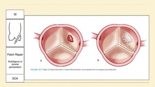
ARTIC VALVE TEE
Dr Hasmukh Patel
NORMAL ADULT AORTIC VALVE
AREA IS 3.0 TO 4.0 CM2.
AORTIC VALVE
WHY THE ORIFICE OF AORTIC ARTERY IS TRIANGULAR
“the more obtuse
angle is stronger
than the right angle
of the square”
ANATOMY - AORTIC ROOT STRUCTURE
Defined as the portion of the left ventricular outflow tract which
supports the leaflets of the aortic valve, delineated by the
sinotubular ridge superiorly and the bases of the valve leaflets
inferiorly.
It comprises the sinuses, the aortic valve leaflets, the
commissures, and the interleaflet triangles.
The sinuses are expanded portions of the aortic root which are
confined proximally by the attachments of the valve leaflets
and distally by the sinotubular junction & named according to
the coronary arteries arising from them right, left, and non-
coronary.
The sino-tubular junction is circular and composed of primarily
elastic tissue, and it supports the peripheral attachments of the
Valve leaflets : portions of the aortic root which separate, haemodynamically, the aorta and
the left ventricle. They are inserted into the wall of the root in a semilunar fashion, and the
base of the aortic root is defined by the nadirs of attachment of these leaflets.
The term annulus is implies to a circular structure and as such is inaccurate for AV.
The posterior aspect of the aortic root (mainly non-coronary leaflet) is supported by fibrous
tissue for approximately 55% of its circumference (membranous part of the septum to the left
fibrous trigone), while the remainder is supported by ventricular muscle, an important
distinction when performing a surgical annuloplasty.
At the midpoint of the free edge of each aortic valve leaflet is a fibrous nodule,
on either side of which is a thin crescent shaped portion of the leaflet called
the lannula which are occasionally fenestrated near the commissures.
The commissures are defined as those structures where the adjacent
attachments of the leaflets run parallel for a short distance.
The diameter of the sinotubular junction is 10±15% smaller
than the diameter of the annulus.
The upper part of the valve commissures are attached just
below the sinotubular junction and the diameter of the aortic
root at this level approximates to the diameter of the annulus.
The non-coronary sinus is the largest of the three sinuses. The
length of the base of the leaflets is approximately 1.5 times
longer than the length of its free margin and the height of the
leaflets ranges from 12±18 mm.
As with the sinuses, the non-coronary leaflet is slightly larger
than the other two.
Aortic root: function
Aortic valve function is, however, much more complicated and the aortic
root complex acts as an individual haemodynamic system.
The upper portion of the aortic root is exposed to aortic pressure
changes so it expands during systole (like aorta) allowing the leaflets to
retract and open.
The proximal part of the root complex is exposed to ventricular pressure
changes and it will expand as the ventricle fills and contracts during
peak systole which decreases the distance the valve leaflets have to
travel to coapt.
However, it is the sinuses which seem to serve a most important
physiological role in aortic valve function, and their importance has been
The sinuses play two very important roles.
They provide space behind the open aortic leaflets so that the leaflets do not occlude
the coronary artery orifices.
Secondly, this space favours the development of eddy currents behind the leaflets
when they are open. These hold the leaflets away from the aortic wall in a position
where they will be promptly caught and closed by blood flow at the end of systole.
After peak systole the currents force the leaflets to
move back away from the aortic wall so that they
almost totally coapt before the end of systole.
Sinus shape is important when considering valve
function, the curvature of the sinus probably being
important in determining the distribution of stress on
the valve leaflets.
The stress on the leaflets in diastole is almost four
times that in the sinuses, and this would result in the
sinus walls being drawn inwards in diastole if the
stress were not shared by the sinuses. Instead the
sinus walls move outward decreasing the stress and
wear on the aortic leaflets.
TEE VIEWS FOR AORTIC VALVE
Mid-esophagial short axis
Mid-esophagial long axis
TG long axis
Deep transgastric long axis
ME AV SAX
ME AV LAX
TG LAX & DEEP TG LAX
AORTIC STENOSIS
Most common :-
Bicuspid aortic valve with calcification
Senile or Degenerative calcific AS
Rheumatic AS
Less common:-
Congenital
Type 2 Hyperlipoproteinemia
Onchronosis
SYMPTOMS
Cardinal Manifestation: Exertional Dyspnea, Angina, Syncope, and ultimately Heart Failure.
Symptoms typically occur at age 50 to 70 years with bicuspid aortic valve stenosis and in
those older than 70 years with calcific stenosis of a trileaflet valve.
The mechanism of exertional dyspnea may be LV diastolic dysfunction.
Angina results from oxygen demand vs supply imbalance, 50% associated significant
coronary artery obstruction. Very rarely, angina results from calcium emboli.
Syncope is most commonly caused by the reduced cerebral perfusion that occurs during
exertion when arterial pressure declines consequent to systemic vasodilation in the presence
of a fixed cardiac output. Syncope has also been attributed to malfunction of the baroreceptor
mechanism in severe AS, as well as to a vasodepressor response to a greatly elevated LV
systolic pressure during exercise.
Survival : 2 years in patients with heart failure, 3 years in those with syncope, and 5 years in
those with angina.
PHYSICAL EXAMINATION
Key features: Palpation of the carotid upstroke, Evaluation of the systolic
murmur, Assessment of splitting of the second heart sound, and Examination
for signs of heart failure.
Carotid upstroke is a slow-rising, late-peaking, low-amplitude carotid pulse, the
parvus and tardus carotid impulse. Radiation of the murmur to the carotids
may result in a palpable thrill.
A systolic thrill is usually best appreciated ( in the second right intercostal
space or suprasternal notch ) when the patient leans forward during full
expiration. A systolic thrill is specific, but not sensitive for AS.
AUSCULTATION
The ejection systolic murmur of AS typically is late peaking and heard best at the base
of the heart, with radiation to the carotids.
In patients with calcified aortic valves, the systolic murmur is loudest at the base of the
heart, but high-frequency components may radiate to the apex (the so-called
Gallavardin phenomenon), in which the murmur may be so prominent that it is mistaken
for the murmur of MR.
Splitting of the second heart sound is helpful in excluding the diagnosis of severe AS.
When the left ventricle fails and stroke volume falls, the systolic murmur of AS becomes
softer.
The murmur of valvular AS is augmented by squatting, which increases stroke volume.
It is reduced in intensity during the strain of the Valsalva maneuver and when standing,
which reduce transvalvular flow.
TEE EVALUATION
The primary haemodynamic parameters recommended for TEE evaluation of AS
severity are:
● AS jet velocity
● Mean transaortic gradient
● Valve area by continuity equation
AVA - CONTINUITY EQUATION
Aortic valve area is calculated based on the continuity-equation concept that the
stroke volume (SV) ejected through the LV outflow tract (LVOT) all passes through
the stenotic orifice (AVA) and thus SV is equal at both sites:
Calculation of continuity-equation valve area requires three measurements:
● AS jet velocity by CWD
● LVOT diameter
● LVOT velocity by PWD
Low-flow low-gradient AS includes the following conditions:
➢Effective orifice area < 1.0 Cm2
➢LV ejection fraction < 50%
➢ Mean pressure gradient < 40 mmHg or Aortic velocity < 4 m/s
DOBUTAMINE STRESS ECHO
Helpful to differentiate two clinical situations
➢Severe AS causing LV systolic dysfunction
➢Moderate AS with another cause of LV dysfunction
●A low dose starting at 2.5 or 5 ủg/kg/min with an incremental increase in the
infusion every 3–5 min to a maximum dose of 10–20 ủg/kg/min
The infusion should be stopped as soon as
➢Positive result is obtained
➢Heart rate begins to rise more than 10–20 bpm over baseline or exceeds
100bpm.
DOBUTAMINE STRESS ECHO
An increase in valve area to a final valve area >1.0 cm2 suggests that stenosis is
not severe.
Severe stenosis is suggested by an AS jet > 4.0 or a mean gradient > 40 mmHg
provided that valve area does not exceed 1.0 cm2 at any flow rate.
Absence of contractile reserve (failure to increase SV or ejection fraction by > 20%)
is a predictor of a high surgical mortality and poor long-term outcome although valve
replacement may improve LV function and outcome even in this subgroup.
AORTIC REGURGITATION
• Congenital
Bicuspid valve
•Aortopathy
Cystic medial necrosis
Collagen disorders (e.g. Marfan’s)
Ehler-Danlos
Osteogenesis imperfecta
Pseudoxanthoma elasticum
Acquired
Rheumatic heart disease
Dilated aorta (e.g. hypertension..)
Degenerative
Connective tissue disorders. E.g. ankylosing
spondylitis, rheumatoid arthritis, Reiter’s syndrome,
Giant-cell arteritis )
Syphilis (chronic aortitis)
Acute AI: aortic dissection, infective endocarditis,
El Khoury Functional Classification for AR
Published in 2005
• Type l : Aortic valve cusp anatomy and function are NORMAL
- la : Dilatation of STJ and ascending aorta
- lb : Dilatation of STJ and sinuses of Valsalva, Typical of degenerative disease, Marfan's disease
- lc : Isolated Dilatation of aortic annulus, Bicuspid Aortic Valve, Chronic AR
- ld : Leaflet perforation, aortic root dimension and leaflet mobility are normal
• Central jet in Type la, lb and lc
• Type ll : Cusp prolapse, Age, Hypertension and Degenerative disease ( late stage of Type lb ),
commisural detachment in Aortic Dissection.
• Type lll : Leaflet restriction Rheumatic disease and elderly ( degenerative calcific disease )
El Khoury refers to the STJ, the AVJ and the anatomic crown–shaped annulus as the functional
aortic annulus (FAA), because of their interdependency.
HEMODYNAMICS OF ACUTE AR
Hemodynamically significant AR of sudden onset, into a LV not previously subjected
to volume overload
•Volume overload is poorly tolerated
Ventricular compliance is normal
LV operating on steep portion of diastolic P/V relation
End diastolic LV pressure markedly increased approaching aortic diastolic pressure
• LV fails to increase stroke voume (not hypertrophied or dilated)-- Decrease in CO
• Increase in LVEDP causes rise in mean LA pressure and PCWP-- Pulmonary
Edema
• Premature closure of MV – early crossover of pressures
• Diastolic MR
• Arterial BP-
Fall in Systolic Pressure
Normal pulse pressure
Diastolic pressure maintained by reflex increase in SVR in failure
HEMODYNAMICS OF AR
SYMPTOMS
Mild to moderate AR often asymptomatic except tachycardia
Dyspnea, Orthopnea, PND
•Chest pain.
Nocturnal angina >> exertional angina
( ↓diastolic aortic pressure and increased LVEDP = ↓coronary artery diastolic flow)
•With extreme reduction in diastolic pressures (e.g. < 40) may see angina.
PHYSICAL EXAMINATION
Widened pulse pressure
The apical impulse is diffuse and hyper-dynamic and is displaced laterally and inferiorly
because of the LV dilatation.
Quincke’s sign: systolic pulsation of the fingernail bed on gentle pressure
Corrigan’s sign: water hammer pulse or collapsing pulse
Carotid pulse is bounding
Bisferiens pulse (AS/AR > AR)
De Musset’s sign: systolic head bobbing
Mueller’s sign: systolic pulsation of uvula
Duroziez sign: femoral retrograde bruits
Traube’s sign: pistol shot femorals
Hill’s sign:BP Lower extremity >BP Upper extremity by
> 20 mm Hg - mild AR
> 40 mm Hg – mod AR
> 60 mm Hg – severe AR
A short midsystolic murmur related to the increased ejection rate and stroke
volume may be audible at the base of the heart and transmitted to the carotid
vessels.
The aortic regurgitant murmur is one of high frequency that begins immediately
after S2, continues to S1, and has a decrescendo intensity. The murmur is best
heard along the left sternal border in the third or fourth intercostal space with
valve leaflet abnormalities, whereas with aortic root disease a selective radiation
along the right sternal border is common.
Another classic finding in patients with severe chronic AR is the Austin-Flint
murmur, a low pitched mid-diastolic rumble that mimics the murmur of mitral
stenosis. Comparisons of Doppler echocardiographic findings with physical
examination suggest that this diastolic murmur is related to the severity of AR
with a jet directed toward the anterior mitral leaflet or LV free wall, causing
vibrations recognized on auscultation as a low-pitched diastolic rumble.
ECG & CXR
The electrocardiogram (ECG) findings shows LVH and T inversion
A strain pattern on the resting ECG correlates strongly with abnormal LV dimensions,
mass, and wall stress.
When the ECG is normal at rest, flat and or downsloping ST depression may develop
with exercise, even in the absence of coronary artery disease, and is associated with
an increased LV systolic dimension.
Ventricular ectopic beats and nonsustained ventricular arrhythmias are also relatively
common in AR, with a significant correlation with LV hypertrophy and function.
The chest radiograph shows an enlarged silhouette due to LV dilatation. Aortic root
enlargement is also frequently present due to primary diseases of the aorta or
dilatation secondary to the increased flow.
ECHOCARDIOGRAPHIC EVALUATION OF
AORTIC REGURGITATION
The cross sectional area of the jet
and its ratio to the LVOT area can
be used to quantify the severity of
AR . This method defines severe
AR as a ratio ≥ 0.6
The ME AV LAX view is the most
useful for quantifying the severity
of AR.
Vena Contracta = The smallest diameter of regurgitant color flow at the level of the
aortic valve.
Measurement in ME AV LAX
Limitations = presence of multiple or abnormally shaped regurgitant jets and the
inability to accurately determine the annular plane of the aortic valve due to
pathology
Pressure half-time
The time required for the peak
regurgitant pressure to decrease to
half of its maximum value and is
measured in milliseconds.
A deep TG LAX or TG LAX view
Color Doppler is used to identify the
location and direction of the Air jet.
While the continuous wave Doppler
cursor is placed within the jet to
obtain the continuous wave spectral
velocity profile.
As a general rule, an AR pressure half-time < 200 ms indicates severe AR, whereas
a pressure half-time > 500 ms suggests mild AR.
Decreased systemic vascular resistance (sepsis) and reduced left ventricular
compliance (ischemia, cardiomyopathy, acute AR) cause a steeper deceleration
slope because aortic and left ventricular pressures equalize more rapidly in these
conditions.
Another limitation to this technique is that measurement of regurgitant jet velocity is
difficult and unreliable in patients with eccentric jets, because it is difficult to align
the Doppler beam with the regurgitant jet.
DIASTOLIC FLOW REVERSAL
Holodiastolic flow reversal in the abdominal aorta is both sensitive and specific for
severe AR. Detection of holodiastolic retrograde flow in the proximal descending
thoracic aorta and aortic arch is a sensitive indicator of AR, but is not specific for
severe AR.
The short-axis TEE view of the descending thoracic aorta is used for placement of
the pulsed wave sample volume distal in the aorta, near the diaphragm.
REG- VOL, REG- FRACTION & ERO
Regurgitant volume is the difference between the systolic flow across the aortic valve and
“net forward” cardiac output.
In the absence of intracardiac shunts and mitral regurgitation, flow through the pulmonary
artery or mitral valve is equivalent to (net) cardiac output. Pulmonary artery blood flow is
reliably measured with TEE by measuring the pulmonary artery diameter (d), calculating
its area [π (d/2)2], and multiplying the area by the pulmonary artery VTI and heart rate.
Aortic valve systolic flow is the product of aortic valve area and VTI.
Regurgitant Volume = Aortic Valve Systolic Flow – Cardiac Output
Regurgitant Fraction = Regurgitant Volume / Aortic Valve Systolic Flow
ERO = AORTIC ROOT area X AORTIC ROOT DIASTOLIC vti / AV Regurgitant JET
vti
LV Size = Serial progression of LV dilation predicts the need for surgery. Because LV
chamber dilation and systolic dysfunction can occur from other causes (ie,
cardiomyopathy), it is important to establish a link between severity of AR and LV
dysfunction.
Cardiac Catheterization = To evaluate coronary anatomy in patients requiring
surgical intervention. Supravalvular aortography to grade AR.
Visual grading = Mild or 1 AR is contrast appearing in the LV but clearing with each
beat. Moderate or 2 AR is faint opacification of the entire LV over several cardiac
cycles. Moderately severe or 3 AR is opacification of the entire LV with the same
intensity as in the aorta. Severe or 4 AR is opacification of the entire LV on the first
heart beat with an intensity higher than in the aorta.
Unfortunately, this method is subjective, depends on the amount of contrast injected
and the size of the LV, and correlates poorly with regurgitant volume, particularly in
patients with dilated LVs.
MEDICAL MANAGEMENT
Treatment of hypertension (systolic BP >140 mmhg) is recommended in patients
with chronic AR with dihydropyredine CCBs or ACE inhibitors.(class 1B)
Medical therapy with ACE inhibitors and B blockers is reasonable in patients with
severe AR who have symptoms and/or LV dysfunction, when surgery is not
performed because of comorbidities.
Vasodilator therapy is not recommended in patients with chronic asymptomatic AR
and normal LV systolic function
REPAIR TECHNIQUES
Aortic valve Repair & Replacement.TEEpptx

Aortic valve Repair & Replacement.TEEpptx

  • 1.
    ARTIC VALVE TEE DrHasmukh Patel
  • 2.
    NORMAL ADULT AORTICVALVE AREA IS 3.0 TO 4.0 CM2. AORTIC VALVE
  • 3.
    WHY THE ORIFICEOF AORTIC ARTERY IS TRIANGULAR “the more obtuse angle is stronger than the right angle of the square”
  • 6.
    ANATOMY - AORTICROOT STRUCTURE Defined as the portion of the left ventricular outflow tract which supports the leaflets of the aortic valve, delineated by the sinotubular ridge superiorly and the bases of the valve leaflets inferiorly. It comprises the sinuses, the aortic valve leaflets, the commissures, and the interleaflet triangles. The sinuses are expanded portions of the aortic root which are confined proximally by the attachments of the valve leaflets and distally by the sinotubular junction & named according to the coronary arteries arising from them right, left, and non- coronary. The sino-tubular junction is circular and composed of primarily elastic tissue, and it supports the peripheral attachments of the
  • 8.
    Valve leaflets :portions of the aortic root which separate, haemodynamically, the aorta and the left ventricle. They are inserted into the wall of the root in a semilunar fashion, and the base of the aortic root is defined by the nadirs of attachment of these leaflets. The term annulus is implies to a circular structure and as such is inaccurate for AV. The posterior aspect of the aortic root (mainly non-coronary leaflet) is supported by fibrous tissue for approximately 55% of its circumference (membranous part of the septum to the left fibrous trigone), while the remainder is supported by ventricular muscle, an important distinction when performing a surgical annuloplasty.
  • 9.
    At the midpointof the free edge of each aortic valve leaflet is a fibrous nodule, on either side of which is a thin crescent shaped portion of the leaflet called the lannula which are occasionally fenestrated near the commissures. The commissures are defined as those structures where the adjacent attachments of the leaflets run parallel for a short distance.
  • 10.
    The diameter ofthe sinotubular junction is 10±15% smaller than the diameter of the annulus. The upper part of the valve commissures are attached just below the sinotubular junction and the diameter of the aortic root at this level approximates to the diameter of the annulus. The non-coronary sinus is the largest of the three sinuses. The length of the base of the leaflets is approximately 1.5 times longer than the length of its free margin and the height of the leaflets ranges from 12±18 mm. As with the sinuses, the non-coronary leaflet is slightly larger than the other two.
  • 11.
    Aortic root: function Aorticvalve function is, however, much more complicated and the aortic root complex acts as an individual haemodynamic system. The upper portion of the aortic root is exposed to aortic pressure changes so it expands during systole (like aorta) allowing the leaflets to retract and open. The proximal part of the root complex is exposed to ventricular pressure changes and it will expand as the ventricle fills and contracts during peak systole which decreases the distance the valve leaflets have to travel to coapt. However, it is the sinuses which seem to serve a most important physiological role in aortic valve function, and their importance has been
  • 13.
    The sinuses playtwo very important roles. They provide space behind the open aortic leaflets so that the leaflets do not occlude the coronary artery orifices. Secondly, this space favours the development of eddy currents behind the leaflets when they are open. These hold the leaflets away from the aortic wall in a position where they will be promptly caught and closed by blood flow at the end of systole.
  • 14.
    After peak systolethe currents force the leaflets to move back away from the aortic wall so that they almost totally coapt before the end of systole. Sinus shape is important when considering valve function, the curvature of the sinus probably being important in determining the distribution of stress on the valve leaflets. The stress on the leaflets in diastole is almost four times that in the sinuses, and this would result in the sinus walls being drawn inwards in diastole if the stress were not shared by the sinuses. Instead the sinus walls move outward decreasing the stress and wear on the aortic leaflets.
  • 15.
    TEE VIEWS FORAORTIC VALVE Mid-esophagial short axis Mid-esophagial long axis TG long axis Deep transgastric long axis
  • 16.
  • 17.
  • 18.
    TG LAX &DEEP TG LAX
  • 19.
    AORTIC STENOSIS Most common:- Bicuspid aortic valve with calcification Senile or Degenerative calcific AS Rheumatic AS Less common:- Congenital Type 2 Hyperlipoproteinemia Onchronosis
  • 21.
    SYMPTOMS Cardinal Manifestation: ExertionalDyspnea, Angina, Syncope, and ultimately Heart Failure. Symptoms typically occur at age 50 to 70 years with bicuspid aortic valve stenosis and in those older than 70 years with calcific stenosis of a trileaflet valve. The mechanism of exertional dyspnea may be LV diastolic dysfunction. Angina results from oxygen demand vs supply imbalance, 50% associated significant coronary artery obstruction. Very rarely, angina results from calcium emboli. Syncope is most commonly caused by the reduced cerebral perfusion that occurs during exertion when arterial pressure declines consequent to systemic vasodilation in the presence of a fixed cardiac output. Syncope has also been attributed to malfunction of the baroreceptor mechanism in severe AS, as well as to a vasodepressor response to a greatly elevated LV systolic pressure during exercise. Survival : 2 years in patients with heart failure, 3 years in those with syncope, and 5 years in those with angina.
  • 22.
    PHYSICAL EXAMINATION Key features:Palpation of the carotid upstroke, Evaluation of the systolic murmur, Assessment of splitting of the second heart sound, and Examination for signs of heart failure. Carotid upstroke is a slow-rising, late-peaking, low-amplitude carotid pulse, the parvus and tardus carotid impulse. Radiation of the murmur to the carotids may result in a palpable thrill. A systolic thrill is usually best appreciated ( in the second right intercostal space or suprasternal notch ) when the patient leans forward during full expiration. A systolic thrill is specific, but not sensitive for AS.
  • 23.
    AUSCULTATION The ejection systolicmurmur of AS typically is late peaking and heard best at the base of the heart, with radiation to the carotids. In patients with calcified aortic valves, the systolic murmur is loudest at the base of the heart, but high-frequency components may radiate to the apex (the so-called Gallavardin phenomenon), in which the murmur may be so prominent that it is mistaken for the murmur of MR. Splitting of the second heart sound is helpful in excluding the diagnosis of severe AS. When the left ventricle fails and stroke volume falls, the systolic murmur of AS becomes softer. The murmur of valvular AS is augmented by squatting, which increases stroke volume. It is reduced in intensity during the strain of the Valsalva maneuver and when standing, which reduce transvalvular flow.
  • 24.
    TEE EVALUATION The primaryhaemodynamic parameters recommended for TEE evaluation of AS severity are: ● AS jet velocity ● Mean transaortic gradient ● Valve area by continuity equation
  • 25.
    AVA - CONTINUITYEQUATION Aortic valve area is calculated based on the continuity-equation concept that the stroke volume (SV) ejected through the LV outflow tract (LVOT) all passes through the stenotic orifice (AVA) and thus SV is equal at both sites:
  • 26.
    Calculation of continuity-equationvalve area requires three measurements: ● AS jet velocity by CWD ● LVOT diameter ● LVOT velocity by PWD
  • 28.
    Low-flow low-gradient ASincludes the following conditions: ➢Effective orifice area < 1.0 Cm2 ➢LV ejection fraction < 50% ➢ Mean pressure gradient < 40 mmHg or Aortic velocity < 4 m/s
  • 29.
    DOBUTAMINE STRESS ECHO Helpfulto differentiate two clinical situations ➢Severe AS causing LV systolic dysfunction ➢Moderate AS with another cause of LV dysfunction ●A low dose starting at 2.5 or 5 ủg/kg/min with an incremental increase in the infusion every 3–5 min to a maximum dose of 10–20 ủg/kg/min The infusion should be stopped as soon as ➢Positive result is obtained ➢Heart rate begins to rise more than 10–20 bpm over baseline or exceeds 100bpm.
  • 30.
    DOBUTAMINE STRESS ECHO Anincrease in valve area to a final valve area >1.0 cm2 suggests that stenosis is not severe. Severe stenosis is suggested by an AS jet > 4.0 or a mean gradient > 40 mmHg provided that valve area does not exceed 1.0 cm2 at any flow rate. Absence of contractile reserve (failure to increase SV or ejection fraction by > 20%) is a predictor of a high surgical mortality and poor long-term outcome although valve replacement may improve LV function and outcome even in this subgroup.
  • 34.
    AORTIC REGURGITATION • Congenital Bicuspidvalve •Aortopathy Cystic medial necrosis Collagen disorders (e.g. Marfan’s) Ehler-Danlos Osteogenesis imperfecta Pseudoxanthoma elasticum Acquired Rheumatic heart disease Dilated aorta (e.g. hypertension..) Degenerative Connective tissue disorders. E.g. ankylosing spondylitis, rheumatoid arthritis, Reiter’s syndrome, Giant-cell arteritis ) Syphilis (chronic aortitis) Acute AI: aortic dissection, infective endocarditis,
  • 35.
    El Khoury FunctionalClassification for AR Published in 2005 • Type l : Aortic valve cusp anatomy and function are NORMAL - la : Dilatation of STJ and ascending aorta - lb : Dilatation of STJ and sinuses of Valsalva, Typical of degenerative disease, Marfan's disease - lc : Isolated Dilatation of aortic annulus, Bicuspid Aortic Valve, Chronic AR - ld : Leaflet perforation, aortic root dimension and leaflet mobility are normal • Central jet in Type la, lb and lc • Type ll : Cusp prolapse, Age, Hypertension and Degenerative disease ( late stage of Type lb ), commisural detachment in Aortic Dissection. • Type lll : Leaflet restriction Rheumatic disease and elderly ( degenerative calcific disease ) El Khoury refers to the STJ, the AVJ and the anatomic crown–shaped annulus as the functional aortic annulus (FAA), because of their interdependency.
  • 36.
    HEMODYNAMICS OF ACUTEAR Hemodynamically significant AR of sudden onset, into a LV not previously subjected to volume overload •Volume overload is poorly tolerated Ventricular compliance is normal LV operating on steep portion of diastolic P/V relation End diastolic LV pressure markedly increased approaching aortic diastolic pressure
  • 37.
    • LV failsto increase stroke voume (not hypertrophied or dilated)-- Decrease in CO • Increase in LVEDP causes rise in mean LA pressure and PCWP-- Pulmonary Edema • Premature closure of MV – early crossover of pressures • Diastolic MR • Arterial BP- Fall in Systolic Pressure Normal pulse pressure Diastolic pressure maintained by reflex increase in SVR in failure
  • 38.
  • 39.
    SYMPTOMS Mild to moderateAR often asymptomatic except tachycardia Dyspnea, Orthopnea, PND •Chest pain. Nocturnal angina >> exertional angina ( ↓diastolic aortic pressure and increased LVEDP = ↓coronary artery diastolic flow) •With extreme reduction in diastolic pressures (e.g. < 40) may see angina.
  • 40.
    PHYSICAL EXAMINATION Widened pulsepressure The apical impulse is diffuse and hyper-dynamic and is displaced laterally and inferiorly because of the LV dilatation. Quincke’s sign: systolic pulsation of the fingernail bed on gentle pressure Corrigan’s sign: water hammer pulse or collapsing pulse Carotid pulse is bounding Bisferiens pulse (AS/AR > AR) De Musset’s sign: systolic head bobbing Mueller’s sign: systolic pulsation of uvula
  • 41.
    Duroziez sign: femoralretrograde bruits Traube’s sign: pistol shot femorals Hill’s sign:BP Lower extremity >BP Upper extremity by > 20 mm Hg - mild AR > 40 mm Hg – mod AR > 60 mm Hg – severe AR
  • 42.
    A short midsystolicmurmur related to the increased ejection rate and stroke volume may be audible at the base of the heart and transmitted to the carotid vessels. The aortic regurgitant murmur is one of high frequency that begins immediately after S2, continues to S1, and has a decrescendo intensity. The murmur is best heard along the left sternal border in the third or fourth intercostal space with valve leaflet abnormalities, whereas with aortic root disease a selective radiation along the right sternal border is common. Another classic finding in patients with severe chronic AR is the Austin-Flint murmur, a low pitched mid-diastolic rumble that mimics the murmur of mitral stenosis. Comparisons of Doppler echocardiographic findings with physical examination suggest that this diastolic murmur is related to the severity of AR with a jet directed toward the anterior mitral leaflet or LV free wall, causing vibrations recognized on auscultation as a low-pitched diastolic rumble.
  • 44.
    ECG & CXR Theelectrocardiogram (ECG) findings shows LVH and T inversion A strain pattern on the resting ECG correlates strongly with abnormal LV dimensions, mass, and wall stress. When the ECG is normal at rest, flat and or downsloping ST depression may develop with exercise, even in the absence of coronary artery disease, and is associated with an increased LV systolic dimension. Ventricular ectopic beats and nonsustained ventricular arrhythmias are also relatively common in AR, with a significant correlation with LV hypertrophy and function. The chest radiograph shows an enlarged silhouette due to LV dilatation. Aortic root enlargement is also frequently present due to primary diseases of the aorta or dilatation secondary to the increased flow.
  • 45.
    ECHOCARDIOGRAPHIC EVALUATION OF AORTICREGURGITATION The cross sectional area of the jet and its ratio to the LVOT area can be used to quantify the severity of AR . This method defines severe AR as a ratio ≥ 0.6 The ME AV LAX view is the most useful for quantifying the severity of AR.
  • 46.
    Vena Contracta =The smallest diameter of regurgitant color flow at the level of the aortic valve. Measurement in ME AV LAX Limitations = presence of multiple or abnormally shaped regurgitant jets and the inability to accurately determine the annular plane of the aortic valve due to pathology
  • 47.
    Pressure half-time The timerequired for the peak regurgitant pressure to decrease to half of its maximum value and is measured in milliseconds. A deep TG LAX or TG LAX view Color Doppler is used to identify the location and direction of the Air jet. While the continuous wave Doppler cursor is placed within the jet to obtain the continuous wave spectral velocity profile.
  • 48.
    As a generalrule, an AR pressure half-time < 200 ms indicates severe AR, whereas a pressure half-time > 500 ms suggests mild AR. Decreased systemic vascular resistance (sepsis) and reduced left ventricular compliance (ischemia, cardiomyopathy, acute AR) cause a steeper deceleration slope because aortic and left ventricular pressures equalize more rapidly in these conditions. Another limitation to this technique is that measurement of regurgitant jet velocity is difficult and unreliable in patients with eccentric jets, because it is difficult to align the Doppler beam with the regurgitant jet.
  • 49.
    DIASTOLIC FLOW REVERSAL Holodiastolicflow reversal in the abdominal aorta is both sensitive and specific for severe AR. Detection of holodiastolic retrograde flow in the proximal descending thoracic aorta and aortic arch is a sensitive indicator of AR, but is not specific for severe AR. The short-axis TEE view of the descending thoracic aorta is used for placement of the pulsed wave sample volume distal in the aorta, near the diaphragm.
  • 50.
    REG- VOL, REG-FRACTION & ERO Regurgitant volume is the difference between the systolic flow across the aortic valve and “net forward” cardiac output. In the absence of intracardiac shunts and mitral regurgitation, flow through the pulmonary artery or mitral valve is equivalent to (net) cardiac output. Pulmonary artery blood flow is reliably measured with TEE by measuring the pulmonary artery diameter (d), calculating its area [π (d/2)2], and multiplying the area by the pulmonary artery VTI and heart rate. Aortic valve systolic flow is the product of aortic valve area and VTI. Regurgitant Volume = Aortic Valve Systolic Flow – Cardiac Output Regurgitant Fraction = Regurgitant Volume / Aortic Valve Systolic Flow ERO = AORTIC ROOT area X AORTIC ROOT DIASTOLIC vti / AV Regurgitant JET vti
  • 51.
    LV Size =Serial progression of LV dilation predicts the need for surgery. Because LV chamber dilation and systolic dysfunction can occur from other causes (ie, cardiomyopathy), it is important to establish a link between severity of AR and LV dysfunction. Cardiac Catheterization = To evaluate coronary anatomy in patients requiring surgical intervention. Supravalvular aortography to grade AR. Visual grading = Mild or 1 AR is contrast appearing in the LV but clearing with each beat. Moderate or 2 AR is faint opacification of the entire LV over several cardiac cycles. Moderately severe or 3 AR is opacification of the entire LV with the same intensity as in the aorta. Severe or 4 AR is opacification of the entire LV on the first heart beat with an intensity higher than in the aorta. Unfortunately, this method is subjective, depends on the amount of contrast injected and the size of the LV, and correlates poorly with regurgitant volume, particularly in patients with dilated LVs.
  • 55.
    MEDICAL MANAGEMENT Treatment ofhypertension (systolic BP >140 mmhg) is recommended in patients with chronic AR with dihydropyredine CCBs or ACE inhibitors.(class 1B) Medical therapy with ACE inhibitors and B blockers is reasonable in patients with severe AR who have symptoms and/or LV dysfunction, when surgery is not performed because of comorbidities. Vasodilator therapy is not recommended in patients with chronic asymptomatic AR and normal LV systolic function
  • 58.

Editor's Notes

  • #17 The RCC at the bottom LCC on the right side of the image and the NCC to the left side on the image adjacent to the interatrial septum.