Downloaded 182 times
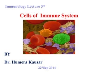


Cells of the immune system can be categorized as cells of the innate immune system or cells of the adaptive immune system. Cells of the innate immune system include phagocytes such as macrophages, neutrophils, dendritic cells, and basophils and mast cells. Cells of the adaptive immune system include lymphocytes such as B cells and T cells. B cells are involved in antibody production while T cells include cytotoxic T cells and helper T cells that activate other immune cells. Natural killer cells are also lymphocytes that help identify and destroy tumor or virus infected cells.


