The prostate is a conical gland that surrounds the urethra in males. It secretes fluid that is slightly acidic and contains substances like zinc that form part of semen. The prostate is located in the pelvis below the bladder and above the urethral sphincter. It has lobes including a median lobe and two lateral lobes. Structures like the urethra and ejaculatory ducts pass through it. The prostate receives blood supply from inferior vesical and internal pudendal arteries and drains into prostatic veins. It is innervated by sympathetic and parasympathetic nerves. The size and structure of the prostate changes with age. Diseases of the prostate include prostat
GROSS ANATOMY
OF PROSTATEGLAND
Dr. Nitin B Masaram
Assistant Professor
Dept. Of Anatomy
Dr. V. M. Govt. Medical College, Solapur
2.
• The Prostateis a conical Fibro-Musculo-Glandular
organ surrounding the proximal part of male urethra.
• Corresponds with paraurethral glands of female
developmentally.
• Secretion of Prostate forms considerable part of semen.
• Is slightly acidic, contains acid phosphatase,
fibrinolysin, prostaglandin and large amount of Zinc.
4.
• Situation:
Lesser pelvisbelow the neck of bladder, above
urogenital diaphragm, behind the lower part of
symphysis pubis, anterior to rectal ampulla and
on each side embraced by levator ani muscle.
• Measurements:
• Chest nut in appearance.
• Transverse (at base): 4 cm.
• Vertical: 3 cm
• Anteroposterior: 2 cm.
• Weight : 8 gm.
5.
Coverings of ProstateGland
• It is invested by Inner True capsule and Outer false
capsule.
• True Capsule: Intimately invest entire prostate and
formed by condensation of fibrous stroma of gland.
• False capsule or Prostatic sheath : derived from
visceral layer of pelvic fascia.
• At bladder neck false capsule is attached to the pubic
bones by medial and lateral Pubo-prostatic ligaments.
• Posteriorly , capsule blends with Rectovesical fascia of
Denonvillier.
• Space between true and false capsule is occupied by
Prostatic venous plexus, except at Posterior surface of
gland.
6.
Presenting Parts ofProstate gland
• Apex: Directed downwards, in contact with Superior fascia
of urogenital diaphragm.
• Base : Directed upwards, surrounding the neck of the
bladder, pierced by urethra in median plane at the junction
of ant. 1/3rd and post. 2/3rd of the gland.
• Anterior surface : narrow and convex and situated about
2cm behind lower part of symphysis pubis separated by
retropubic fat, prostatic venous plexus and deep dorsal vein
of penis.
• Posterior surface: broad, flat, related to the ampulla of
rectum separated by rectovesical fascia.
• This surface is palpable by rectal examination about 4 cm
above anus.
7.
• Posteror surfaceis subdivided by transverse groove
into upper small and lower large areas. It is pierced
by ejaculatory ducts on each side.
• The upper area forms median lobe; lower area is
subdivided by a median sulcus into two lateral
lobes.
• Each of two Infero-lateral surfaces, related to the
anterior fibres of levator ani which acts as levator
prostate; anterior recess of the ischio-rectal fossa
lies outside the levator ani.
9.
LOBES OFPROSTATE GLAND
Anatomically divided into 3 lobes: Median and 2 lateral
Surgically: 5 lobes: Median, anterior, posterior and two
lateral.
The median lobe: is wedge shaped, apex directed below
towards colliculus seminalis, base forms uvula vesicae at the
apex of trigonum vesicae.
Bounded anteriorly by urethra, behind and on each side by
the ejaculatory duct, behind and in the median plane by
prostatic utricle.
This lobe is predominantly fibro-muscular with mucus glands.
10.
The twolateral lobes are separated superficially by
posterior median sulcus , but deep to the sulcus and behind
urethra both the lobes are continuous.
This continuity is described as posterior lobe surgically.
Each lateral lobe covers sides of urethra and in front of
urethra are connected by fibro-muscular isthmus,which is
known as anterior lobe in foetal life containing glands.(may
persist upto 6 years after birth)
12.
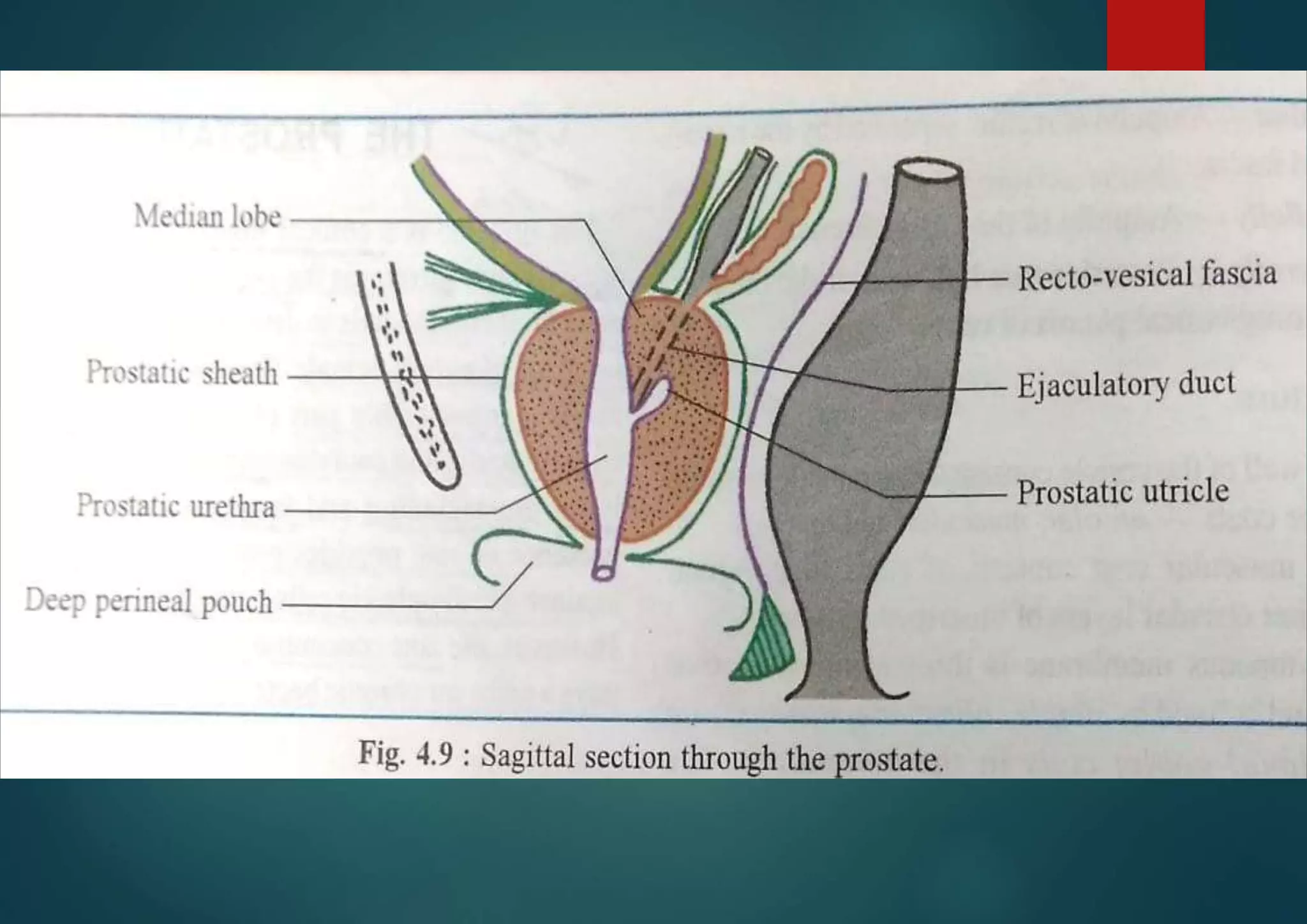
Structures traversingthe Prostate gland
Prostatic urethra: runs vertically downwards from
base to slightly in front of apex, at the junction of ant.
1/3rd and post. 2/3rd of gland
Pair of ejaculatory ducts: each passes postero-lateral
to median lobe, opens at the colliculus on each side of
prostatic utricle.
Prostatic utricle: is mucus cul –de- sac, about 6mm
long, extends upwards, and backwards from colliculus
behind the median lobe.
13.
STRUCTURE OFPROSTATE GLAND
Consist of 1/4th fibrous, 1/4th muscular and 2/4th glandular
tissue.
Fibrous tissue: forms true capsule at the periphery, postero-
median fibrous septum connects capsule with urethral
crest.
Muscular tissue: smooth muscle, continuous with detrusor
muscle, arranged in outer and inner sheets, connected by
radiating fibres. Spaces between these fibres occupied by
follicles of the gland.
There are transversely oriented arched striated muscle
fibres within the prostate anterior to the urethra, blending
with fibrous capsule postero-laterally and with
posteromedian septum.
14.
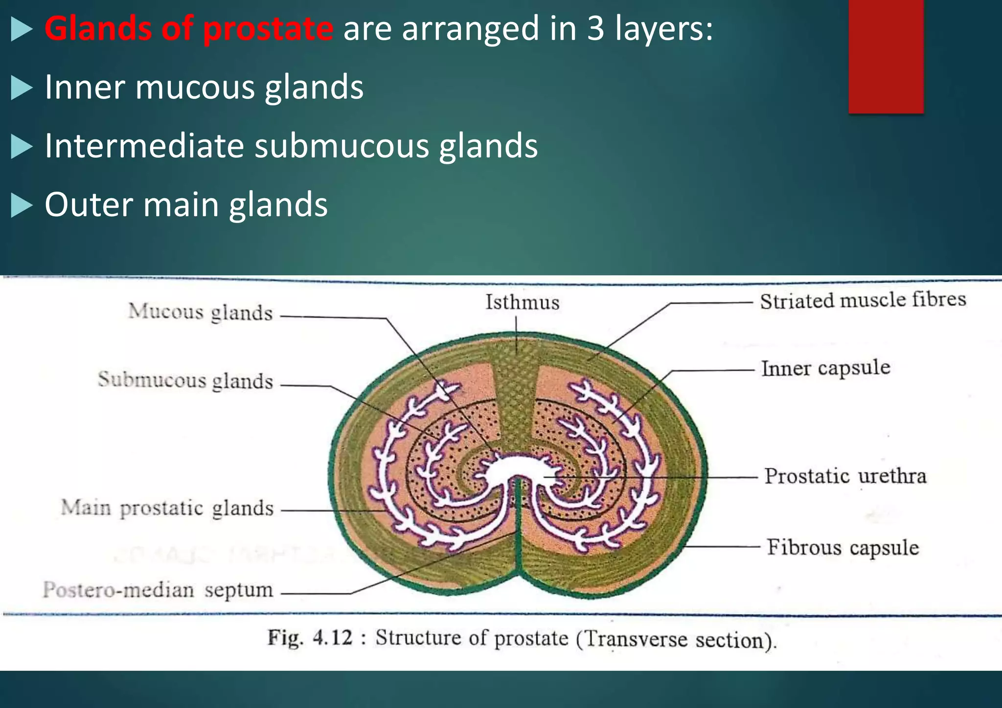
Glands ofprostate are arranged in 3 layers:
Inner mucous glands
Intermediate submucous glands
Outer main glands
15.
Arterial supply: 1. inferior vesical artery
2. Middle rectal artery
3. internal pudendal arteries
Venous drainage: veins form prostatic plexus in between
true and false capsules. Plexus receives deep dorsal vein of
penis anteriorly and communicates above with the vesical
venous plexus. Finally draining into internal iliac vein.
Few veins from prostate pass backwards through anterior
sacral foramina , draining into internal vertebral venous
plexus, known as paravertebral veins of Batson. (metastatic
spread of cancer prostate to vertebrae)
16.
Lymphatic drainage:1. internal iliac group of lymph nodes.
2. external iliac group of lymph nodes
3. sacral group of lymph nodes.
Nerve supply: 1. superior Hypogastric plexus conveys
sympathetic nerves(L1,L2 preganglionic
fibres)
2. Parasympathetic fibres derived from pelvic
spanchnic nerves conveying preganglionic
fibres from S2, S3, and S4.(secretomotor to
gland)
17.
AGE CHANGESIN PROSTATE GLAND
In newborn: consist basically of duct system in fibro-
muscular stroma. Before puberty grows slowly and
rudimentary follicles bud out from sides of ducts.
At Puberty: shows sudden growth, doubles in size. Follicles
shows infoldings. Above 45 years age mucous folds
disappears and follicles contains corpora amylacea.
In old age: It may atrophy or show hypertrophy.
18.
APPLIED ANATOMY
Prostatitis
Benign hypertrophy of prostate.
Prostatectomy: i) Suprapubic approach
ii) Transurethral resection of prostate
(TURP)
Carcinoma of Prostate.
Digital per rectal examination.
19.
References:
Essentialsof Human Anatomy(Thorax and abdomen) by A K Dutta.
Human Anatomy Vol.2 BD Chaurasia.
Thank You!!!!!!!!!