Downloaded 272 times


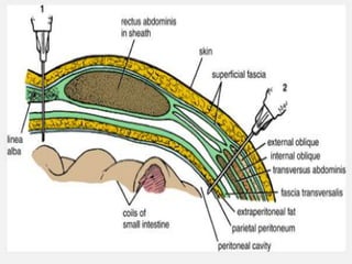
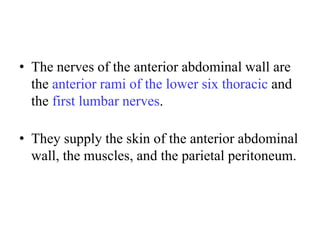
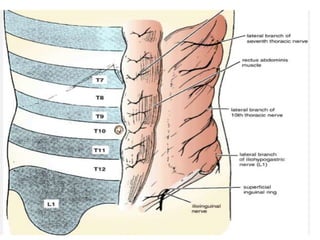
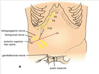
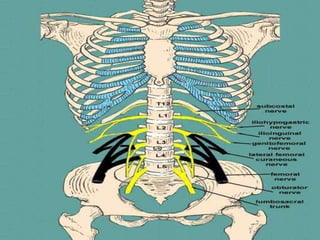

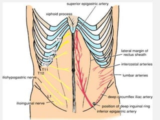

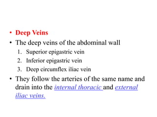


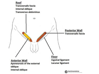
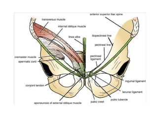
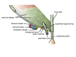
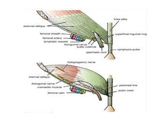
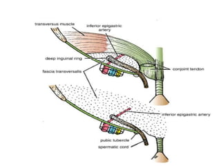
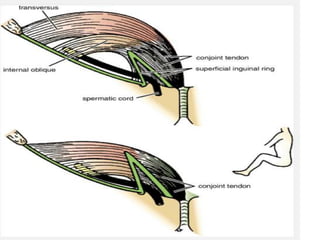
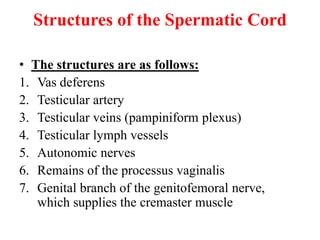
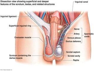
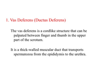
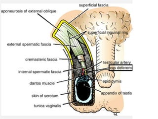
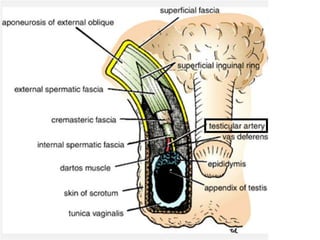
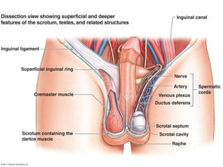
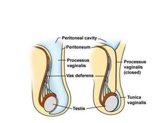
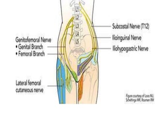

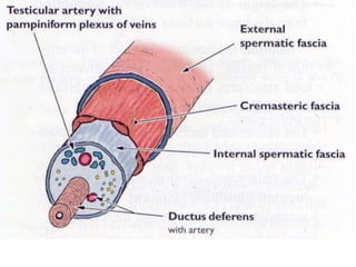
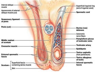
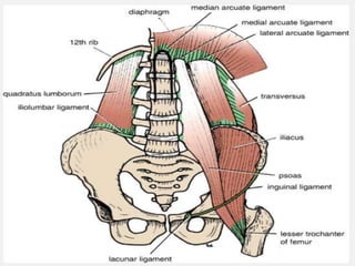


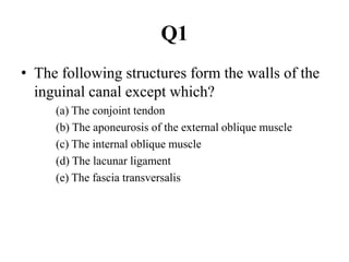
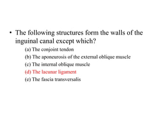
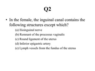
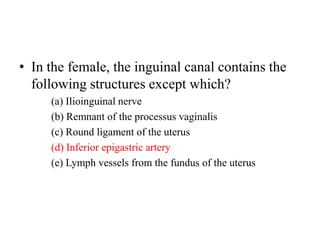
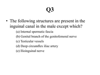
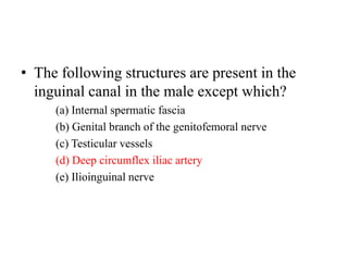


The document describes the anatomy of the anterior abdominal wall, including nerves, arteries, veins, and lymph drainage. It also discusses the inguinal canal, spermatic cord, and posterior abdominal wall. Key points include: - The anterior abdominal wall is supplied by thoracic and lumbar nerves and arteries like the superior and inferior epigastric arteries. - The inguinal canal allows structures like the spermatic cord to pass from the abdomen into the scrotum in males. It has walls formed by muscles like the internal oblique. - The spermatic cord contains structures like the vas deferens, testicular vessels, and remnants of the processus vaginalis in males.


![ANATOMY OF THE LOWER URINARY TRACT AND MALE [Autosaved] [Autosaved].pptx](https://cdn.slidesharecdn.com/ss_thumbnails/anatomyofthelowerurinarytractandmaleautosavedautosaved-240526080531-9d6371e3-thumbnail.jpg?width=640&height=640&fit=bounds)