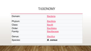
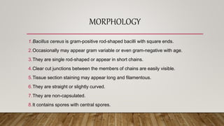
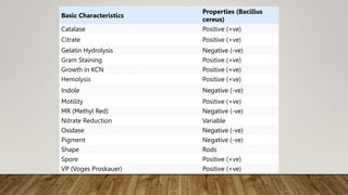
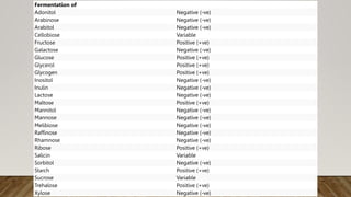
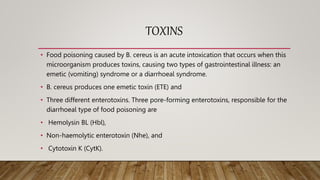
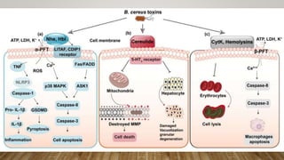
MICROBIOLOGY
BACILLUS CEREUS
MADE BY :- HARSHIKA RIZANI 226A
SUBMITTED TO :- PROFESSOR KOSTINA YULIA
CONTENTS
• INTRODUCTION 3
• TAXONOMY 5
• MORPHOLOGY 6
• CULTURAL PROPERTIES 8
• BIOCHEMICAL PROPERTIES 13
• TOXINS 18
• VIRULENCE FACTORS 20
• EPIDEMIOLOGY 22
• CLINICAL SYMPTOMS 24
• LABORATORY DIAGNOSTICS 25
• TREATMENT 27
• PREVENTION AND CONTROL 28
• REFERENCES 29
INTRODUCTION
• Bacillus cereus is a Gram-positive rod-shaped bacterium commonly found in soil, food, and
marine sponges.
• The specific name, cereus, meaning "waxy" in Latin, refers to the appearance of colonies grown
on blood agar.
• Some strains are harmful to humans and cause foodborne illness due to their spore-forming
nature, while other strains can be beneficial as probiotics for animals, and even exhibit
mutualism with certain plants.
• B. cereus bacteria may be anaerobes or facultative anaerobes, and like other members of the
genus Bacillus, can produce protective endospores.
• They have a wide range of virulence factors, including phospholipase
C, cereulide, sphingomyelinase, metalloproteases, and cytotoxin K,etc.
• B. cereus strains exhibit flagellar motility.
TAXONOMY
Domain: Bacteria
Phylum: Bacillota
Class: Bacilli
Order: Bacillales
Family: Bacillaceae
Genus: Bacillus
Species: B. cereus
MORPHOLOGY
1.Bacillus cereus is gram-positive rod-shaped bacilli with square ends.
2.Occasionally may appear gram variable or even gram-negative with age.
3.They are single rod-shaped or appear in short chains.
4.Clear cut junctions between the members of chains are easily visible.
5.Tissue section staining may appear long and filamentous.
6.They are straight or slightly curved.
7.They are non-capsulated.
8.It contains spores with central spores.
1.Spores are oval (ellipsoidal) and not formed in the animal’s blood and tissues or
in aerobic culture.
2.It is 1×3-4 µm in size.
3.It is motile and flagellated with peritrichous flagella.
4.Endospores are able to survive long periods of exposure to air and other adverse
environmental conditions.
5.It is a beta-hemolytic bacterium that causes foodborne disease.
6.Its virulence factors include cerolysin and phospholipase.
CULTURAL PROPERTIES
1.Growth on 5% sheep blood agar, chocolate agar, routine blood culture media, and
nutrient broths.
2.Detectable growth within 24 hours following incubation on media incubated at 35° C,
in ambient air, or in 5% carbon dioxide (CO2).
3.Colony character on blood agar: Large, feathery, spreading, dull, gray, granular,
spreading colonies and opaque with a rough matted surface and irregular perimeters,
beta-hemolytic.
4.Bacillus cereus can be isolated from feces by using selective media such as MYPA
(mannitol, egg yolk, polymyxin, phenol red, and agar), PEMBA (polymyxin, egg yolk,
mannitol, bromothymol, blue agar).
5.These media take advantage of the phospholipase C positive reaction on egg yolk
agar, no production of acid from mannitol, and incorporation of pyruvate or polymyxin
as the selective agents.
1.Most Bacillus spp grow readily on nutrient agar or peptone media.
2.The optimum temperature for growth varies from 20°C to 40°C, mostly 37°C.
3.B. cereus is mesophilic and is capable of adapting to a wide range of
environmental conditions.
4.On Nutrient Agar at 37°C, it forms large (2-5 mm) grey-white, granular colonies
with a less wavy edge and less membranous consistency.
5.On 5% sheep blood agar at 37°C, B. cereus colonies are large, feathery, dull,
gray, granular, spreading colonies, and opaque with a rough matted surface and
irregular perimeters.
1.On blood agar, it is beta-hemolytic.
2.In some instances, smooth colonies develop either alone or in the midst of rough
colonies.
3.When grown apart from the initial inoculum, smooth colonies are surrounded by a
uniform zone of beta-hemolysis framing the centrally situated colony.
4.The MYP (Mannitol egg Yolk Polymyxin) agar has been the standard media for
plating B. cereus, but it has little selectivity so background flora is not inhibited
and can mask the presence of B. cereus.
5.B. cereus colonies are usually lecithinase-positive and mannitol-negative on MYP
agar.
1.Bacara is a chromogenic selective and differential agar that promotes the growth
and identification of B. cereus but inhibits the growth of background flora.
2.Bacillus cereus colonies turn pink-orange with an opaque halo.
3.The chromogenic agar has been suggested for the enumeration of the B.
cereus group as a substitute for MYP.
4.Typical colonies will grow as pink-orange uniform colonies surrounded by a zone
of precipitation.
BIOCHEMICAL PROPERTIES
1.Catalase: positive
2.Oxidase: negative
3.OF test: fermentative
4.Indole: negative
5.Methyl red: positive
6.Vogues proskauer: positive
7.Glucose: fermentative, production of acid
8.Sucrose: fermentative, production of acid
9.Lactose: no fermentation
10.Starch hydrolysis: positive
11.Nitrate reduction: positive
12.Gelatin hydrolysis: positive
13.Spore staining: endospore-forming bacteria
14.Motility: motile
Basic Characteristics
Properties (Bacillus
cereus)
Catalase Positive (+ve)
Citrate Positive (+ve)
Gelatin Hydrolysis Negative (-ve)
Gram Staining Positive (+ve)
Growth in KCN Positive (+ve)
Hemolysis Positive (+ve)
Indole Negative (-ve)
Motility Positive (+ve)
MR (Methyl Red) Negative (-ve)
Nitrate Reduction Variable
Oxidase Negative (-ve)
Pigment Negative (-ve)
Shape Rods
Spore Positive (+ve)
VP (Voges Proskauer) Positive (+ve)
Fermentation of
Adonitol Negative (-ve)
Arabinose Negative (-ve)
Arabitol Negative (-ve)
Cellobiose Variable
Fructose Positive (+ve)
Galactose Negative (-ve)
Glucose Positive (+ve)
Glycerol Positive (+ve)
Glycogen Positive (+ve)
Inositol Negative (-ve)
Inulin Negative (-ve)
Lactose Negative (-ve)
Maltose Positive (+ve)
Mannitol Negative (-ve)
Mannose Negative (-ve)
Melibiose Negative (-ve)
Raffinose Negative (-ve)
Rhamnose Negative (-ve)
Ribose Positive (+ve)
Salicin Variable
Sorbitol Negative (-ve)
Starch Positive (+ve)
Sucrose Variable
Trehalose Positive (+ve)
Xylose Negative (-ve)
Enzymatic Reactions
Acetate Utilization Variable
Arginine Dehydrolase Variable
Casein Hydrolysis Positive (+ve)
Esculin Hydrolysis Positive (+ve)
Lecithinase Positive (+ve)
Lysine Negative (-ve)
Ornithine Decarboxylase Negative (-ve)
Phenylalanine Deaminase Negative (-ve)
Tyrosine Hydrolysis Positive (+ve)
TOXINS
• Food poisoning caused by B. cereus is an acute intoxication that occurs when this
microorganism produces toxins, causing two types of gastrointestinal illness: an
emetic (vomiting) syndrome or a diarrhoeal syndrome.
• B. cereus produces one emetic toxin (ETE) and
• Three different enterotoxins. Three pore-forming enterotoxins, responsible for the
diarrhoeal type of food poisoning are
• Hemolysin BL (Hbl),
• Non-haemolytic enterotoxin (Nhe), and
• Cytotoxin K (CytK).
VIRULENCE FACTORS
• Bacillus cereus is mainly known for causing food poisoning and
severe non-gastrointestinal tract infections.
• The intestinal and non intestinal pathogenicity of this microorganism
is mainly due to the synergistic effects of a number of virulence
products that promote intestinal cell destruction and/or resistance to
the host immune system.
• The various substances produced by B. cereus are
mainly enterotoxins, hemolysins, phospholipases, and emetic toxin,
whose activity may overlap in causing human disease.
• Virulence factors of B. cereus include
• Cytotoxin K,
• Cereulide,
• Metalloproteases,
• Sphingomyelinase, and
• Phospholipases
EPIDEMIOLOGY
• The natural environmental reservoir for B. cereus consists of decaying organic
matter, fresh and marine waters, vegetables and fomites, and the intestinal tract
of invertebrates, from which soil and food products may become contaminated,
leading to the transient colonization of the human intestine.
• Spores germinate when they come into contact with organic matter or within an
insect or animal host.
• A multicellular filamentous growth pattern containing refractile inclusions, termed
arthromitus (rooted), has been observed in the guts of certain arthropods, which
is regarded as the normal intestinal stage in soil-dwelling insects.
• In this setting, as long rod-shaped bacteria, the bacilli lose their flagella, attach to
the arthropod intestinal epithelium, and sporulate .
• B. cereus also has a saprophytic life cycle in which spores germinate in soil, with
the production of a vegetative bacillus, which could then sporulate, maintaining
the life cycle .
• Defecation by or death of the host releases cells and spores into the soil, where
vegetative cells may sporulate and survive until their uptake by another host .
• Furthermore, when B. cereus grows in soil, it undergoes a switching from a
single-cell to a multicellular phenotype, which allows it to translocate through the
soil .
CLINICAL SYMPTOMS
• B. cereus causes self-limiting (24-48 hours) food-poisoning syndromes (a diarrheal
type and an emetic type), opportunistic infections and is associated with clinical
infections such as endophthalmitis and other ocular infections.
• The diarrheal form of B. cereus food poisoning is characterized by abdominal cramps,
profuse watery diarrhea, and rectal tenesmus, and, occasionally, fever and vomiting.
• The emetic form of B. cereus food poisoning is characterized by nausea, vomiting, and
malaise, occasionally with diarrhea.
• B. cereus can cause wound infections, bacteremia, septicaemia, meningitis,
pneumonia, central nervous system infections, endocarditis, pericarditis, respiratory
infections, and peripheral infections.
• Infection in immunocompromised individuals can be life-threatening.
LABORATORY DIAGNOSTICS
• SPECIMENS: Faeces, vomitus, remaining food (if any), eye specimen
(corneal swab)
• DIRECT DETECTION METHODS
1.Microscopically the organisms appear as large gram-positive rods in
singles, pairs, or serpentine with square ends after Gram staining.
2.Endospores formation are seen as an unstained oval or round region
within the center of the cell. Spores are oval (ellipsoidal) and not swelling
of the mother cell.
• SERODIAGNOSIS
1.Serologic methods are available for the detection of B. cereus toxin
in food and feces.
2.Microslide gel diffusion test is generally used as a toxin detection
system.
• MOLECULAR METHOD
1.The toxigenic potential of B. cereus isolates, genes encoding
emetic-toxin cereulide (ces), and enterotoxins (nhe, hbl and cytK)
can be analyzed by multiplex PCR.
TREATMENT
1.Persons with B. cereus food poisoning require only supportive
treatment.
2.Oral rehydration or, occasionally, intravenous fluid and electrolyte
replacement for patients with severe dehydration is indicated.
Antibiotics are not indicated.
3.Patients with the invasive disease require antibiotic
therapy. Bacillus cereus is susceptible to clindamycin, erythromycin,
vancomycin, aminoglycosides, and tetracycline. It is resistant to
penicillin and trimethoprim.
PREVENTION AND CONTROL
1.Diarrheal and vomiting intoxications by this organism are readily preventable by
appropriate food-handling procedures.
2.Meat and vegetables should not be held at temperatures between 10 and 45 °C
for long periods, and rice held overnight after cooking should be refrigerated and
not held at room temperature.
3.Prevention of infection in patients following surgery or in those who are
immunocompromised or who are otherwise predisposed to infection depends on
good practice.
REFERENCES
• https://microbenotes.com/bacillus-
cereus/#:~:text=is%20considered%20diagnostic.-
,Morphology%20of%20Bacillus%20cereus,of%20chains%20are%20easily%20visibl
e.
• https://microbenotes.com/bacillus-cereus/#:~:text=kb%20in%20size.-
,Cultural%20Characteristics%20of%20Bacillus%20cereus,wide%20range%20of%20
environmental%20conditions.
• https://microbiologyinfo.com/biochemical-test-identification-bacillus-cereus/
• https://www.ncbi.nlm.nih.gov/pmc/articles/PMC4397285/#:~:text=Toxins-
,B.,and%20Cytotoxin%20K%20(CytK).
• https://pubmed.ncbi.nlm.nih.gov/33004259/#:~:text=Other%20putative%20virule
nce%20factors%20of,metalloproteases%2C%20sphingomyelinase%2C%20and%2
0phospholipases.
• https://www.canada.ca/en/public-health/services/laboratory-biosafety-
biosecurity/pathogen-safety-data-sheets-risk-assessment/bacillus-cereus.html
• https://microbenotes.com/bacillus-cereus/
• https://en.wikipedia.org/wiki/Bacillus_cereus
THANK YOU !!!

Microbiology bacillus cereus.pptx

  • 1.
    MICROBIOLOGY BACILLUS CEREUS MADE BY:- HARSHIKA RIZANI 226A SUBMITTED TO :- PROFESSOR KOSTINA YULIA
  • 2.
    CONTENTS • INTRODUCTION 3 •TAXONOMY 5 • MORPHOLOGY 6 • CULTURAL PROPERTIES 8 • BIOCHEMICAL PROPERTIES 13 • TOXINS 18 • VIRULENCE FACTORS 20 • EPIDEMIOLOGY 22 • CLINICAL SYMPTOMS 24 • LABORATORY DIAGNOSTICS 25 • TREATMENT 27 • PREVENTION AND CONTROL 28 • REFERENCES 29
  • 3.
    INTRODUCTION • Bacillus cereusis a Gram-positive rod-shaped bacterium commonly found in soil, food, and marine sponges. • The specific name, cereus, meaning "waxy" in Latin, refers to the appearance of colonies grown on blood agar. • Some strains are harmful to humans and cause foodborne illness due to their spore-forming nature, while other strains can be beneficial as probiotics for animals, and even exhibit mutualism with certain plants. • B. cereus bacteria may be anaerobes or facultative anaerobes, and like other members of the genus Bacillus, can produce protective endospores. • They have a wide range of virulence factors, including phospholipase C, cereulide, sphingomyelinase, metalloproteases, and cytotoxin K,etc. • B. cereus strains exhibit flagellar motility.
  • 5.
    TAXONOMY Domain: Bacteria Phylum: Bacillota Class:Bacilli Order: Bacillales Family: Bacillaceae Genus: Bacillus Species: B. cereus
  • 6.
    MORPHOLOGY 1.Bacillus cereus isgram-positive rod-shaped bacilli with square ends. 2.Occasionally may appear gram variable or even gram-negative with age. 3.They are single rod-shaped or appear in short chains. 4.Clear cut junctions between the members of chains are easily visible. 5.Tissue section staining may appear long and filamentous. 6.They are straight or slightly curved. 7.They are non-capsulated. 8.It contains spores with central spores.
  • 7.
    1.Spores are oval(ellipsoidal) and not formed in the animal’s blood and tissues or in aerobic culture. 2.It is 1×3-4 µm in size. 3.It is motile and flagellated with peritrichous flagella. 4.Endospores are able to survive long periods of exposure to air and other adverse environmental conditions. 5.It is a beta-hemolytic bacterium that causes foodborne disease. 6.Its virulence factors include cerolysin and phospholipase.
  • 8.
  • 9.
    1.Growth on 5%sheep blood agar, chocolate agar, routine blood culture media, and nutrient broths. 2.Detectable growth within 24 hours following incubation on media incubated at 35° C, in ambient air, or in 5% carbon dioxide (CO2). 3.Colony character on blood agar: Large, feathery, spreading, dull, gray, granular, spreading colonies and opaque with a rough matted surface and irregular perimeters, beta-hemolytic. 4.Bacillus cereus can be isolated from feces by using selective media such as MYPA (mannitol, egg yolk, polymyxin, phenol red, and agar), PEMBA (polymyxin, egg yolk, mannitol, bromothymol, blue agar). 5.These media take advantage of the phospholipase C positive reaction on egg yolk agar, no production of acid from mannitol, and incorporation of pyruvate or polymyxin as the selective agents.
  • 10.
    1.Most Bacillus sppgrow readily on nutrient agar or peptone media. 2.The optimum temperature for growth varies from 20°C to 40°C, mostly 37°C. 3.B. cereus is mesophilic and is capable of adapting to a wide range of environmental conditions. 4.On Nutrient Agar at 37°C, it forms large (2-5 mm) grey-white, granular colonies with a less wavy edge and less membranous consistency. 5.On 5% sheep blood agar at 37°C, B. cereus colonies are large, feathery, dull, gray, granular, spreading colonies, and opaque with a rough matted surface and irregular perimeters.
  • 11.
    1.On blood agar,it is beta-hemolytic. 2.In some instances, smooth colonies develop either alone or in the midst of rough colonies. 3.When grown apart from the initial inoculum, smooth colonies are surrounded by a uniform zone of beta-hemolysis framing the centrally situated colony. 4.The MYP (Mannitol egg Yolk Polymyxin) agar has been the standard media for plating B. cereus, but it has little selectivity so background flora is not inhibited and can mask the presence of B. cereus. 5.B. cereus colonies are usually lecithinase-positive and mannitol-negative on MYP agar.
  • 12.
    1.Bacara is achromogenic selective and differential agar that promotes the growth and identification of B. cereus but inhibits the growth of background flora. 2.Bacillus cereus colonies turn pink-orange with an opaque halo. 3.The chromogenic agar has been suggested for the enumeration of the B. cereus group as a substitute for MYP. 4.Typical colonies will grow as pink-orange uniform colonies surrounded by a zone of precipitation.
  • 13.
  • 14.
    1.Catalase: positive 2.Oxidase: negative 3.OFtest: fermentative 4.Indole: negative 5.Methyl red: positive 6.Vogues proskauer: positive 7.Glucose: fermentative, production of acid 8.Sucrose: fermentative, production of acid 9.Lactose: no fermentation 10.Starch hydrolysis: positive 11.Nitrate reduction: positive 12.Gelatin hydrolysis: positive 13.Spore staining: endospore-forming bacteria 14.Motility: motile
  • 15.
    Basic Characteristics Properties (Bacillus cereus) CatalasePositive (+ve) Citrate Positive (+ve) Gelatin Hydrolysis Negative (-ve) Gram Staining Positive (+ve) Growth in KCN Positive (+ve) Hemolysis Positive (+ve) Indole Negative (-ve) Motility Positive (+ve) MR (Methyl Red) Negative (-ve) Nitrate Reduction Variable Oxidase Negative (-ve) Pigment Negative (-ve) Shape Rods Spore Positive (+ve) VP (Voges Proskauer) Positive (+ve)
  • 16.
    Fermentation of Adonitol Negative(-ve) Arabinose Negative (-ve) Arabitol Negative (-ve) Cellobiose Variable Fructose Positive (+ve) Galactose Negative (-ve) Glucose Positive (+ve) Glycerol Positive (+ve) Glycogen Positive (+ve) Inositol Negative (-ve) Inulin Negative (-ve) Lactose Negative (-ve) Maltose Positive (+ve) Mannitol Negative (-ve) Mannose Negative (-ve) Melibiose Negative (-ve) Raffinose Negative (-ve) Rhamnose Negative (-ve) Ribose Positive (+ve) Salicin Variable Sorbitol Negative (-ve) Starch Positive (+ve) Sucrose Variable Trehalose Positive (+ve) Xylose Negative (-ve)
  • 17.
    Enzymatic Reactions Acetate UtilizationVariable Arginine Dehydrolase Variable Casein Hydrolysis Positive (+ve) Esculin Hydrolysis Positive (+ve) Lecithinase Positive (+ve) Lysine Negative (-ve) Ornithine Decarboxylase Negative (-ve) Phenylalanine Deaminase Negative (-ve) Tyrosine Hydrolysis Positive (+ve)
  • 18.
    TOXINS • Food poisoningcaused by B. cereus is an acute intoxication that occurs when this microorganism produces toxins, causing two types of gastrointestinal illness: an emetic (vomiting) syndrome or a diarrhoeal syndrome. • B. cereus produces one emetic toxin (ETE) and • Three different enterotoxins. Three pore-forming enterotoxins, responsible for the diarrhoeal type of food poisoning are • Hemolysin BL (Hbl), • Non-haemolytic enterotoxin (Nhe), and • Cytotoxin K (CytK).
  • 20.
    VIRULENCE FACTORS • Bacilluscereus is mainly known for causing food poisoning and severe non-gastrointestinal tract infections. • The intestinal and non intestinal pathogenicity of this microorganism is mainly due to the synergistic effects of a number of virulence products that promote intestinal cell destruction and/or resistance to the host immune system. • The various substances produced by B. cereus are mainly enterotoxins, hemolysins, phospholipases, and emetic toxin, whose activity may overlap in causing human disease.
  • 21.
    • Virulence factorsof B. cereus include • Cytotoxin K, • Cereulide, • Metalloproteases, • Sphingomyelinase, and • Phospholipases
  • 22.
    EPIDEMIOLOGY • The naturalenvironmental reservoir for B. cereus consists of decaying organic matter, fresh and marine waters, vegetables and fomites, and the intestinal tract of invertebrates, from which soil and food products may become contaminated, leading to the transient colonization of the human intestine. • Spores germinate when they come into contact with organic matter or within an insect or animal host. • A multicellular filamentous growth pattern containing refractile inclusions, termed arthromitus (rooted), has been observed in the guts of certain arthropods, which is regarded as the normal intestinal stage in soil-dwelling insects.
  • 23.
    • In thissetting, as long rod-shaped bacteria, the bacilli lose their flagella, attach to the arthropod intestinal epithelium, and sporulate . • B. cereus also has a saprophytic life cycle in which spores germinate in soil, with the production of a vegetative bacillus, which could then sporulate, maintaining the life cycle . • Defecation by or death of the host releases cells and spores into the soil, where vegetative cells may sporulate and survive until their uptake by another host . • Furthermore, when B. cereus grows in soil, it undergoes a switching from a single-cell to a multicellular phenotype, which allows it to translocate through the soil .
  • 24.
    CLINICAL SYMPTOMS • B.cereus causes self-limiting (24-48 hours) food-poisoning syndromes (a diarrheal type and an emetic type), opportunistic infections and is associated with clinical infections such as endophthalmitis and other ocular infections. • The diarrheal form of B. cereus food poisoning is characterized by abdominal cramps, profuse watery diarrhea, and rectal tenesmus, and, occasionally, fever and vomiting. • The emetic form of B. cereus food poisoning is characterized by nausea, vomiting, and malaise, occasionally with diarrhea. • B. cereus can cause wound infections, bacteremia, septicaemia, meningitis, pneumonia, central nervous system infections, endocarditis, pericarditis, respiratory infections, and peripheral infections. • Infection in immunocompromised individuals can be life-threatening.
  • 25.
    LABORATORY DIAGNOSTICS • SPECIMENS:Faeces, vomitus, remaining food (if any), eye specimen (corneal swab) • DIRECT DETECTION METHODS 1.Microscopically the organisms appear as large gram-positive rods in singles, pairs, or serpentine with square ends after Gram staining. 2.Endospores formation are seen as an unstained oval or round region within the center of the cell. Spores are oval (ellipsoidal) and not swelling of the mother cell.
  • 26.
    • SERODIAGNOSIS 1.Serologic methodsare available for the detection of B. cereus toxin in food and feces. 2.Microslide gel diffusion test is generally used as a toxin detection system. • MOLECULAR METHOD 1.The toxigenic potential of B. cereus isolates, genes encoding emetic-toxin cereulide (ces), and enterotoxins (nhe, hbl and cytK) can be analyzed by multiplex PCR.
  • 27.
    TREATMENT 1.Persons with B.cereus food poisoning require only supportive treatment. 2.Oral rehydration or, occasionally, intravenous fluid and electrolyte replacement for patients with severe dehydration is indicated. Antibiotics are not indicated. 3.Patients with the invasive disease require antibiotic therapy. Bacillus cereus is susceptible to clindamycin, erythromycin, vancomycin, aminoglycosides, and tetracycline. It is resistant to penicillin and trimethoprim.
  • 28.
    PREVENTION AND CONTROL 1.Diarrhealand vomiting intoxications by this organism are readily preventable by appropriate food-handling procedures. 2.Meat and vegetables should not be held at temperatures between 10 and 45 °C for long periods, and rice held overnight after cooking should be refrigerated and not held at room temperature. 3.Prevention of infection in patients following surgery or in those who are immunocompromised or who are otherwise predisposed to infection depends on good practice.
  • 29.
    REFERENCES • https://microbenotes.com/bacillus- cereus/#:~:text=is%20considered%20diagnostic.- ,Morphology%20of%20Bacillus%20cereus,of%20chains%20are%20easily%20visibl e. • https://microbenotes.com/bacillus-cereus/#:~:text=kb%20in%20size.- ,Cultural%20Characteristics%20of%20Bacillus%20cereus,wide%20range%20of%20 environmental%20conditions. •https://microbiologyinfo.com/biochemical-test-identification-bacillus-cereus/ • https://www.ncbi.nlm.nih.gov/pmc/articles/PMC4397285/#:~:text=Toxins- ,B.,and%20Cytotoxin%20K%20(CytK). • https://pubmed.ncbi.nlm.nih.gov/33004259/#:~:text=Other%20putative%20virule nce%20factors%20of,metalloproteases%2C%20sphingomyelinase%2C%20and%2 0phospholipases.
  • 30.
  • 31.