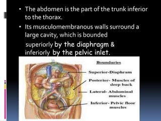
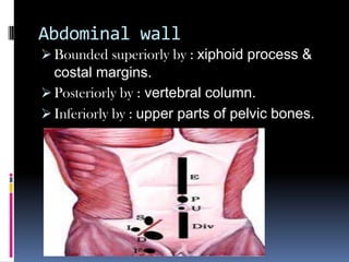
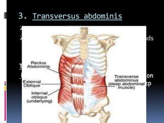
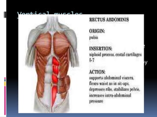
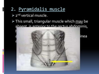
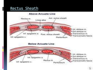
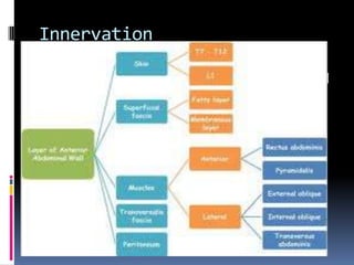
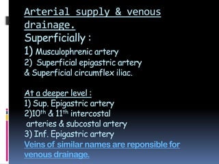
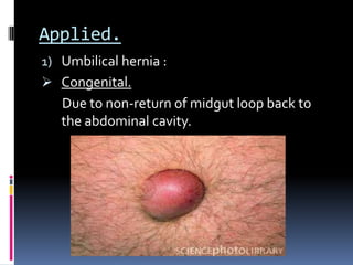
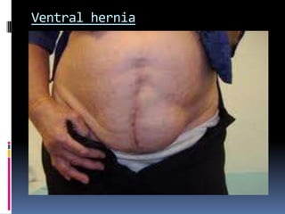
PRESENTED BY : SUNTOO IRMA
ROLL NO : 102
DATE : 21/09/12
ANTEROLATERAL ABDOMINAL
WALL.
Table of Content
 Introduction to Abdomen
 Abdominal wall
 Anterolateral muscles
 Innervation
 Arterial supply &Venous drainage
 Lymphatic drainage
 Applied
 References
• The abdomen is the part of the trunk inferior
to the thorax.
• Its musculomembranous walls surround a
large cavity, which is bounded
superiorly by the diaphragm &
inferiorly by the pelvic inlet.
Abdominal wall
 Bounded superiorly by : xiphoid process &
costal margins.
 Posteriorly by : vertebral column.
 Inferiorly by : upper parts of pelvic bones.
Anterolateral Muscles
o There are 5 muscles in the anterolateral
group of abdominal wall muscles.
o 3 flat muscles whose fibres begin
posterolaterally, pass anteriorly, & are
replaced by an aponeurosis as the muscle
continues towards the midline.
1. External oblique
2. Internal oblique
3. Transversus abdominis
o 2 vertical muscles, near the mildline which
are enclosed within a tendinous sheath
formed by aponeuroses of flat muscles.
1. Rectus abdominis
2. Pyramidalis.
1. External oblique muscle:
 Most superficial out of the three flat muscles.
 Immediately superficial to superficial
fascia.
 Its laterally placed muscle fibres pass in an
inferomedial direction,while its large
aponeurotic component covers the anterior part
to the midline.
 At the midline, aponeuroses are entwined
forming the linea alba, which extends from
xiphoid process to pubic symphysis.
2. Internal oblique muscle
 Deep to external oblique muscle.
 2nd of the 3 flat muscles.
 Most of its muscle fibres pass in a
superolateral direction.
 Its lat muscular components end anteriorly as
an aponeurosis that blends into linea alba at
the midline.
3. Transversus abdominis
 Deep to internal oblique.
 Ends in an anterior aponeurosis, which blends
with the linea alba at the midline.
Transversalis fascia
Each of the three flat muscles is covered on
the ant & post surfaces by a layer of deep
fascia. Only the layer deep to the
transversus abdominis is remarkable.
Vertical muscles
1. Rectus abdominis
 long, flat muscle and extends the length of the
anterior abd wall.
 It is a paired muscle, separated in the midline by
linea alba.
 It widens & thins as it ascends from the pubic
symphysis to costal margin.
2. Pyramidalis muscle
 2nd vertical muscle.
 This small, triangular muscle which may be
absent, is anterior to the rectus abdominis,
has its base on the pubis, and apex is
attached superiorly & medially to the linea
alba.
Rectus Sheath
 Aponeurotic sheath covering the rectus
abdominis.
 Completely encloses the upper ¾ of rectus
abdominis & covers the anterior surface of
lower ¼ of the muscle
 The posterior surface of lower quarter of
rectus abdominis is in direct contact with the
transversalis fascia.
Innervation
 The skin, muscles and parietal peritoneum of
the anterolateral abdominal wall are supplied
byT7 toT12 and L1 spinal nerves.
 NervesT7 toT9 supply skin from xiphoid
process to just above umbilicus.
 T10 supplies skin around umbilicus.
 T11,T12 &L1 supply skin from just below the
umbilicus to, and including the pubic region.
Arterial supply & venous
drainage.
Superficially :
1) Musculophrenic artery
2) Superficial epigastric artery
& Superficial circumflex iliac.
At a deeper level :
1) Sup. Epigastric artery
2)10th & 11th intercostal
arteries & subcostal artery
3) Inf. Epigastric artery
Veins of similar names are reponsible for
venous drainage.
Lymphatic drainage
 Superficial lymphatics :
1) Axillary nodes
2) Superficial inguinal nodes
 Deep lymphatic drainage follows the deep
arteries back to parasternal nodes along with
internal thoracic artery.
Applied.
1) Umbilical hernia :
 Congenital.
Due to non-return of midgut loop back to
the abdominal cavity.
2)Acquired infantile umbilical
hernia.
 Due to weakness of umbilical scar, a part of
the gut may be seen protruding out. It
disappears as the infant grows.
3) Paraumbilical hernia
 Loop of intestine protrude through the linea
alba around the region of umbilicus.
4) Femoral hernia
 Occurs more in females due to larger pelvis,
smaller blood vessels and larger femoral
canal.
 Surgery is essential for its treatment.
5) Epigastric hernia
 Occurs through the upper part of wide linea
alba.
6) Incisional hernia
 Occurs through the anterolateral abdominal
wall when incisions are made for the surgery,
involving cutting of spinal nerves.
•Internal Hernia
 Remnants of the vitellointestinal duct may
form a tumour at the umbilicus (raspberry red
tumour)
 Persistance of a patent vitellointestine duct
results in a faecal fistula at the umbilicus.
Ventral hernia
 Supraumbilical median incisions through
the linea alba have various advantages
as being
1) Bloodless
2) Safety to muscles & nerves
However, It tends to leave a
postoperative weakness through which
a ventral hernia may develop.
Infraumbilical Median Incision
 Safer, because the close approx. of recti
prevents formation of any ventral hernia.
 Paramedian incisions are more sound than
median incisions.
 The rectus muscle is retracted laterally to
protect the nerves supplying it from any
injury.
References:
 GRAY’S ANATOMY.
 Wikipedia.
 B D Chaurasia’s Human Anatomy.
Anterolateral abdominal wall (rectus sheath) & hernia

Anterolateral abdominal wall (rectus sheath) & hernia

  • 1.
    PRESENTED BY :SUNTOO IRMA ROLL NO : 102 DATE : 21/09/12 ANTEROLATERAL ABDOMINAL WALL.
  • 2.
    Table of Content Introduction to Abdomen  Abdominal wall  Anterolateral muscles  Innervation  Arterial supply &Venous drainage  Lymphatic drainage  Applied  References
  • 3.
    • The abdomenis the part of the trunk inferior to the thorax. • Its musculomembranous walls surround a large cavity, which is bounded superiorly by the diaphragm & inferiorly by the pelvic inlet.
  • 4.
    Abdominal wall  Boundedsuperiorly by : xiphoid process & costal margins.  Posteriorly by : vertebral column.  Inferiorly by : upper parts of pelvic bones.
  • 5.
    Anterolateral Muscles o Thereare 5 muscles in the anterolateral group of abdominal wall muscles. o 3 flat muscles whose fibres begin posterolaterally, pass anteriorly, & are replaced by an aponeurosis as the muscle continues towards the midline. 1. External oblique 2. Internal oblique 3. Transversus abdominis
  • 6.
    o 2 verticalmuscles, near the mildline which are enclosed within a tendinous sheath formed by aponeuroses of flat muscles. 1. Rectus abdominis 2. Pyramidalis.
  • 7.
    1. External obliquemuscle:  Most superficial out of the three flat muscles.  Immediately superficial to superficial fascia.  Its laterally placed muscle fibres pass in an inferomedial direction,while its large aponeurotic component covers the anterior part to the midline.  At the midline, aponeuroses are entwined forming the linea alba, which extends from xiphoid process to pubic symphysis.
  • 8.
    2. Internal obliquemuscle  Deep to external oblique muscle.  2nd of the 3 flat muscles.  Most of its muscle fibres pass in a superolateral direction.  Its lat muscular components end anteriorly as an aponeurosis that blends into linea alba at the midline.
  • 9.
    3. Transversus abdominis Deep to internal oblique.  Ends in an anterior aponeurosis, which blends with the linea alba at the midline. Transversalis fascia Each of the three flat muscles is covered on the ant & post surfaces by a layer of deep fascia. Only the layer deep to the transversus abdominis is remarkable.
  • 10.
    Vertical muscles 1. Rectusabdominis  long, flat muscle and extends the length of the anterior abd wall.  It is a paired muscle, separated in the midline by linea alba.  It widens & thins as it ascends from the pubic symphysis to costal margin.
  • 11.
    2. Pyramidalis muscle 2nd vertical muscle.  This small, triangular muscle which may be absent, is anterior to the rectus abdominis, has its base on the pubis, and apex is attached superiorly & medially to the linea alba.
  • 12.
    Rectus Sheath  Aponeuroticsheath covering the rectus abdominis.  Completely encloses the upper ¾ of rectus abdominis & covers the anterior surface of lower ¼ of the muscle  The posterior surface of lower quarter of rectus abdominis is in direct contact with the transversalis fascia.
  • 13.
    Innervation  The skin,muscles and parietal peritoneum of the anterolateral abdominal wall are supplied byT7 toT12 and L1 spinal nerves.  NervesT7 toT9 supply skin from xiphoid process to just above umbilicus.  T10 supplies skin around umbilicus.  T11,T12 &L1 supply skin from just below the umbilicus to, and including the pubic region.
  • 14.
    Arterial supply &venous drainage. Superficially : 1) Musculophrenic artery 2) Superficial epigastric artery & Superficial circumflex iliac. At a deeper level : 1) Sup. Epigastric artery 2)10th & 11th intercostal arteries & subcostal artery 3) Inf. Epigastric artery Veins of similar names are reponsible for venous drainage.
  • 15.
    Lymphatic drainage  Superficiallymphatics : 1) Axillary nodes 2) Superficial inguinal nodes  Deep lymphatic drainage follows the deep arteries back to parasternal nodes along with internal thoracic artery.
  • 16.
    Applied. 1) Umbilical hernia:  Congenital. Due to non-return of midgut loop back to the abdominal cavity.
  • 17.
    2)Acquired infantile umbilical hernia. Due to weakness of umbilical scar, a part of the gut may be seen protruding out. It disappears as the infant grows.
  • 18.
    3) Paraumbilical hernia Loop of intestine protrude through the linea alba around the region of umbilicus.
  • 19.
    4) Femoral hernia Occurs more in females due to larger pelvis, smaller blood vessels and larger femoral canal.  Surgery is essential for its treatment.
  • 20.
    5) Epigastric hernia Occurs through the upper part of wide linea alba.
  • 22.
    6) Incisional hernia Occurs through the anterolateral abdominal wall when incisions are made for the surgery, involving cutting of spinal nerves.
  • 23.
    •Internal Hernia  Remnantsof the vitellointestinal duct may form a tumour at the umbilicus (raspberry red tumour)  Persistance of a patent vitellointestine duct results in a faecal fistula at the umbilicus.
  • 24.
    Ventral hernia  Supraumbilicalmedian incisions through the linea alba have various advantages as being 1) Bloodless 2) Safety to muscles & nerves However, It tends to leave a postoperative weakness through which a ventral hernia may develop.
  • 25.
    Infraumbilical Median Incision Safer, because the close approx. of recti prevents formation of any ventral hernia.  Paramedian incisions are more sound than median incisions.  The rectus muscle is retracted laterally to protect the nerves supplying it from any injury.
  • 26.
    References:  GRAY’S ANATOMY. Wikipedia.  B D Chaurasia’s Human Anatomy.