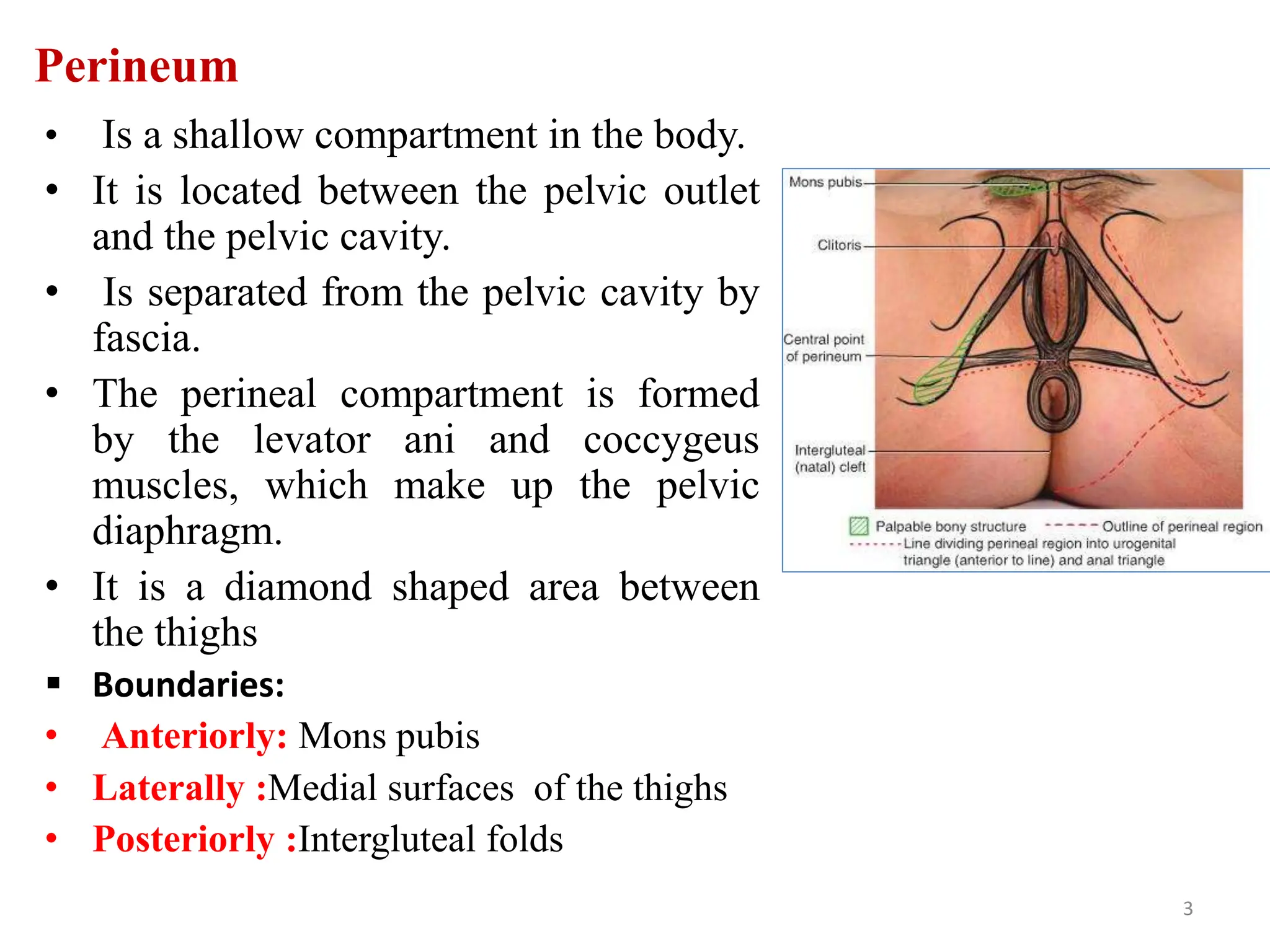
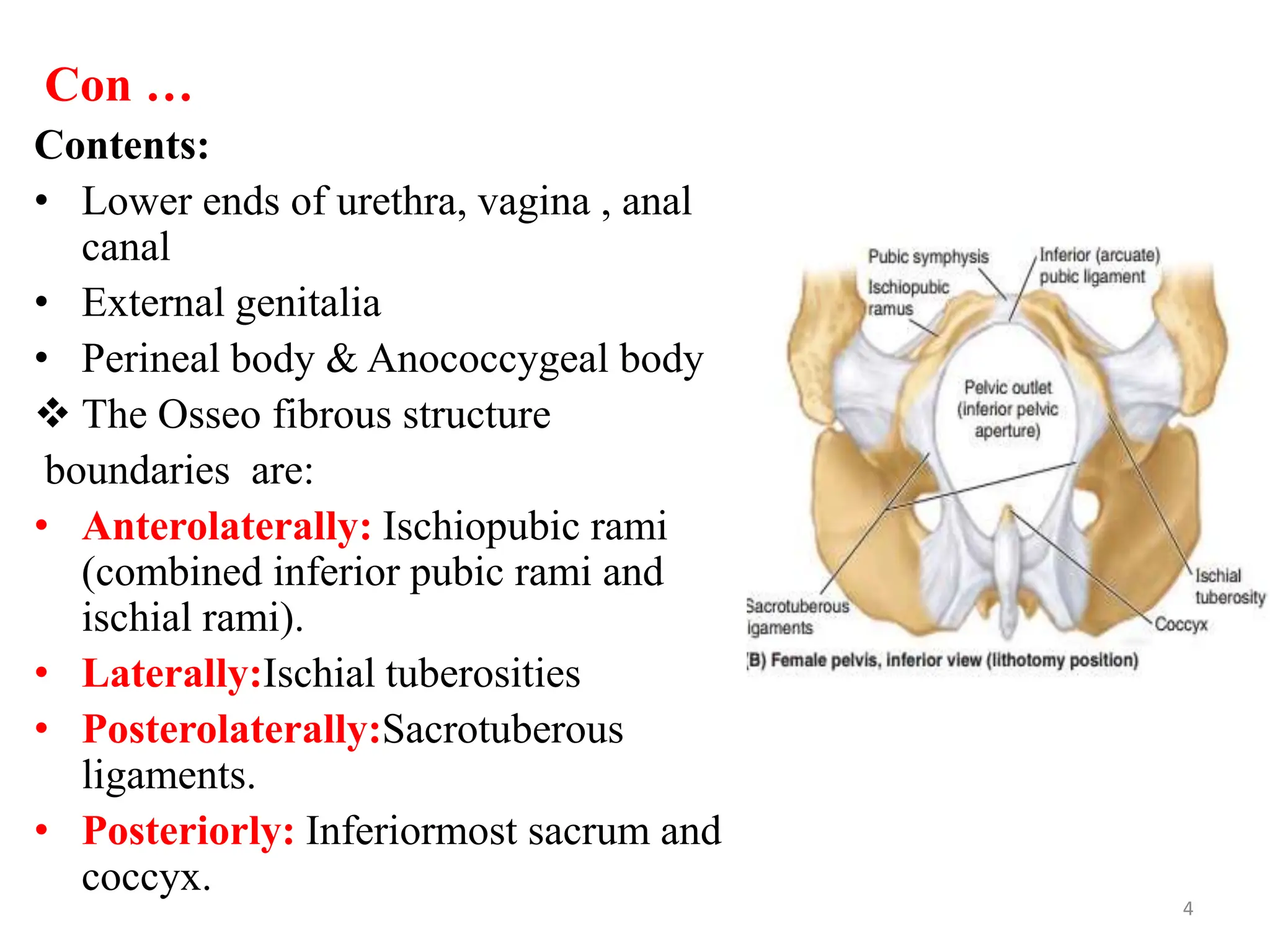
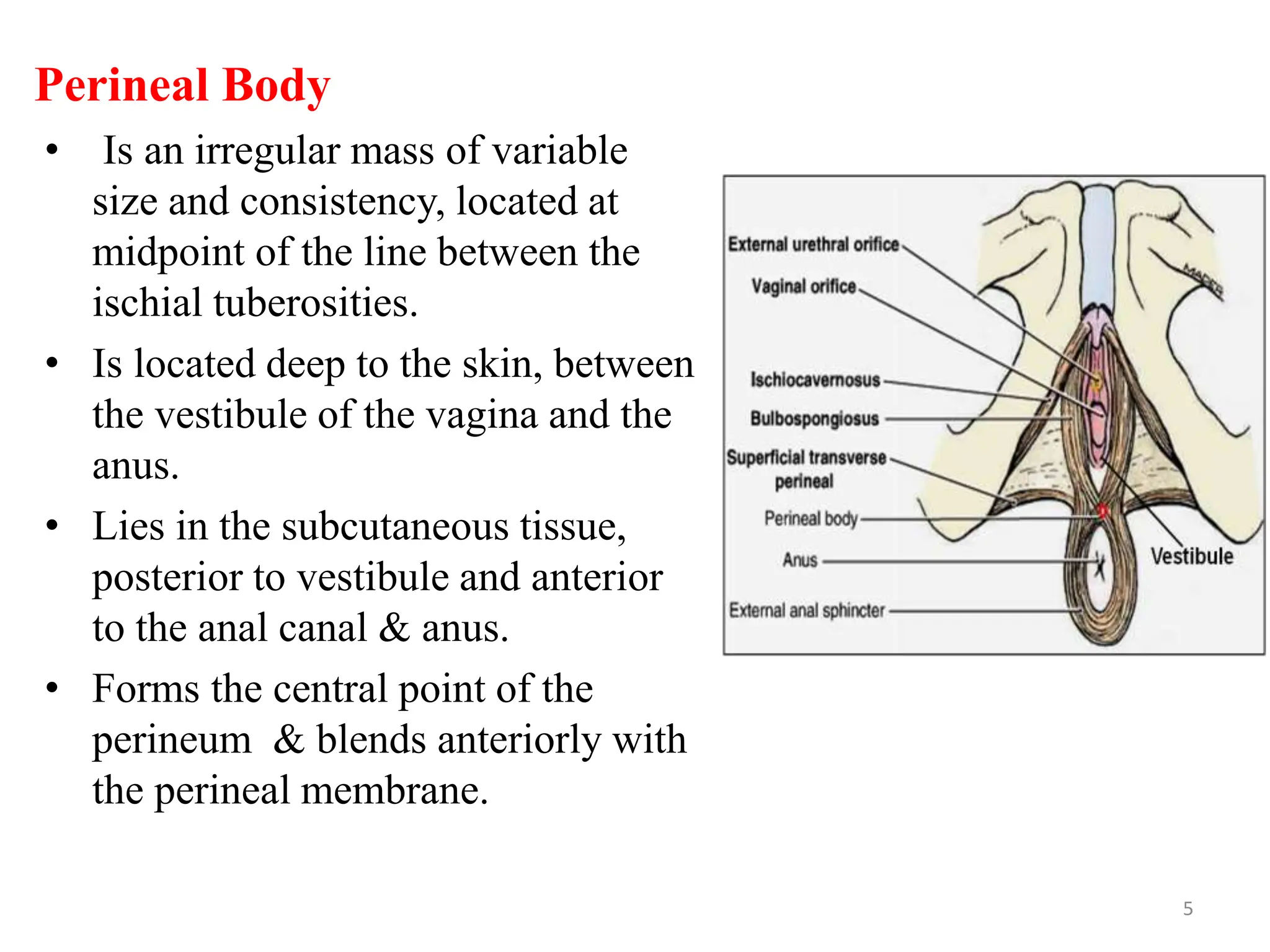
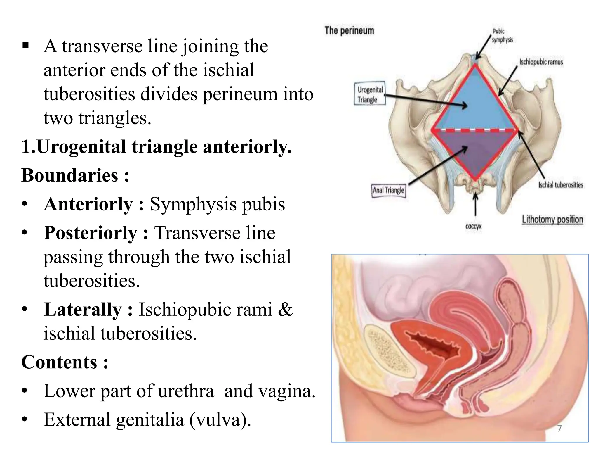
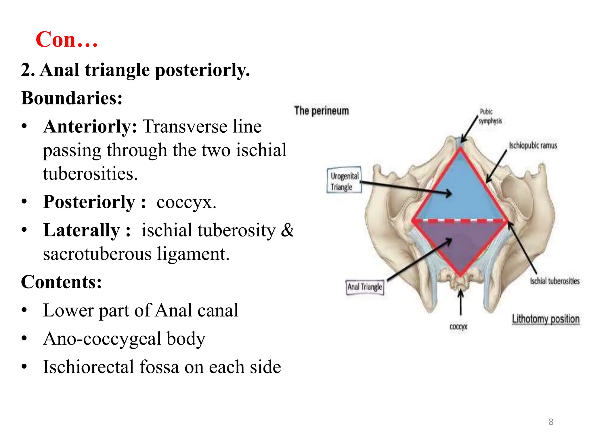
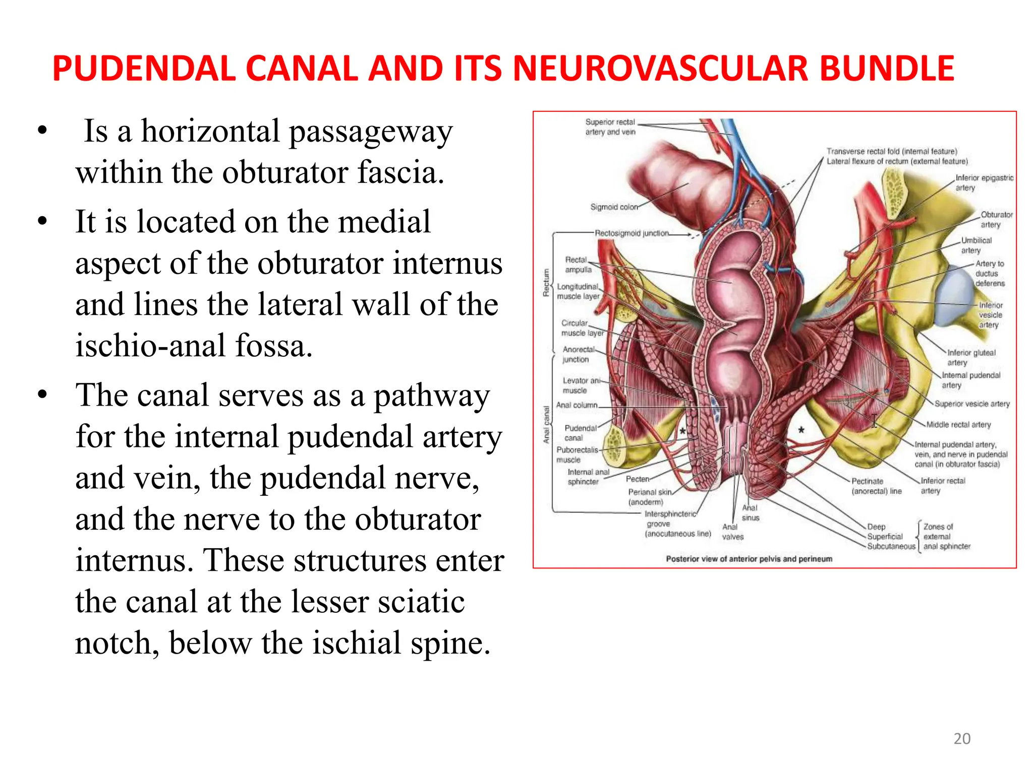
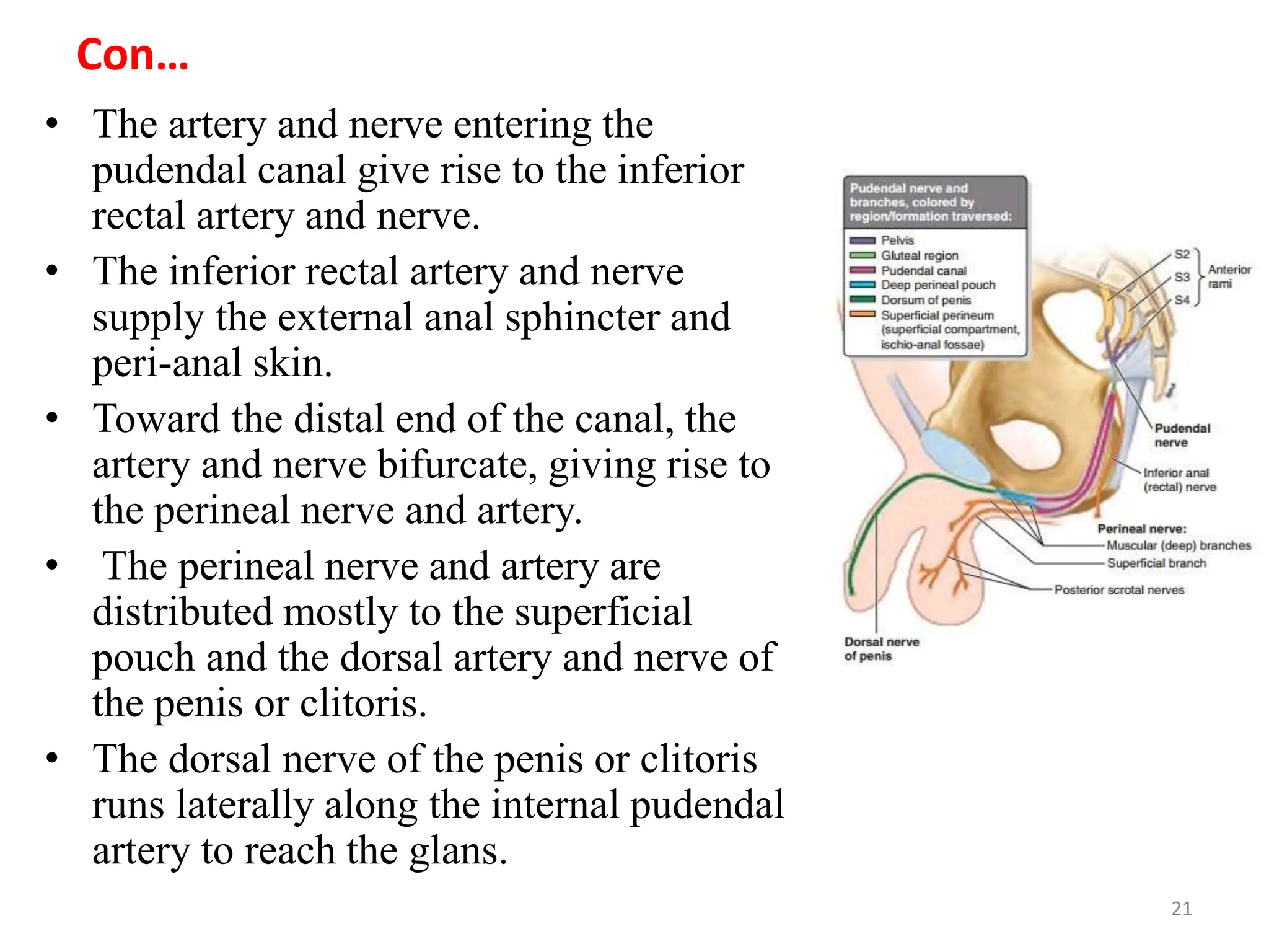
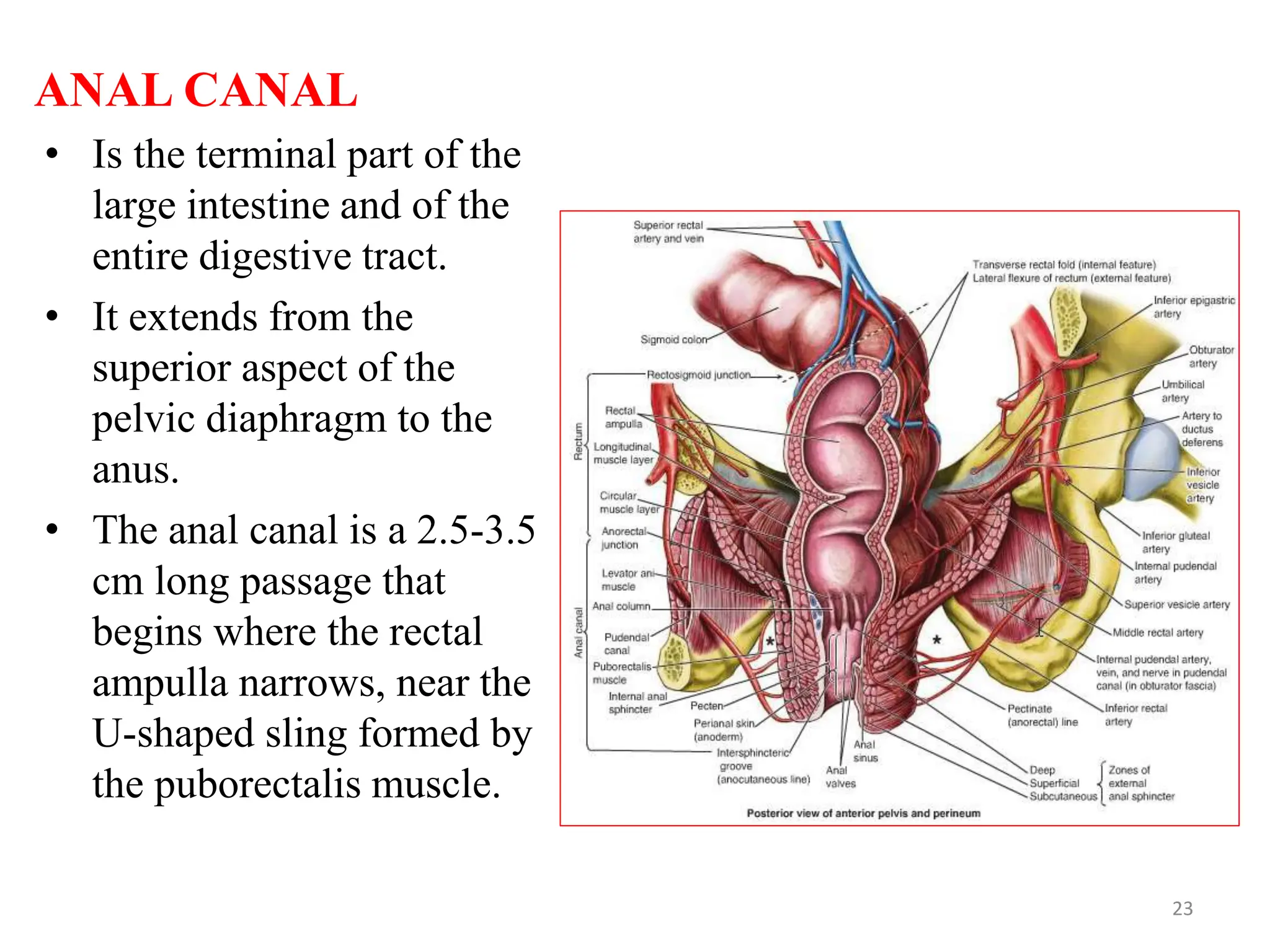
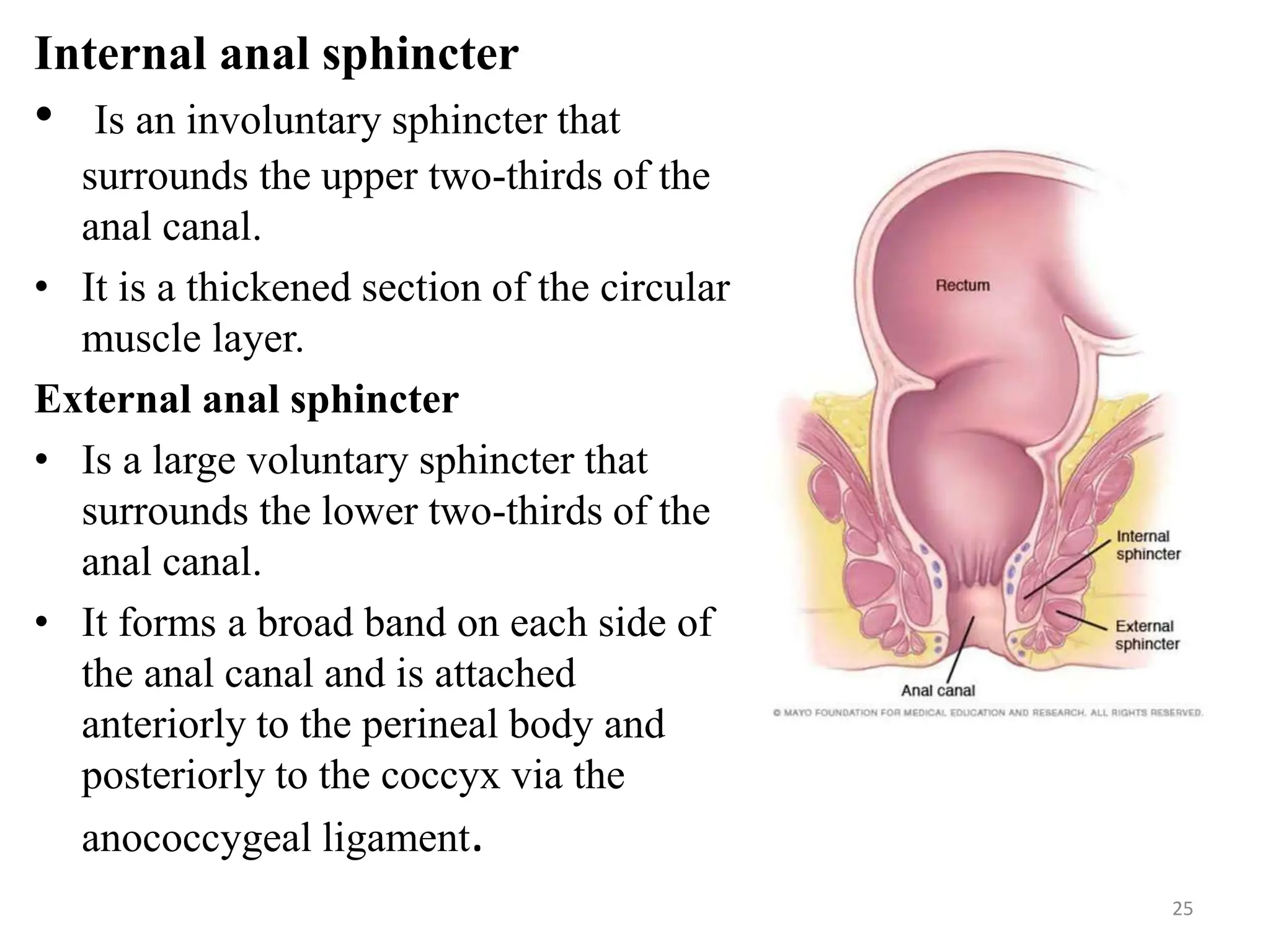
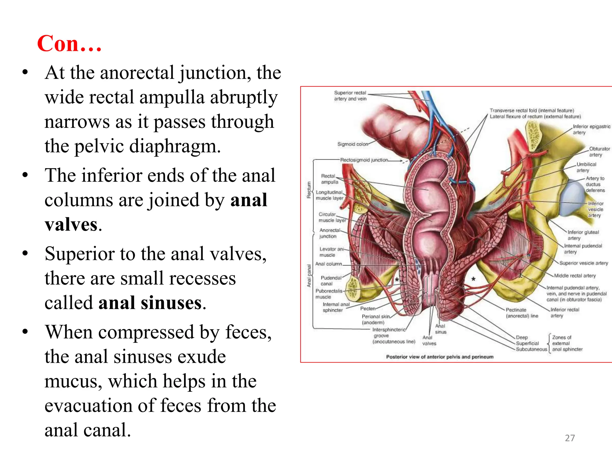
JIMMA UNIVERSITY
INSTITUTE OF HEALTH
COLLEGE OF MEDICAL SCIENCES
DEPARTEMENT OF BIOMEDICAL SCIENCES
ANATOMY COURSE TEAM
MASTERS PROGRAM IN CLINICAL ANATOMY
Course :Pelvis and Perineum
TOPIC – The anatomy of female perineum
PRPARED BY: Nigatu Woldekidan
outline
Perienum
Bounderies of female perienum
Perineal Body
Urogenital Diaphragm
Fasciae and Pouches of Urogenital Triangle
Features of Anal Triangle
Female urogenital triangle
Innervation, blood supply and lympatic drainage
of female perineum
2
Perineum
• Is a shallow compartment in the body.
• It is located between the pelvic outlet
and the pelvic cavity.
• Is separated from the pelvic cavity by
fascia.
• The perineal compartment is formed
by the levator ani and coccygeus
muscles, which make up the pelvic
diaphragm.
• It is a diamond shaped area between
the thighs
 Boundaries:
• Anteriorly: Mons pubis
• Laterally :Medial surfaces of the thighs
• Posteriorly :Intergluteal folds
3
Con …
Contents:
• Lower ends of urethra, vagina , anal
canal
• External genitalia
• Perineal body & Anococcygeal body
 The Osseo fibrous structure
boundaries are:
• Anterolaterally: Ischiopubic rami
(combined inferior pubic rami and
ischial rami).
• Laterally:Ischial tuberosities
• Posterolaterally:Sacrotuberous
ligaments.
• Posteriorly: Inferiormost sacrum and
coccyx.
4
Perineal Body
• Is an irregular mass of variable
size and consistency, located at
midpoint of the line between the
ischial tuberosities.
• Is located deep to the skin, between
the vestibule of the vagina and the
anus.
• Lies in the subcutaneous tissue,
posterior to vestibule and anterior
to the anal canal & anus.
• Forms the central point of the
perineum & blends anteriorly with
the perineal membrane.
5
Con…
• It is the convergence point of several muscles, including the
bulbospongiosus, external anal sphincter, superficial and deep
transverse perineal muscles.
• It also receives fibers from the external urethral sphincter, levator
ani, and muscular coats of the rectum.
Function:
• Gives attachment to perineal muscles
• Plays an important role in visceral support especially in female
• It plays a crucial role in supporting and coordinating the muscles
of the pelvic floor.
6
 A transverse line joining the
anterior ends of the ischial
tuberosities divides perineum into
two triangles.
1.Urogenital triangle anteriorly.
Boundaries :
• Anteriorly : Symphysis pubis
• Posteriorly : Transverse line
passing through the two ischial
tuberosities.
• Laterally : Ischiopubic rami &
ischial tuberosities.
Contents :
• Lower part of urethra and vagina.
• External genitalia (vulva). 7
Con…
2. Anal triangle posteriorly.
Boundaries:
• Anteriorly: Transverse line
passing through the two ischial
tuberosities.
• Posteriorly : coccyx.
• Laterally : ischial tuberosity &
sacrotuberous ligament.
Contents:
• Lower part of Anal canal
• Ano-coccygeal body
• Ischiorectal fossa on each side
8
Urogenital Diaphragm
• A triangular musculofascial
diaphragm located in the anterior
part of the perineum.
• Fills in the gap between the pubic
arch.
• Composed of: Sphincter urethrae
and the deep transverse perineal
muscles enclosed within the
superior and inferior layers of
fascia of the urogenital
diaphragm.
• The inferior layer of the fascia is
formed by the perineal
membrane.
9
Fasciae and Pouches of Urogenital Triangle
PERINEAL FASCIAE
• The perineal fascia is continuous
anteriorly with the fascia of
abdomen and consists of
superficial and deep layers.
Superficial perineal fascia:
consists of:
 Superficial fatty layer (Camper’s
fascia)
• Makes up the substance of mons
pubis & labia majora and
extends into the anal region.
10
 Deep membranous layer (Colle’s fascia ):
• Does not extend to anal region. Becomes fused with the posterior
margin of the perineal membrane and peritomial body.
Deep perineal fascia
• Intimately invests in the muscle superficial perineal pouch
(ischiocavernosus, bulbospongiosus, and superficial transverse
perineal).
• It is also attached laterally to the ischiopubic rami.
• Is fused with the suspensory ligament of the clitoris.
11
Perineal Pouches
12
Perineal Pouches
Superficial Perineal Pouch
• Is a potential space between
the perineal fascia and the
perineal membrane.
Boundaries:
Inferiorly: membranous layer of
superficial fascia
Superiorly: perineal membrane
Laterally: ischiopubic rami
13
Contents of the superficial perineal pouch
 Clitoris and associated muscle
(ischiocavernosus).
 Bulbs of the vestibule and
surrounding muscle
(bulbospongiosus).
 Greater vestibular glands.
 Superficial transverse perineal
muscles.
 Related vessels and nerves (deep
perineal branches of the internal
pudendal vessels and pudendal
nerves).
14
Deep Perineal Pouch
• It is a completely closed
space deep to the perineal
membrane
BOUNDARIES:
 Inferiorly: the perineal
membrane,
 Superiorly : the inferior
fascia of the pelvic
diaphragm, and
 Laterally : inferior portion
of the obturator fascia
(covering the obturator
internus muscle).
15
Contents of Deep Perineal Pouch
• Proximal part of the urethra.
• A mass of smooth muscle in
the place of deep transverse
perineal muscles on the
posterior edge of the perineal
membrane, associated with
the perineal body.
• Internal pudental vessels
• Spincter uretherae
muscle,which is pirched by
urethera and vagina.
• Dorsal neurovasculature of
the clitoris.
16
Features of Anal Triangle
ISCHIO-ANAL FOSSAE
• Are large fascia-lined, wedge
shaped spaces between the
skin of the anal region and the
pelvic diaphragm. .
• The apex of each fossa lies
superiorly where the levator
ani muscle arises from the
obturator fascia.
• Wide inferiorly and narrow
superiorly, are filled with fat
and loose connective tissue.
17
Con…
 The two ischio-anal fossae communicate by means of the
deep postanal space over the anococcygeal ligament (body),
a fibrous mass located between the anal canal and the tip of
the coccyx.
Boundaries:
 Laterally: ischium and overlapping inferior part of the obturator
internus, covered with obturator fascia.
 Medially: external anal sphincter, with a sloping
superior medial wall or roof formed by the levator ani as it descends
to blend with the sphincter; both structures surround the anal canal.
18
Con…
 Posteriorly : sacrotuberous
ligament and gluteus
maximus.
 Anteriorly: bodies of the
pubic bones, inferior to the
origin of the puborectalis.
 Each ischio-anal fossa is
filled with a fat body of the
ischio-anal fossa.
• The fat bodies support the
anal canal but they are readily
displaced to permit descent
and expansion of the anal
canal during the passage of
feces.
19
PUDENDAL CANAL AND ITS NEUROVASCULAR BUNDLE
• Is a horizontal passageway
within the obturator fascia.
• It is located on the medial
aspect of the obturator internus
and lines the lateral wall of the
ischio-anal fossa.
• The canal serves as a pathway
for the internal pudendal artery
and vein, the pudendal nerve,
and the nerve to the obturator
internus. These structures enter
the canal at the lesser sciatic
notch, below the ischial spine.
20
Con…
• The artery and nerve entering the
pudendal canal give rise to the inferior
rectal artery and nerve.
• The inferior rectal artery and nerve
supply the external anal sphincter and
peri-anal skin.
• Toward the distal end of the canal, the
artery and nerve bifurcate, giving rise to
the perineal nerve and artery.
• The perineal nerve and artery are
distributed mostly to the superficial
pouch and the dorsal artery and nerve of
the penis or clitoris.
• The dorsal nerve of the penis or clitoris
runs laterally along the internal pudendal
artery to reach the glans.
21
Con…
• The perineal nerve has two branches: the superficial perineal
nerve and the deep perineal nerve.
• Superficial perineal nerve gives rise to posterior scrotal or
labial branches.
• Deep perineal nerve supplies the muscles of the deep and
superficial perineal pouches, the vestibule skin, and the lower
part of the vagina.
• The inferior rectal nerve communicates with the posterior
scrotal or labial and perineal nerves.
• The dorsal nerve of the penis or clitoris is the primary sensory
nerve for the glans.
22
ANAL CANAL
• Is the terminal part of the
large intestine and of the
entire digestive tract.
• It extends from the
superior aspect of the
pelvic diaphragm to the
anus.
• The anal canal is a 2.5-3.5
cm long passage that
begins where the rectal
ampulla narrows, near the
U-shaped sling formed by
the puborectalis muscle.
23
Con…
• It extends to the anus, which is
the external opening of the
digestive tract.
• Is surrounded by internal and
external anal sphincters and
descends in a postero-inferior
direction between the
anococcygeal ligament and the
perineal body.
Relations (In female):
• Anteriorly: Perineal body,
urogenital diaphragm, and
lower part of vagina
• Posteriorly: Anococcygeal
body.
• Laterally: Ischiorectal fossae. 24
Internal anal sphincter
• Is an involuntary sphincter that
surrounds the upper two-thirds of the
anal canal.
• It is a thickened section of the circular
muscle layer.
External anal sphincter
• Is a large voluntary sphincter that
surrounds the lower two-thirds of the
anal canal.
• It forms a broad band on each side of
the anal canal and is attached
anteriorly to the perineal body and
posteriorly to the coccyx via the
anococcygeal ligament.
25
Con…
• The main nerve supply to the external anal sphincter is from the
S4 spinal segment through the inferior rectal nerve.
• The deep part of the external anal sphincter also receives fibers
from the nerve to the levator ani muscle.
• Contracts in unison with the puborectalis muscle to maintain
continence when the internal sphincter is relaxed, except during
defecation.
• The superior half of the mucous membrane of the anal canal has
longitudinal ridges called anal columns.
• The anal columns contain the terminal branches of the superior
rectal artery and vein.
• The anorectal junction, where the rectum joins the anal canal, is
indicated by the superior ends of the anal columns.
26
Con…
• At the anorectal junction, the
wide rectal ampulla abruptly
narrows as it passes through
the pelvic diaphragm.
• The inferior ends of the anal
columns are joined by anal
valves.
• Superior to the anal valves,
there are small recesses
called anal sinuses.
• When compressed by feces,
the anal sinuses exude
mucus, which helps in the
evacuation of feces from the
anal canal. 27
Con…
• The inferior boundary of the anal
valves forms an irregular line
known as the pectinate line.
• .The pectinate line marks the
junction between the superior part
of the anal canal (visceral) and the
inferior part (somatic).
• The anal canal above the pectinate
line and below the pectinate line
differ in terms of their arterial
supply, innervation, and venous
and lymphatic drainage.
• These differences are a result of the
distinct embryological origins of
the superior and inferior parts of
the anal canal.
28
Arterial Supply of Anal Canal
• The superior rectal artery supplies
the anal canal above the pectinate
line.
• The two inferior rectal arteries
supply the anal canal below the
pectinate line, as well as the
surrounding muscles and peri-
anal skin.
• The middle rectal arteries
contribute to the blood supply of
the anal canal by forming
connections (anastomoses) with
the superior and inferior rectal
arteries.
29
Venous and Lymphatic Drainage of Anal Canal
• The internal rectal venous plexus
drains in both directions from the
pectinate line.
• Above the pectinate line, the
internal rectal plexus mainly
drains into the superior rectal
vein and the portal system.
• Below the pectinate line, the
internal rectal plexus drains into
the inferior rectal veins around
the external anal sphincter.
30
Con…
• The middle rectal veins primarily drain the muscularis externa of
the ampulla and connect with the superior and inferior rectal
veins.
• The rectal plexuses have numerous arteriovenous anastomoses
(AVAs) from the superior and middle rectal arteries.
• Above the pectinate line, lymphatic vessels drain deeply into the
internal iliac lymph nodes, which then lead to the common iliac
and lumbar lymph nodes.
• Below the pectinate line, lymphatic vessels drain superficially
into the superficial inguinal lymph nodes.
• Most of the perineum also drains into the superficial inguinal
lymph nodes.
31
Innervation of Anal Canal
• The nerve supply to the anal canal above the pectinate line is
visceral innervation from the inferior hypogastric plexus.
• Sympathetic fibers maintain the tonus of the internal anal
sphincter.
• Parasympathetic fibers inhibit the tonus of the internal
sphincter and promote peristaltic contraction for defecation.
• The superior part of the anal canal, like the rectum above it, is
sensitive to stretching.
• The nerve supply to the anal canal below the pectinate line is
somatic innervation from the inferior anal (rectal) nerves,
which are branches of the pudendal nerve.
• Somatic efferent fibers stimulate contraction of the voluntary
external anal sphincter.
32
Female urogenital triangle
 Includes the female external genitalia and perineal muscles.
FEMALE EXTERNAL GENITALIA
 Include the mons pubis, labia majora, labia minora, clitoris,
bulbs of vestibule, and greater and lesser vestibular glands.
 They serve as sensory and erectile tissue for sexual arousal,
direct the flow of urine, and prevent the entry of foreign
material into the urogenital tract.
33
34
Mons Pubis
• Is a rounded, fatty prominence
located in front of the pubic
symphysis, pubic tubercles, and
superior pubic rami.
• It is formed by a mass of fatty
subcutaneous tissue.
• The amount of fat in the mons
pubis increases during puberty and
decreases after menopause.
• The surface of the mons pubis is
continuous with the anterior
abdominal wall.
• After puberty, the mons pubis is
covered with coarse pubic hairs.
35
Labia Majora
• Are prominent folds of skin in
the external genitalia of females.
• They serve as protective
structures for the clitoris,
urethral orifice, and vaginal
orifice.
• Each labium majus contains a
finger-like "digital process"
made up of loose subcutaneous
tissue that contains smooth
muscle.
• It also houses the termination of
the round ligament of the uterus.
.
36
Con…
• Extends inferoposteriorly from the mons pubis, which is the fatty
tissue over the pubic symphysis, towards the anus.
• Located on the sides of a central depression called the pudendal
cleft. This cleft appears as a narrow slit when the thighs are
adducted (pressed together).
• Within the pudendal cleft, the labia minora and vestibule can be
found.
• In adult females, the external aspects of the labia majora are
covered with pigmented skin that contains numerous sebaceous
glands. They are also covered with pubic hair, which is typically
coarse and crisp.
• Internally, the labia majora have smooth, pink, and hairless
surfaces.
• Are thicker at the front where they join to form the anterior
commissure. 37
Con…
• In nulliparous women, they merge at the back to create a ridge called the
posterior commissure, which overlays the perineal body and serves as
the posterior boundary of the vulva.
• The posterior commissure typically disappears after the first vaginal
birth.
Labia Minora
• Are rounded folds of fat-free, hairless skin.
• They surround and close over the vestibule of the vagina.
• They contain erectile tissue and blood vessels.
• Anteriorly, they form two laminae.
• Medial laminae unite as the frenulum of the clitoris.
• Lateral laminae unite to form the prepuce of the clitoris.
• In young women, they are connected by the frenulum of the labia
minora.
• The internal surface has thin moist skin with pink color and contains
sebaceous glands and sensory nerve endings.
38
Clitoris
• Is an erectile organ located at
the anterior meeting point of the
labia minora.
• It consists of a root, a small
cylindrical body, two crura, two
corpora cavernosa, and the
glans clitoris.
• The crura attach to the inferior
pubic rami and perineal
membrane.
• The body of the clitoris is
covered by the prepuce.
39
Con…
• It is approximately 2 cm in length and <1 cm in diameter.
• is not functionally related to the urethra or urination.
• It functions solely as an organ of sexual arousal.
• The glans clitoris is highly innervated and densely supplied with
sensory endings.
Vestibule of Vagina
• Is the space surrounded by the labia minora.
• It contains the openings of the urethra, vagina, and ducts of the
greater and lesser vestibular glands.
• The external urethral orifice is located 2-3 cm postero-inferior to
the glans clitoris and anterior to the vaginal orifice.
• The para-urethral glands open on each side of the external
urethral orifice.
40
Con…
• The ducts of the greater vestibular
glands open on the upper, medial
aspects of the labia minora.
• The size and appearance of the
vaginal orifice vary based on the
condition of the hymen.
• After hymen rupture, only
remnants called hymenal
caruncles are visible.
• The hymen is considered a
developmental vestige with no
established physiological
function.
41
Bulbs of Vestibule
• Are paired masses of elongated
erectile tissue.
• They are approximately 3 cm in
length.
• The bulbs are located along the
sides of the vaginal orifice,
superior or deep to the labia
minora.
• They are positioned immediately
inferior to the perineal
membrane.
• The bulbs are covered inferiorly
and laterally by the
bulbospongiosus muscles.
• They are homologous to the bulb
of the penis.
42
Vestibular Glands (Bartholin glands)
• Are approximately 0.5 cm in diameter.
• They are located in the superficial perineal pouch.
• The glands lie on each side of the vestibule of the vagina,
posterolateral to the vaginal orifice and inferior to the perineal
membrane.
• They are round or oval in shape and are partly overlapped by the
bulbs of the vestibule.
• They secrete mucus into the vestibule during sexual arousal
• The lesser vestibular glands are small glands located on each side
of the vestibule of the vagina.
• They open into the vestibule between the urethral and vaginal
orifices.
• These glands secrete mucus into the vestibule.
• The mucus helps moisturize the labia and vestibule. 43
Arterial Supply and Venous Drainage of Vulva
• The vulva receives an abundant arterial
supply from the external and internal
pudendal arteries.
• The internal pudendal artery supplies
the skin, external genitalia, and
perineal muscles.
• The labial arteries and clitoral arteries
are branches of the internal pudendal
artery.
• The labial veins are tributaries of the
internal pudendal veins and
accompanying veins of the internal
pudendal artery.
44
Innervation of Vulva
• The anterior aspect of the vulva (mons pubis, anterior labia) is supplied
by the ilio-inguinal nerve (anterior labial nerves) and the genital branch of
the genitofemoral nerve.
• The posterior aspect of the vulva is supplied by the perineal branch of the
posterior cutaneous nerve of the thigh (laterally) and the pudendal nerve
(centrally).
• The pudendal nerve is the primary nerve of the perineum.
• The posterior labial nerves (terminal superficial branches of the perineal
nerve) supply the labia.
• Deep and muscular branches of the perineal nerve supply the vaginal
orifice and superficial perineal muscles.
• The dorsal nerve of the clitoris supplies deep perineal muscles and
sensation to the clitoris.
• The bulb of the vestibule and erectile bodies of the clitoris receive
parasympathetic fibers via cavernous nerves from the uterovaginal nerve
plexus.
• Parasympathetic stimulation leads to increased vaginal secretion, erection
of the clitoris, and engorgement of erectile tissue in the bulbs of the
vestibule.
45
46
LYMPHATIC DRAINAGE OF FEMALE PERINEUM
• The vulva contains a rich network of lymphatic vessels.
• Lymph from the skin of the perineum, including the area below
the pectinate line of the anorectum, the lowermost vagina, vaginal
orifice, and vestibule, initially drains to the superficial inguinal
lymph nodes.
• Lymph from the clitoris, vestibular bulb, and anterior labia
minora drains to the deep inguinal lymph nodes or directly to the
internal iliac lymph nodes.
• Lymph from the urethra drains to the internal iliac or sacral
lymph nodes.
47
48
PERINEAL MUSCLES OF FEMALE
 The perineal muscles in females include:
• Bulbospongiosus muscle surrounds the vestibule of the
vagina and the base of the clitoris, contributing to vaginal
constriction and clitoral compression.
• Ischiocavernosus muscle helps in erecting and maintaining
rigidity of the clitoris during sexual arousal.
• Superficial transverse perineal muscle provides support to
the perineal body and stabilizes perineal structures.
• Levator ani muscles, including the puborectalis,
pubococcygeus, and iliococcygeus muscles, support the pelvic
organs and contribute to continence.
• External anal sphincter surrounds the anal canal and helps
in maintaining bowel continence by closing the anus.
49
50
Referance
• Moore, K. L., Dalley, A. F., & Agur, A. M. R. (2014). Moore's
Clinical Oriented Anatomy (7th ed.). Wolters
Kluwer/Lippincott Williams & Wilkins.
51
THANK YOU!!
52

FEMALEperineum.pptxAssignment Presentation

  • 1.
    JIMMA UNIVERSITY INSTITUTE OFHEALTH COLLEGE OF MEDICAL SCIENCES DEPARTEMENT OF BIOMEDICAL SCIENCES ANATOMY COURSE TEAM MASTERS PROGRAM IN CLINICAL ANATOMY Course :Pelvis and Perineum TOPIC – The anatomy of female perineum PRPARED BY: Nigatu Woldekidan
  • 2.
    outline Perienum Bounderies of femaleperienum Perineal Body Urogenital Diaphragm Fasciae and Pouches of Urogenital Triangle Features of Anal Triangle Female urogenital triangle Innervation, blood supply and lympatic drainage of female perineum 2
  • 3.
    Perineum • Is ashallow compartment in the body. • It is located between the pelvic outlet and the pelvic cavity. • Is separated from the pelvic cavity by fascia. • The perineal compartment is formed by the levator ani and coccygeus muscles, which make up the pelvic diaphragm. • It is a diamond shaped area between the thighs  Boundaries: • Anteriorly: Mons pubis • Laterally :Medial surfaces of the thighs • Posteriorly :Intergluteal folds 3
  • 4.
    Con … Contents: • Lowerends of urethra, vagina , anal canal • External genitalia • Perineal body & Anococcygeal body  The Osseo fibrous structure boundaries are: • Anterolaterally: Ischiopubic rami (combined inferior pubic rami and ischial rami). • Laterally:Ischial tuberosities • Posterolaterally:Sacrotuberous ligaments. • Posteriorly: Inferiormost sacrum and coccyx. 4
  • 5.
    Perineal Body • Isan irregular mass of variable size and consistency, located at midpoint of the line between the ischial tuberosities. • Is located deep to the skin, between the vestibule of the vagina and the anus. • Lies in the subcutaneous tissue, posterior to vestibule and anterior to the anal canal & anus. • Forms the central point of the perineum & blends anteriorly with the perineal membrane. 5
  • 6.
    Con… • It isthe convergence point of several muscles, including the bulbospongiosus, external anal sphincter, superficial and deep transverse perineal muscles. • It also receives fibers from the external urethral sphincter, levator ani, and muscular coats of the rectum. Function: • Gives attachment to perineal muscles • Plays an important role in visceral support especially in female • It plays a crucial role in supporting and coordinating the muscles of the pelvic floor. 6
  • 7.
     A transverseline joining the anterior ends of the ischial tuberosities divides perineum into two triangles. 1.Urogenital triangle anteriorly. Boundaries : • Anteriorly : Symphysis pubis • Posteriorly : Transverse line passing through the two ischial tuberosities. • Laterally : Ischiopubic rami & ischial tuberosities. Contents : • Lower part of urethra and vagina. • External genitalia (vulva). 7
  • 8.
    Con… 2. Anal triangleposteriorly. Boundaries: • Anteriorly: Transverse line passing through the two ischial tuberosities. • Posteriorly : coccyx. • Laterally : ischial tuberosity & sacrotuberous ligament. Contents: • Lower part of Anal canal • Ano-coccygeal body • Ischiorectal fossa on each side 8
  • 9.
    Urogenital Diaphragm • Atriangular musculofascial diaphragm located in the anterior part of the perineum. • Fills in the gap between the pubic arch. • Composed of: Sphincter urethrae and the deep transverse perineal muscles enclosed within the superior and inferior layers of fascia of the urogenital diaphragm. • The inferior layer of the fascia is formed by the perineal membrane. 9
  • 10.
    Fasciae and Pouchesof Urogenital Triangle PERINEAL FASCIAE • The perineal fascia is continuous anteriorly with the fascia of abdomen and consists of superficial and deep layers. Superficial perineal fascia: consists of:  Superficial fatty layer (Camper’s fascia) • Makes up the substance of mons pubis & labia majora and extends into the anal region. 10
  • 11.
     Deep membranouslayer (Colle’s fascia ): • Does not extend to anal region. Becomes fused with the posterior margin of the perineal membrane and peritomial body. Deep perineal fascia • Intimately invests in the muscle superficial perineal pouch (ischiocavernosus, bulbospongiosus, and superficial transverse perineal). • It is also attached laterally to the ischiopubic rami. • Is fused with the suspensory ligament of the clitoris. 11
  • 12.
  • 13.
    Perineal Pouches Superficial PerinealPouch • Is a potential space between the perineal fascia and the perineal membrane. Boundaries: Inferiorly: membranous layer of superficial fascia Superiorly: perineal membrane Laterally: ischiopubic rami 13
  • 14.
    Contents of thesuperficial perineal pouch  Clitoris and associated muscle (ischiocavernosus).  Bulbs of the vestibule and surrounding muscle (bulbospongiosus).  Greater vestibular glands.  Superficial transverse perineal muscles.  Related vessels and nerves (deep perineal branches of the internal pudendal vessels and pudendal nerves). 14
  • 15.
    Deep Perineal Pouch •It is a completely closed space deep to the perineal membrane BOUNDARIES:  Inferiorly: the perineal membrane,  Superiorly : the inferior fascia of the pelvic diaphragm, and  Laterally : inferior portion of the obturator fascia (covering the obturator internus muscle). 15
  • 16.
    Contents of DeepPerineal Pouch • Proximal part of the urethra. • A mass of smooth muscle in the place of deep transverse perineal muscles on the posterior edge of the perineal membrane, associated with the perineal body. • Internal pudental vessels • Spincter uretherae muscle,which is pirched by urethera and vagina. • Dorsal neurovasculature of the clitoris. 16
  • 17.
    Features of AnalTriangle ISCHIO-ANAL FOSSAE • Are large fascia-lined, wedge shaped spaces between the skin of the anal region and the pelvic diaphragm. . • The apex of each fossa lies superiorly where the levator ani muscle arises from the obturator fascia. • Wide inferiorly and narrow superiorly, are filled with fat and loose connective tissue. 17
  • 18.
    Con…  The twoischio-anal fossae communicate by means of the deep postanal space over the anococcygeal ligament (body), a fibrous mass located between the anal canal and the tip of the coccyx. Boundaries:  Laterally: ischium and overlapping inferior part of the obturator internus, covered with obturator fascia.  Medially: external anal sphincter, with a sloping superior medial wall or roof formed by the levator ani as it descends to blend with the sphincter; both structures surround the anal canal. 18
  • 19.
    Con…  Posteriorly :sacrotuberous ligament and gluteus maximus.  Anteriorly: bodies of the pubic bones, inferior to the origin of the puborectalis.  Each ischio-anal fossa is filled with a fat body of the ischio-anal fossa. • The fat bodies support the anal canal but they are readily displaced to permit descent and expansion of the anal canal during the passage of feces. 19
  • 20.
    PUDENDAL CANAL ANDITS NEUROVASCULAR BUNDLE • Is a horizontal passageway within the obturator fascia. • It is located on the medial aspect of the obturator internus and lines the lateral wall of the ischio-anal fossa. • The canal serves as a pathway for the internal pudendal artery and vein, the pudendal nerve, and the nerve to the obturator internus. These structures enter the canal at the lesser sciatic notch, below the ischial spine. 20
  • 21.
    Con… • The arteryand nerve entering the pudendal canal give rise to the inferior rectal artery and nerve. • The inferior rectal artery and nerve supply the external anal sphincter and peri-anal skin. • Toward the distal end of the canal, the artery and nerve bifurcate, giving rise to the perineal nerve and artery. • The perineal nerve and artery are distributed mostly to the superficial pouch and the dorsal artery and nerve of the penis or clitoris. • The dorsal nerve of the penis or clitoris runs laterally along the internal pudendal artery to reach the glans. 21
  • 22.
    Con… • The perinealnerve has two branches: the superficial perineal nerve and the deep perineal nerve. • Superficial perineal nerve gives rise to posterior scrotal or labial branches. • Deep perineal nerve supplies the muscles of the deep and superficial perineal pouches, the vestibule skin, and the lower part of the vagina. • The inferior rectal nerve communicates with the posterior scrotal or labial and perineal nerves. • The dorsal nerve of the penis or clitoris is the primary sensory nerve for the glans. 22
  • 23.
    ANAL CANAL • Isthe terminal part of the large intestine and of the entire digestive tract. • It extends from the superior aspect of the pelvic diaphragm to the anus. • The anal canal is a 2.5-3.5 cm long passage that begins where the rectal ampulla narrows, near the U-shaped sling formed by the puborectalis muscle. 23
  • 24.
    Con… • It extendsto the anus, which is the external opening of the digestive tract. • Is surrounded by internal and external anal sphincters and descends in a postero-inferior direction between the anococcygeal ligament and the perineal body. Relations (In female): • Anteriorly: Perineal body, urogenital diaphragm, and lower part of vagina • Posteriorly: Anococcygeal body. • Laterally: Ischiorectal fossae. 24
  • 25.
    Internal anal sphincter •Is an involuntary sphincter that surrounds the upper two-thirds of the anal canal. • It is a thickened section of the circular muscle layer. External anal sphincter • Is a large voluntary sphincter that surrounds the lower two-thirds of the anal canal. • It forms a broad band on each side of the anal canal and is attached anteriorly to the perineal body and posteriorly to the coccyx via the anococcygeal ligament. 25
  • 26.
    Con… • The mainnerve supply to the external anal sphincter is from the S4 spinal segment through the inferior rectal nerve. • The deep part of the external anal sphincter also receives fibers from the nerve to the levator ani muscle. • Contracts in unison with the puborectalis muscle to maintain continence when the internal sphincter is relaxed, except during defecation. • The superior half of the mucous membrane of the anal canal has longitudinal ridges called anal columns. • The anal columns contain the terminal branches of the superior rectal artery and vein. • The anorectal junction, where the rectum joins the anal canal, is indicated by the superior ends of the anal columns. 26
  • 27.
    Con… • At theanorectal junction, the wide rectal ampulla abruptly narrows as it passes through the pelvic diaphragm. • The inferior ends of the anal columns are joined by anal valves. • Superior to the anal valves, there are small recesses called anal sinuses. • When compressed by feces, the anal sinuses exude mucus, which helps in the evacuation of feces from the anal canal. 27
  • 28.
    Con… • The inferiorboundary of the anal valves forms an irregular line known as the pectinate line. • .The pectinate line marks the junction between the superior part of the anal canal (visceral) and the inferior part (somatic). • The anal canal above the pectinate line and below the pectinate line differ in terms of their arterial supply, innervation, and venous and lymphatic drainage. • These differences are a result of the distinct embryological origins of the superior and inferior parts of the anal canal. 28
  • 29.
    Arterial Supply ofAnal Canal • The superior rectal artery supplies the anal canal above the pectinate line. • The two inferior rectal arteries supply the anal canal below the pectinate line, as well as the surrounding muscles and peri- anal skin. • The middle rectal arteries contribute to the blood supply of the anal canal by forming connections (anastomoses) with the superior and inferior rectal arteries. 29
  • 30.
    Venous and LymphaticDrainage of Anal Canal • The internal rectal venous plexus drains in both directions from the pectinate line. • Above the pectinate line, the internal rectal plexus mainly drains into the superior rectal vein and the portal system. • Below the pectinate line, the internal rectal plexus drains into the inferior rectal veins around the external anal sphincter. 30
  • 31.
    Con… • The middlerectal veins primarily drain the muscularis externa of the ampulla and connect with the superior and inferior rectal veins. • The rectal plexuses have numerous arteriovenous anastomoses (AVAs) from the superior and middle rectal arteries. • Above the pectinate line, lymphatic vessels drain deeply into the internal iliac lymph nodes, which then lead to the common iliac and lumbar lymph nodes. • Below the pectinate line, lymphatic vessels drain superficially into the superficial inguinal lymph nodes. • Most of the perineum also drains into the superficial inguinal lymph nodes. 31
  • 32.
    Innervation of AnalCanal • The nerve supply to the anal canal above the pectinate line is visceral innervation from the inferior hypogastric plexus. • Sympathetic fibers maintain the tonus of the internal anal sphincter. • Parasympathetic fibers inhibit the tonus of the internal sphincter and promote peristaltic contraction for defecation. • The superior part of the anal canal, like the rectum above it, is sensitive to stretching. • The nerve supply to the anal canal below the pectinate line is somatic innervation from the inferior anal (rectal) nerves, which are branches of the pudendal nerve. • Somatic efferent fibers stimulate contraction of the voluntary external anal sphincter. 32
  • 33.
    Female urogenital triangle Includes the female external genitalia and perineal muscles. FEMALE EXTERNAL GENITALIA  Include the mons pubis, labia majora, labia minora, clitoris, bulbs of vestibule, and greater and lesser vestibular glands.  They serve as sensory and erectile tissue for sexual arousal, direct the flow of urine, and prevent the entry of foreign material into the urogenital tract. 33
  • 34.
  • 35.
    Mons Pubis • Isa rounded, fatty prominence located in front of the pubic symphysis, pubic tubercles, and superior pubic rami. • It is formed by a mass of fatty subcutaneous tissue. • The amount of fat in the mons pubis increases during puberty and decreases after menopause. • The surface of the mons pubis is continuous with the anterior abdominal wall. • After puberty, the mons pubis is covered with coarse pubic hairs. 35
  • 36.
    Labia Majora • Areprominent folds of skin in the external genitalia of females. • They serve as protective structures for the clitoris, urethral orifice, and vaginal orifice. • Each labium majus contains a finger-like "digital process" made up of loose subcutaneous tissue that contains smooth muscle. • It also houses the termination of the round ligament of the uterus. . 36
  • 37.
    Con… • Extends inferoposteriorlyfrom the mons pubis, which is the fatty tissue over the pubic symphysis, towards the anus. • Located on the sides of a central depression called the pudendal cleft. This cleft appears as a narrow slit when the thighs are adducted (pressed together). • Within the pudendal cleft, the labia minora and vestibule can be found. • In adult females, the external aspects of the labia majora are covered with pigmented skin that contains numerous sebaceous glands. They are also covered with pubic hair, which is typically coarse and crisp. • Internally, the labia majora have smooth, pink, and hairless surfaces. • Are thicker at the front where they join to form the anterior commissure. 37
  • 38.
    Con… • In nulliparouswomen, they merge at the back to create a ridge called the posterior commissure, which overlays the perineal body and serves as the posterior boundary of the vulva. • The posterior commissure typically disappears after the first vaginal birth. Labia Minora • Are rounded folds of fat-free, hairless skin. • They surround and close over the vestibule of the vagina. • They contain erectile tissue and blood vessels. • Anteriorly, they form two laminae. • Medial laminae unite as the frenulum of the clitoris. • Lateral laminae unite to form the prepuce of the clitoris. • In young women, they are connected by the frenulum of the labia minora. • The internal surface has thin moist skin with pink color and contains sebaceous glands and sensory nerve endings. 38
  • 39.
    Clitoris • Is anerectile organ located at the anterior meeting point of the labia minora. • It consists of a root, a small cylindrical body, two crura, two corpora cavernosa, and the glans clitoris. • The crura attach to the inferior pubic rami and perineal membrane. • The body of the clitoris is covered by the prepuce. 39
  • 40.
    Con… • It isapproximately 2 cm in length and <1 cm in diameter. • is not functionally related to the urethra or urination. • It functions solely as an organ of sexual arousal. • The glans clitoris is highly innervated and densely supplied with sensory endings. Vestibule of Vagina • Is the space surrounded by the labia minora. • It contains the openings of the urethra, vagina, and ducts of the greater and lesser vestibular glands. • The external urethral orifice is located 2-3 cm postero-inferior to the glans clitoris and anterior to the vaginal orifice. • The para-urethral glands open on each side of the external urethral orifice. 40
  • 41.
    Con… • The ductsof the greater vestibular glands open on the upper, medial aspects of the labia minora. • The size and appearance of the vaginal orifice vary based on the condition of the hymen. • After hymen rupture, only remnants called hymenal caruncles are visible. • The hymen is considered a developmental vestige with no established physiological function. 41
  • 42.
    Bulbs of Vestibule •Are paired masses of elongated erectile tissue. • They are approximately 3 cm in length. • The bulbs are located along the sides of the vaginal orifice, superior or deep to the labia minora. • They are positioned immediately inferior to the perineal membrane. • The bulbs are covered inferiorly and laterally by the bulbospongiosus muscles. • They are homologous to the bulb of the penis. 42
  • 43.
    Vestibular Glands (Bartholinglands) • Are approximately 0.5 cm in diameter. • They are located in the superficial perineal pouch. • The glands lie on each side of the vestibule of the vagina, posterolateral to the vaginal orifice and inferior to the perineal membrane. • They are round or oval in shape and are partly overlapped by the bulbs of the vestibule. • They secrete mucus into the vestibule during sexual arousal • The lesser vestibular glands are small glands located on each side of the vestibule of the vagina. • They open into the vestibule between the urethral and vaginal orifices. • These glands secrete mucus into the vestibule. • The mucus helps moisturize the labia and vestibule. 43
  • 44.
    Arterial Supply andVenous Drainage of Vulva • The vulva receives an abundant arterial supply from the external and internal pudendal arteries. • The internal pudendal artery supplies the skin, external genitalia, and perineal muscles. • The labial arteries and clitoral arteries are branches of the internal pudendal artery. • The labial veins are tributaries of the internal pudendal veins and accompanying veins of the internal pudendal artery. 44
  • 45.
    Innervation of Vulva •The anterior aspect of the vulva (mons pubis, anterior labia) is supplied by the ilio-inguinal nerve (anterior labial nerves) and the genital branch of the genitofemoral nerve. • The posterior aspect of the vulva is supplied by the perineal branch of the posterior cutaneous nerve of the thigh (laterally) and the pudendal nerve (centrally). • The pudendal nerve is the primary nerve of the perineum. • The posterior labial nerves (terminal superficial branches of the perineal nerve) supply the labia. • Deep and muscular branches of the perineal nerve supply the vaginal orifice and superficial perineal muscles. • The dorsal nerve of the clitoris supplies deep perineal muscles and sensation to the clitoris. • The bulb of the vestibule and erectile bodies of the clitoris receive parasympathetic fibers via cavernous nerves from the uterovaginal nerve plexus. • Parasympathetic stimulation leads to increased vaginal secretion, erection of the clitoris, and engorgement of erectile tissue in the bulbs of the vestibule. 45
  • 46.
  • 47.
    LYMPHATIC DRAINAGE OFFEMALE PERINEUM • The vulva contains a rich network of lymphatic vessels. • Lymph from the skin of the perineum, including the area below the pectinate line of the anorectum, the lowermost vagina, vaginal orifice, and vestibule, initially drains to the superficial inguinal lymph nodes. • Lymph from the clitoris, vestibular bulb, and anterior labia minora drains to the deep inguinal lymph nodes or directly to the internal iliac lymph nodes. • Lymph from the urethra drains to the internal iliac or sacral lymph nodes. 47
  • 48.
  • 49.
    PERINEAL MUSCLES OFFEMALE  The perineal muscles in females include: • Bulbospongiosus muscle surrounds the vestibule of the vagina and the base of the clitoris, contributing to vaginal constriction and clitoral compression. • Ischiocavernosus muscle helps in erecting and maintaining rigidity of the clitoris during sexual arousal. • Superficial transverse perineal muscle provides support to the perineal body and stabilizes perineal structures. • Levator ani muscles, including the puborectalis, pubococcygeus, and iliococcygeus muscles, support the pelvic organs and contribute to continence. • External anal sphincter surrounds the anal canal and helps in maintaining bowel continence by closing the anus. 49
  • 50.
  • 51.
    Referance • Moore, K.L., Dalley, A. F., & Agur, A. M. R. (2014). Moore's Clinical Oriented Anatomy (7th ed.). Wolters Kluwer/Lippincott Williams & Wilkins. 51
  • 52.