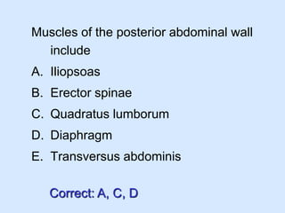
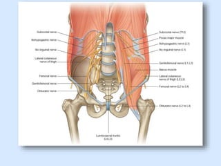
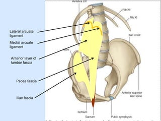
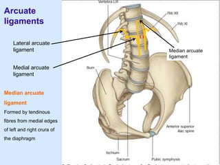
The document describes the anatomy of the posterior abdominal wall. It contains the following key points:
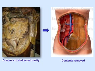
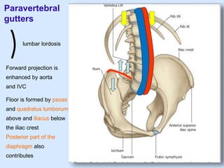
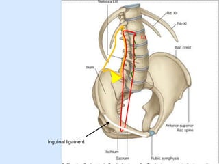
1) The posterior abdominal wall is formed by muscles, fascia, and bones between the rib cage and pelvic brim. It forms the posterior boundary of the abdominal cavity.
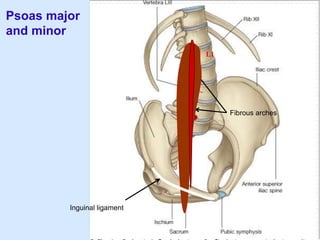
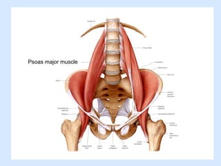
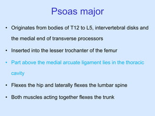
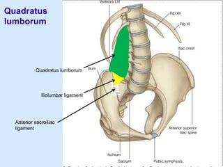
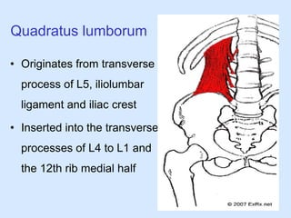
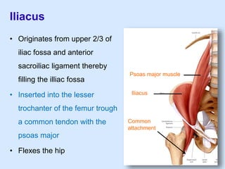
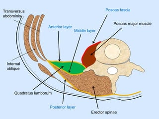
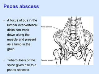
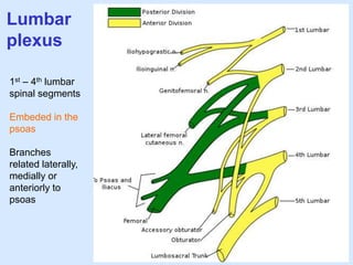
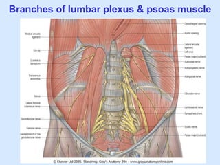
2) Major muscles include the psoas major, which flexes the hip and spine, and quadratus lumborum, which fixes the 12th rib and lumbar vertebrae.
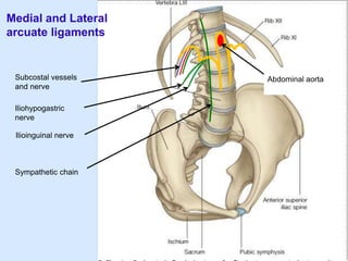
3) Fascia include the psoas fascia, iliac fascia, and lumbar part of the thoracolumbar fascia.
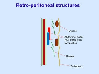
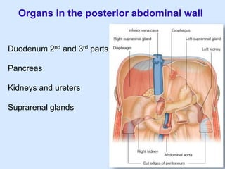
4) Retroperitoneal structures in the posterior wall include the duod