Downloaded 112 times


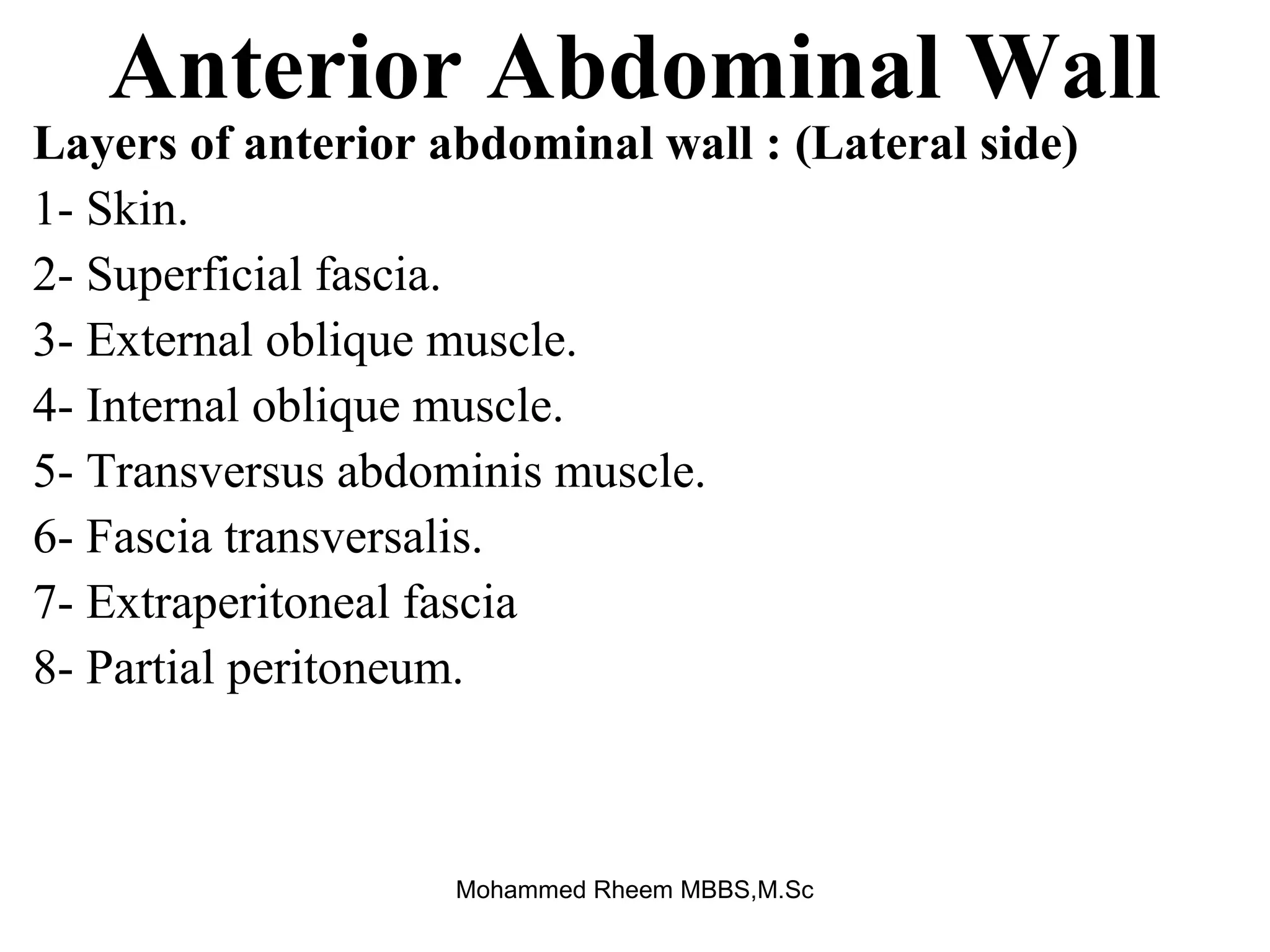


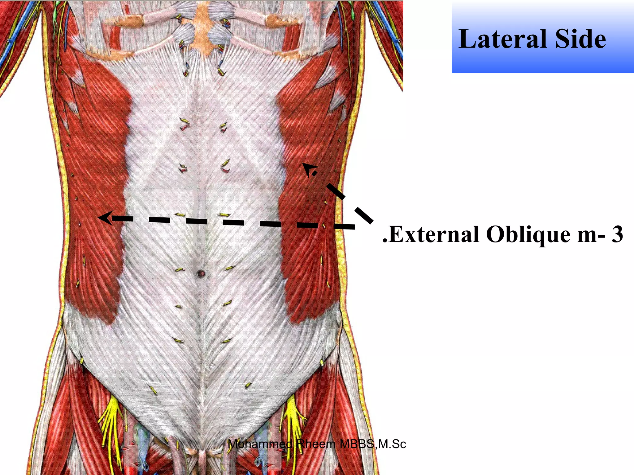


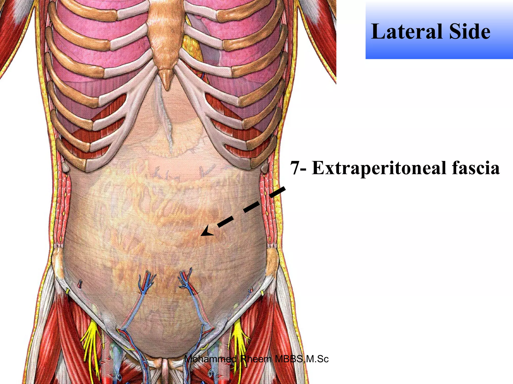

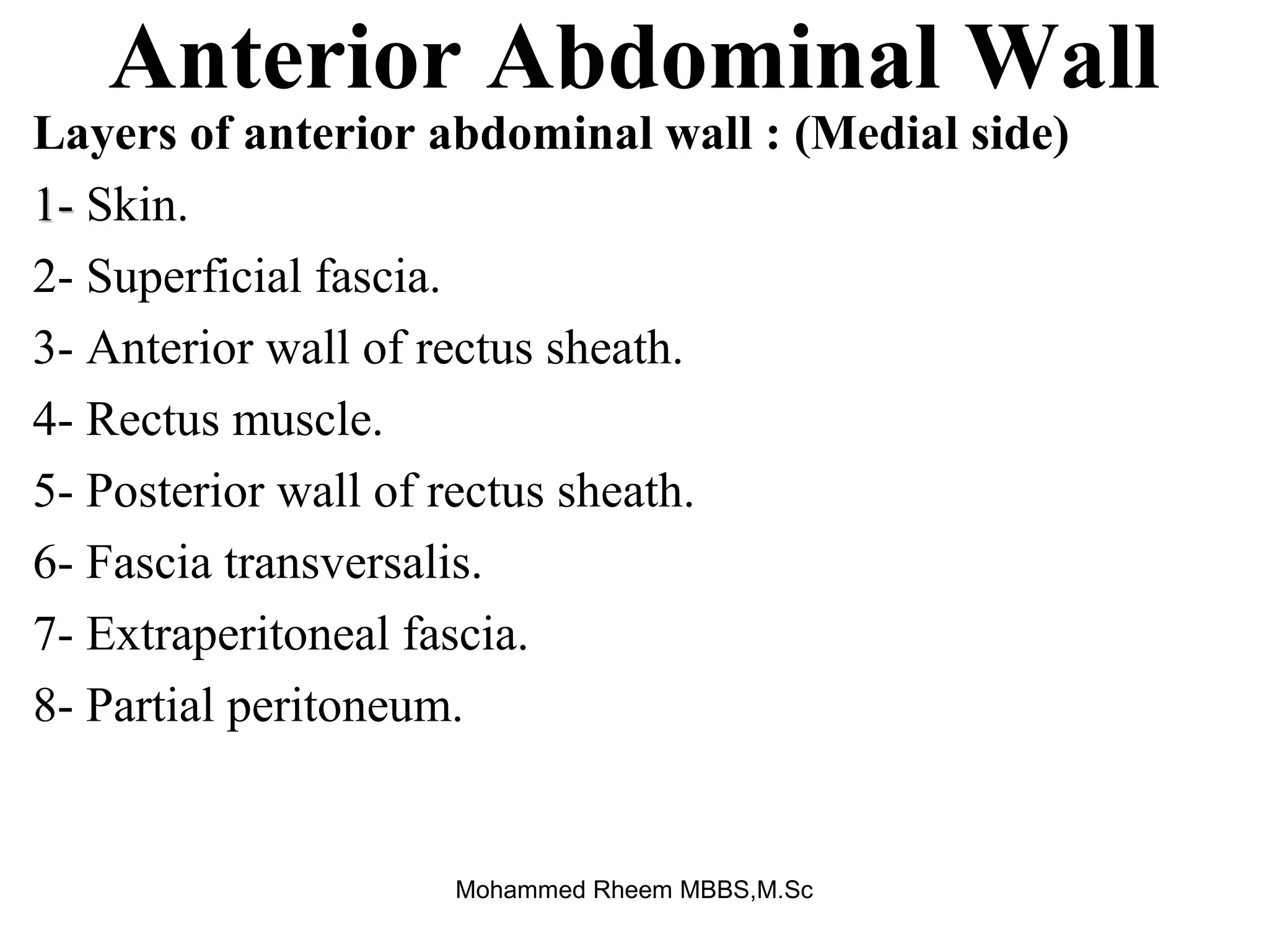


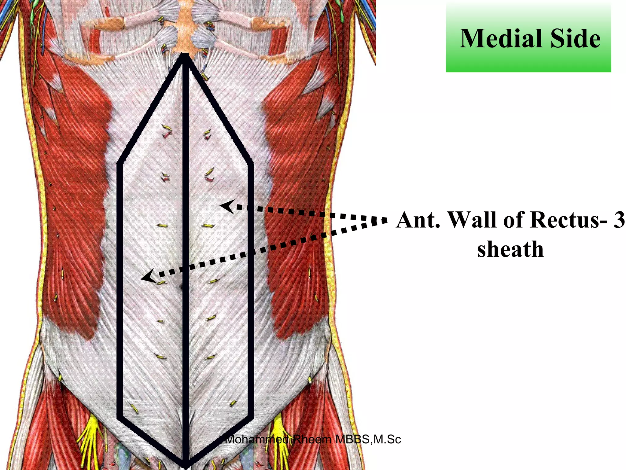

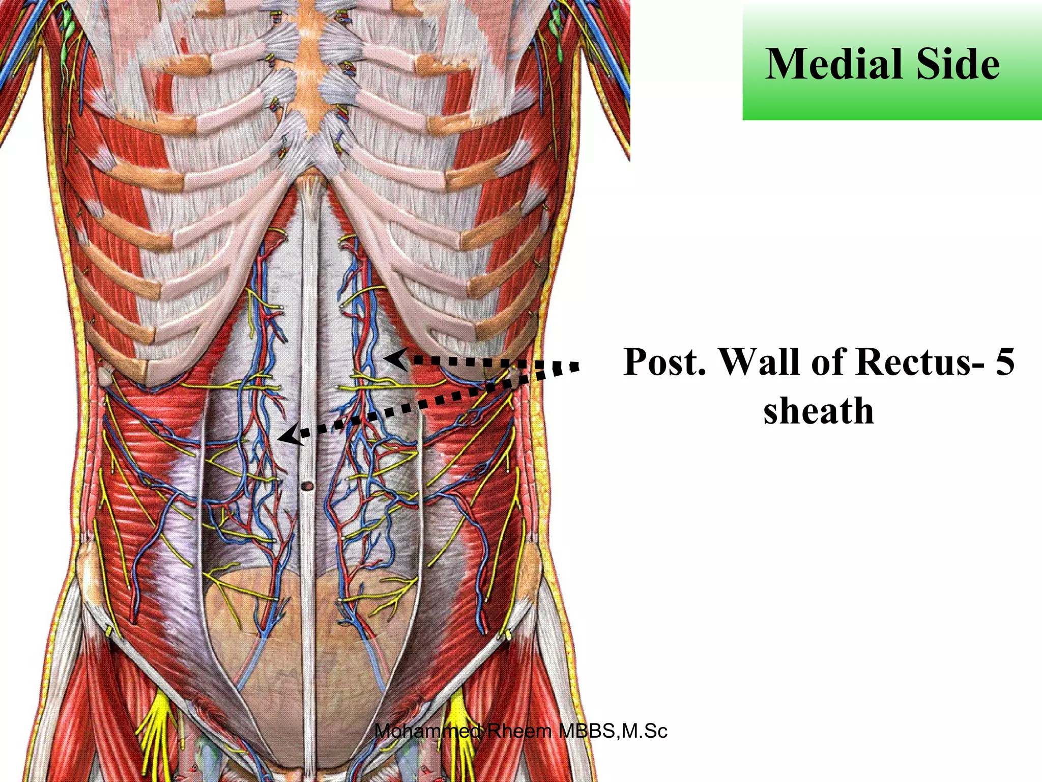


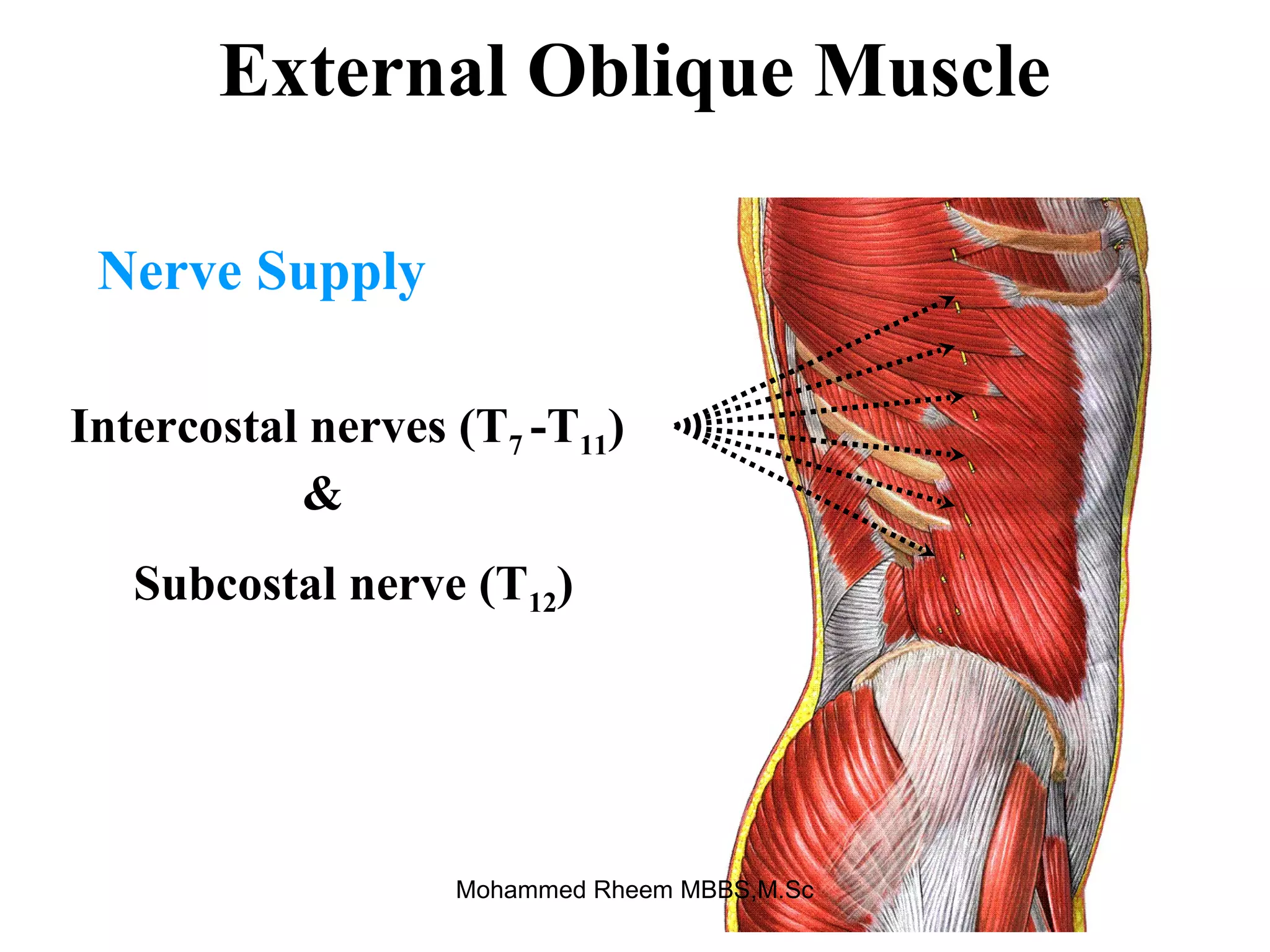


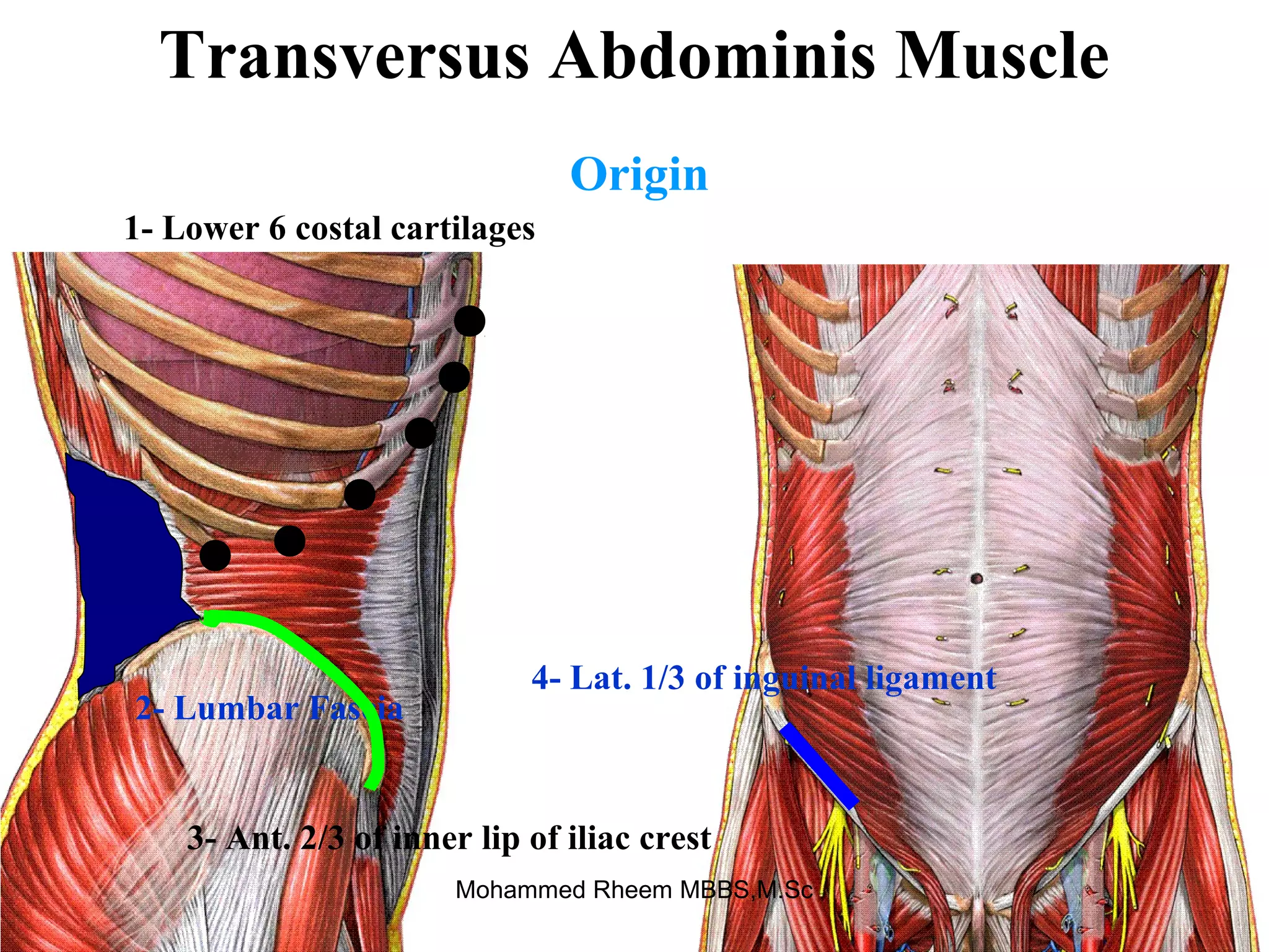


This document provides information about the anatomy of the anterior abdominal wall. It begins with an introductory case study about a new doctor struggling with surgical notes. It then covers the layers of the anterior abdominal wall, abdominal muscles and fascia, blood vessels and nerves, abdominal regions, hernias, and the inguinal canal. Diagrams are provided to illustrate key anatomical structures like the rectus sheath, abdominal wall incisions, and inguinal ring locations. The document aims to equip readers with knowledge of anterior abdominal wall anatomy.