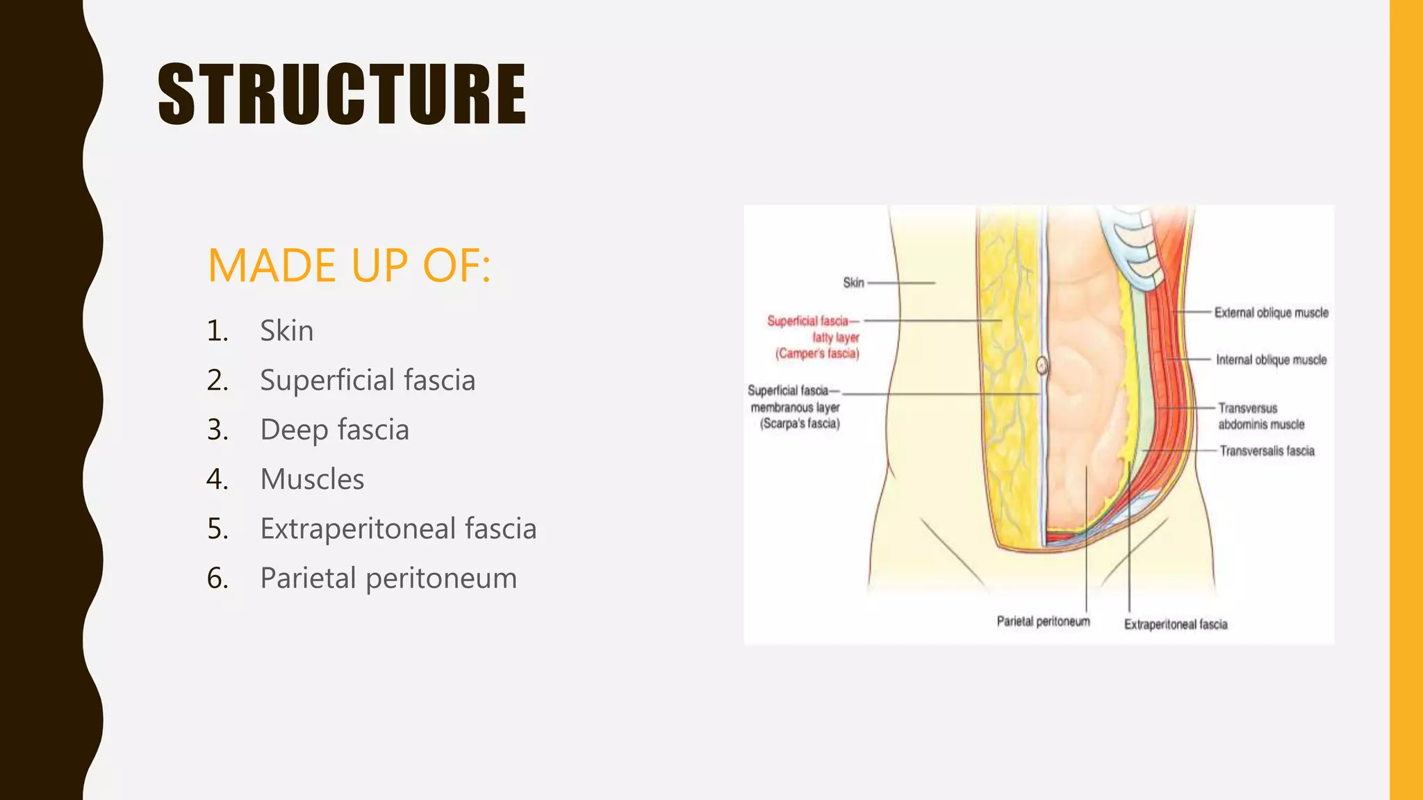
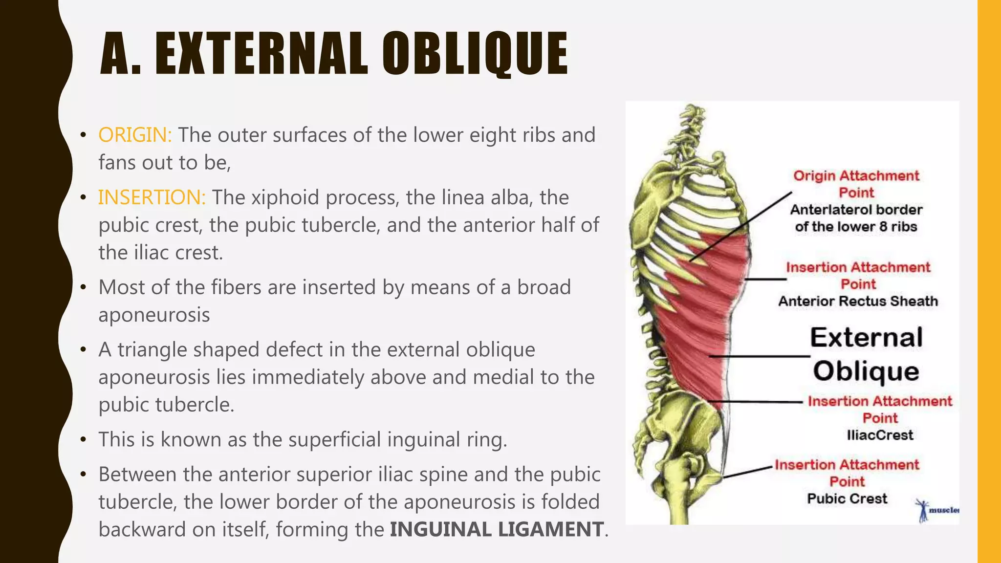
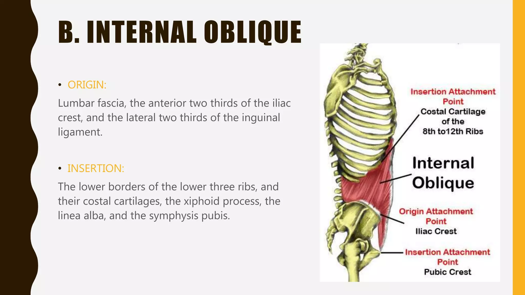
ANTERIOR
ABDOMINAL
WALL
D R . TA N YA R A S H I D
A N AT O M Y D E P T
STRUCTURE
MADE UP OF:
1. Skin
2. Superficial fascia
3. Deep fascia
4. Muscles
5. Extraperitoneal fascia
6. Parietal peritoneum
SKIN
• Skin is loosely attached to the underlying
structures except at the umbilicus, where it is
tethered to the scar tissue.
• The umbilicus is a scar representing the site of
attachment of the umbilical cord in the fetus, it
is situated in linea alba.
DERMATOME:
• The cutaneous nerve supply is derived from
the anterior rami of the lower six thoracic and
the first lumbar nerves.
• The dermatome of T7 is located over the
xiphoid process
• The dermatome of T10 includes the umbilicus
• That of L1 lies just above the inguinal ligament
and the symphysis pubis.
SUPERFICIAL FASCIA
• THE SUPERFICIAL FASCIA IS DIVIDED INTO:
– Superficial fatty layer (Camper’s fascia)
– Deep membranous layer (Scarpa’s fascia)
• The FATTY LAYER is continuous with the superficial
fat over the rest of the body and may be extremely
thick (8cm) or more in obese patients.
• The MEMBRANOUS LAYER is thin and fades out
laterally and above, where it becomes continuous
with the superficial fascia of the back and the thorax,
respectively.
DEEP FASCIA
• It lies immediately deep to the membranous
layer of superficial fascia
MUSCLES
• The muscles of anterior abdominal wall consists of
three broad thin sheets.
FROM EXTERIOR TO INTERIOR THEY ARE:
• External oblique
• Internal oblique
• Transversus abdominis
• On either side of the midline anteriorly is, in
addition, a wide vertical muscle, the RECTUS
ABDOMINUS.
• As the aponeuroses of the three sheets pass
forward, they enclose the rectus abdominis to form
the RECTUS SHEATH.
• The lower part of the rectus sheath might contain
a small muscle called the PYRAMIDALIS.
A. EXTERNAL OBLIQUE
• ORIGIN: The outer surfaces of the lower eight ribs and
fans out to be,
• INSERTION: The xiphoid process, the linea alba, the
pubic crest, the pubic tubercle, and the anterior half of
the iliac crest.
• Most of the fibers are inserted by means of a broad
aponeurosis
• A triangle shaped defect in the external oblique
aponeurosis lies immediately above and medial to the
pubic tubercle.
• This is known as the superficial inguinal ring.
• Between the anterior superior iliac spine and the pubic
tubercle, the lower border of the aponeurosis is folded
backward on itself, forming the INGUINAL LIGAMENT.
B. INTERNAL OBLIQUE
• ORIGIN:
Lumbar fascia, the anterior two thirds of the iliac
crest, and the lateral two thirds of the inguinal
ligament.
• INSERTION:
The lower borders of the lower three ribs, and
their costal cartilages, the xiphoid process, the
linea alba, and the symphysis pubis.
• EXTERNAL OBLIQUE MUSCLE FIBERS move downwards and
medially.
• INTERNAL OBLIQUE MUSCLE FIBERS move upwards and medially.
C. TRANSVERSUS ABDOMINUS
• ORIGIN:
The deep surface of the lower six costal
cartilages, the lumbar fascia, the anterior two
thirds of the iliac crest, and the lateral third of
the inguinal ligament.
• INSERTION:
The xiphoid process, the linea alba, and the
symphysis pubis.
D. RECTUS ABDOMINIS
• The rectus abdominis is a long strap
muscle that extends along the whole
length of the anterior abdominal wall.
• It is broaded above and lies close to the
midline, being separated from its fellow
by the linea alba.
• ORIGIN: pubic crest
• INSERTION: ribs (5th, 6th, 7th), xiphoid
process.
• The rectus abdominis muscle is
divided into distinct segments by
three transverse tendinous
intersections:
• 1. one at the level of xiphoid process
• 2. one at the level of the umbilicus
• 3. one halfway between these two
• These insertions are strongly attached
to the anterior wall of the rectus
sheath.
E. PYRAMIDALIS
• The pyramidalis muscle is often absent.
• ORIGIN: anterior surface of the pubis
• INSERTION: linea alba
• It lies infront of the lower part of the
rectus abdominis.
RECTUS SHEATH
• The rectus sheath is a long fibrous sheath
that encloses the rectus abdominis muscle
and pyramidalis muscle (if present)
• It is formed mainly by the aponeuroses of
the three lateral abdominal muscles.
• Between the costal margin and the level
of the anterior superior iliac spine, the
aponeurosis of the internal oblique splits
to enclose the rectus muscle; the external
oblique aponeurosis is directed in front of
the muscle, and the transversus
aponeurosis is directed behind the
muscle.
FUNCTIONS OF MUSCLES OF
ANTERIOR ABDOMINAL WALL
• OBLIQUE MUSCLE: laterally flex and route the trunk
• RECTUS ABDOMINIS: flexes the trunk and stabilizes the pelvis
• PYRAMIDALIS: keeps the linea alba taut during the process.
• The muscles of the anterior and lateral abdominal walls assist the diaphragm during
inspiration by relaxing as the diaphragm descends so that the abdominal viscera can
be accommodated.
• The muscles assist in the act of forces expiration that occurs during coughing and
sneezing by pulling down the ribs and sternum.
• Their tone plays an important part in supporting and protecting the abdominal viscera.
• By contracting simultaneously with the diaphragm, with the glottis of the
larynx closed, they increase the intra abdominal pressure and help in:
1. Micturation
2. Defecation
3. Vomiting
4. Parturition
NERVE SUPPLY OF MUSCLES OF
ANTERIOR ABDOMINAL WALL
• The oblique and transversus
abdominis muscles are supplied by
the lower six thoracic nerves and the
iliohypogastric and ilioinguinal nerves
(L1).
• The rectus muscle is supplied by the
lower six thoracic nerves.
• The pyramidalis is supplied by the 12th
thoracic nerve.
• FASCIA TRANSVERSALIS
• The fascia transversalis is a thin layer of fascia that lines the transversus
abdominis muscle.
• EXTRAPERITONEAL FAT:
• The extraperitoneal fat is a thin layer of connective tissue that contains a variable
amount of fat and lies between the fascia tranversalis and the parietal
peritoneum.
• PARIETAL PERITONEUM:
• The walls of abdomen are lined with parietal peritoneum
• This is a thin serous membrane and is continuous below with the parietal
peritoneum lining the pelvis.
BLOOD SUPPLY OF
ANTERIOR ABDOMINAL WALL
• ARTERIAL SUPPLY:
– Superior Epigastric artery: branch of Internal
Mammary artery.
– Inferior Epigastric artery: branch of External
Iliac artery
– Two Posterior Intercostal art.: Desc. Thor.
Aorta
– Four Lumbar arteries: Desc. Thor. Aorta
– Deep Circumflex iliac artery: branch of Ext.
Iliac artery
• VEINS:
• Above the umbilicus mainly into the axillary
vein via the lateral thoracic vein.
• Below the umbilicus into the femoral vein
through the superficial epigastric and the
great saphenous veins.
NERVE SUPPLY OF
ANTERIOR ABDOMINAL WALL
• Anterior rami of Lower 6 thoracic nerves
– T7 – T11 intercostal nerves
– T12: Subcostal nerve
• Anterior ramus of the 1st lumbar nerve
– Ilioinguinal nerve
– Iliohypogastric nerve
• Pass forward between the internal oblique
and transversus abdominis muscle.
LYMPH DRAINAGE OF
ANTERIOR ABDOMINAL WALL
SUPERFICIAL LYMPH VESSELS:
• Above umbilicus: anterior group of axillary
nodes
• Below umbilicus: superficial inguinal lymph
nodes
DEEP YMPH VESSELS:
Drain into the:
Internal thoracic, external iliac, posterior
mediastinal and para aortic (lumbar) nodes.
MULTIPLE CHOICE QUESTIONS
1. SUPERIOR EPIGASTRIC ARTERY IS A BRANCH OF:
• A. Descending thoracic aorta
• B. Abdominal aorta
• C. Internal thoracic artery
• D. External iliac artery
2. MUSCLE FIBERS OF THE EXTERNAL OBLIQUE MOVE IN WHICH DIRECTION:
• A. upwards and medially
• B. upwards and laterally
• C. downwards and laterally
• D. downwards and medially
3. INSERTION OF RECTUS ABDOMINIS:
• A. linea alba
• B. pubic crest
• C. xyphoid process
• D. anterosuperior iliac spine
Anterior abdominal wall

Anterior abdominal wall

  • 1.
    ANTERIOR ABDOMINAL WALL D R .TA N YA R A S H I D A N AT O M Y D E P T
  • 2.
    STRUCTURE MADE UP OF: 1.Skin 2. Superficial fascia 3. Deep fascia 4. Muscles 5. Extraperitoneal fascia 6. Parietal peritoneum
  • 3.
    SKIN • Skin isloosely attached to the underlying structures except at the umbilicus, where it is tethered to the scar tissue. • The umbilicus is a scar representing the site of attachment of the umbilical cord in the fetus, it is situated in linea alba. DERMATOME: • The cutaneous nerve supply is derived from the anterior rami of the lower six thoracic and the first lumbar nerves. • The dermatome of T7 is located over the xiphoid process • The dermatome of T10 includes the umbilicus • That of L1 lies just above the inguinal ligament and the symphysis pubis.
  • 4.
    SUPERFICIAL FASCIA • THESUPERFICIAL FASCIA IS DIVIDED INTO: – Superficial fatty layer (Camper’s fascia) – Deep membranous layer (Scarpa’s fascia) • The FATTY LAYER is continuous with the superficial fat over the rest of the body and may be extremely thick (8cm) or more in obese patients. • The MEMBRANOUS LAYER is thin and fades out laterally and above, where it becomes continuous with the superficial fascia of the back and the thorax, respectively. DEEP FASCIA • It lies immediately deep to the membranous layer of superficial fascia
  • 5.
    MUSCLES • The musclesof anterior abdominal wall consists of three broad thin sheets. FROM EXTERIOR TO INTERIOR THEY ARE: • External oblique • Internal oblique • Transversus abdominis • On either side of the midline anteriorly is, in addition, a wide vertical muscle, the RECTUS ABDOMINUS. • As the aponeuroses of the three sheets pass forward, they enclose the rectus abdominis to form the RECTUS SHEATH. • The lower part of the rectus sheath might contain a small muscle called the PYRAMIDALIS.
  • 6.
    A. EXTERNAL OBLIQUE •ORIGIN: The outer surfaces of the lower eight ribs and fans out to be, • INSERTION: The xiphoid process, the linea alba, the pubic crest, the pubic tubercle, and the anterior half of the iliac crest. • Most of the fibers are inserted by means of a broad aponeurosis • A triangle shaped defect in the external oblique aponeurosis lies immediately above and medial to the pubic tubercle. • This is known as the superficial inguinal ring. • Between the anterior superior iliac spine and the pubic tubercle, the lower border of the aponeurosis is folded backward on itself, forming the INGUINAL LIGAMENT.
  • 7.
    B. INTERNAL OBLIQUE •ORIGIN: Lumbar fascia, the anterior two thirds of the iliac crest, and the lateral two thirds of the inguinal ligament. • INSERTION: The lower borders of the lower three ribs, and their costal cartilages, the xiphoid process, the linea alba, and the symphysis pubis.
  • 8.
    • EXTERNAL OBLIQUEMUSCLE FIBERS move downwards and medially. • INTERNAL OBLIQUE MUSCLE FIBERS move upwards and medially.
  • 9.
    C. TRANSVERSUS ABDOMINUS •ORIGIN: The deep surface of the lower six costal cartilages, the lumbar fascia, the anterior two thirds of the iliac crest, and the lateral third of the inguinal ligament. • INSERTION: The xiphoid process, the linea alba, and the symphysis pubis.
  • 10.
    D. RECTUS ABDOMINIS •The rectus abdominis is a long strap muscle that extends along the whole length of the anterior abdominal wall. • It is broaded above and lies close to the midline, being separated from its fellow by the linea alba. • ORIGIN: pubic crest • INSERTION: ribs (5th, 6th, 7th), xiphoid process.
  • 11.
    • The rectusabdominis muscle is divided into distinct segments by three transverse tendinous intersections: • 1. one at the level of xiphoid process • 2. one at the level of the umbilicus • 3. one halfway between these two • These insertions are strongly attached to the anterior wall of the rectus sheath.
  • 12.
    E. PYRAMIDALIS • Thepyramidalis muscle is often absent. • ORIGIN: anterior surface of the pubis • INSERTION: linea alba • It lies infront of the lower part of the rectus abdominis.
  • 13.
    RECTUS SHEATH • Therectus sheath is a long fibrous sheath that encloses the rectus abdominis muscle and pyramidalis muscle (if present) • It is formed mainly by the aponeuroses of the three lateral abdominal muscles. • Between the costal margin and the level of the anterior superior iliac spine, the aponeurosis of the internal oblique splits to enclose the rectus muscle; the external oblique aponeurosis is directed in front of the muscle, and the transversus aponeurosis is directed behind the muscle.
  • 14.
    FUNCTIONS OF MUSCLESOF ANTERIOR ABDOMINAL WALL • OBLIQUE MUSCLE: laterally flex and route the trunk • RECTUS ABDOMINIS: flexes the trunk and stabilizes the pelvis • PYRAMIDALIS: keeps the linea alba taut during the process. • The muscles of the anterior and lateral abdominal walls assist the diaphragm during inspiration by relaxing as the diaphragm descends so that the abdominal viscera can be accommodated. • The muscles assist in the act of forces expiration that occurs during coughing and sneezing by pulling down the ribs and sternum. • Their tone plays an important part in supporting and protecting the abdominal viscera.
  • 15.
    • By contractingsimultaneously with the diaphragm, with the glottis of the larynx closed, they increase the intra abdominal pressure and help in: 1. Micturation 2. Defecation 3. Vomiting 4. Parturition
  • 16.
    NERVE SUPPLY OFMUSCLES OF ANTERIOR ABDOMINAL WALL • The oblique and transversus abdominis muscles are supplied by the lower six thoracic nerves and the iliohypogastric and ilioinguinal nerves (L1). • The rectus muscle is supplied by the lower six thoracic nerves. • The pyramidalis is supplied by the 12th thoracic nerve.
  • 17.
    • FASCIA TRANSVERSALIS •The fascia transversalis is a thin layer of fascia that lines the transversus abdominis muscle. • EXTRAPERITONEAL FAT: • The extraperitoneal fat is a thin layer of connective tissue that contains a variable amount of fat and lies between the fascia tranversalis and the parietal peritoneum. • PARIETAL PERITONEUM: • The walls of abdomen are lined with parietal peritoneum • This is a thin serous membrane and is continuous below with the parietal peritoneum lining the pelvis.
  • 18.
    BLOOD SUPPLY OF ANTERIORABDOMINAL WALL • ARTERIAL SUPPLY: – Superior Epigastric artery: branch of Internal Mammary artery. – Inferior Epigastric artery: branch of External Iliac artery – Two Posterior Intercostal art.: Desc. Thor. Aorta – Four Lumbar arteries: Desc. Thor. Aorta – Deep Circumflex iliac artery: branch of Ext. Iliac artery • VEINS: • Above the umbilicus mainly into the axillary vein via the lateral thoracic vein. • Below the umbilicus into the femoral vein through the superficial epigastric and the great saphenous veins.
  • 19.
    NERVE SUPPLY OF ANTERIORABDOMINAL WALL • Anterior rami of Lower 6 thoracic nerves – T7 – T11 intercostal nerves – T12: Subcostal nerve • Anterior ramus of the 1st lumbar nerve – Ilioinguinal nerve – Iliohypogastric nerve • Pass forward between the internal oblique and transversus abdominis muscle.
  • 20.
    LYMPH DRAINAGE OF ANTERIORABDOMINAL WALL SUPERFICIAL LYMPH VESSELS: • Above umbilicus: anterior group of axillary nodes • Below umbilicus: superficial inguinal lymph nodes DEEP YMPH VESSELS: Drain into the: Internal thoracic, external iliac, posterior mediastinal and para aortic (lumbar) nodes.
  • 21.
    MULTIPLE CHOICE QUESTIONS 1.SUPERIOR EPIGASTRIC ARTERY IS A BRANCH OF: • A. Descending thoracic aorta • B. Abdominal aorta • C. Internal thoracic artery • D. External iliac artery
  • 22.
    2. MUSCLE FIBERSOF THE EXTERNAL OBLIQUE MOVE IN WHICH DIRECTION: • A. upwards and medially • B. upwards and laterally • C. downwards and laterally • D. downwards and medially
  • 23.
    3. INSERTION OFRECTUS ABDOMINIS: • A. linea alba • B. pubic crest • C. xyphoid process • D. anterosuperior iliac spine