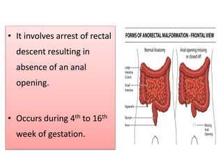
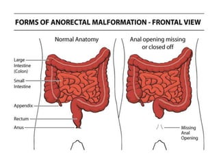
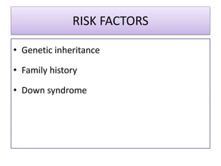
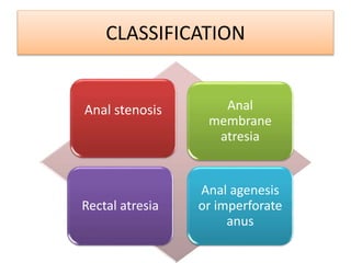
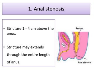
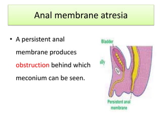
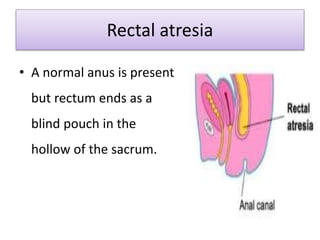
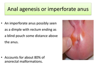
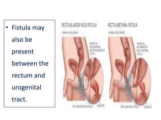
ANORECTAL
MALFORMATION
INTRODUCTION
Comprises a wide spectrum of defects involving
distal anus and rectum as well as urogenital
tract.
• It involves arrest of rectal
descent resulting in
absence of an anal
opening.
• Occurs during 4th to 16th
week of gestation.
INCIDENCE
• Boys are more affected than girls.
• Minor abnormalities occurs in about 1/500 live births
while major abnormalities occur in 1/5000 live
births.
RISK FACTORS
• Genetic inheritance
• Family history
• Down syndrome
CLASSIFICATION
Anal stenosis Anal
membrane
atresia
Rectal atresia
Anal agenesis
or imperforate
anus
1. Anal stenosis
• Stricture 1 - 4 cm above the
anus.
• Stricture may extends
through the entire length
of anus.
Anal membrane atresia
• A persistent anal
membrane produces
obstruction behind which
meconium can be seen.
Rectal atresia
• A normal anus is present
but rectum ends as a
blind pouch in the
hollow of the sacrum.
Anal agenesis or imperforate anus
• An imperforate anus possibly seen
as a dimple with rectum ending as
a blind pouch some distance above
the anus.
• Accounts for about 80% of
anorectal malformations.
• Fistula may
also be
present
between the
rectum and
urogenital
tract.
DIAGNOSTIC EVALUATION
Through newborn assessment:
• No anal opening
• NG tube or thermometer cannot be inserted into the
infant’s rectum.
• No history of passage of meconium
• Presence of meconium in urine (rectovaginal fistula)
• Abdominal USG
• Intravenouspyleogram (IVP)
• Cystourethrogram
DIAGNOSTIC EVALUATION
MANAGEMENT
SURGICAL PROCEDURE:
ANAL STENOSIS:
• Manual dilatation is done by Hegar’s dilators
for increasing the diameter.
ANAL MEMBRANE ATRESIA:
• Incision of membrane or perforation with a
blunt instrument followed by anal dilatation.
IMPERFORATE ANUS:
• Two stage surgery is done.
• First stage: creation of a temporary colostomy.
• Second stage: Intestinal repair and colostomy
closure.
MANAGEMENT
NURSING MANAGEMENT
PRE OPERATIVE:
• Hold oral feeding as soon as the anomaly is noticed.
• IV fluids
• Measure abdominal girth
• Nasogastric aspiration for gastric decompression.
• Monitor vital signs
• Psychological preparation of family members.
POST OPERATIVE
• General post anesthesia care
• IV fluids
• Nasogastric suctioning
• Start oral feeding as the peristalsis begins.
• Care of colostomy.
THANK
YOU

Anorectal Malformation

  • 1.
  • 2.
    INTRODUCTION Comprises a widespectrum of defects involving distal anus and rectum as well as urogenital tract.
  • 3.
    • It involvesarrest of rectal descent resulting in absence of an anal opening. • Occurs during 4th to 16th week of gestation.
  • 5.
    INCIDENCE • Boys aremore affected than girls. • Minor abnormalities occurs in about 1/500 live births while major abnormalities occur in 1/5000 live births.
  • 6.
    RISK FACTORS • Geneticinheritance • Family history • Down syndrome
  • 7.
    CLASSIFICATION Anal stenosis Anal membrane atresia Rectalatresia Anal agenesis or imperforate anus
  • 8.
    1. Anal stenosis •Stricture 1 - 4 cm above the anus. • Stricture may extends through the entire length of anus.
  • 9.
    Anal membrane atresia •A persistent anal membrane produces obstruction behind which meconium can be seen.
  • 10.
    Rectal atresia • Anormal anus is present but rectum ends as a blind pouch in the hollow of the sacrum.
  • 11.
    Anal agenesis orimperforate anus • An imperforate anus possibly seen as a dimple with rectum ending as a blind pouch some distance above the anus. • Accounts for about 80% of anorectal malformations.
  • 12.
    • Fistula may alsobe present between the rectum and urogenital tract.
  • 13.
    DIAGNOSTIC EVALUATION Through newbornassessment: • No anal opening • NG tube or thermometer cannot be inserted into the infant’s rectum. • No history of passage of meconium • Presence of meconium in urine (rectovaginal fistula)
  • 14.
    • Abdominal USG •Intravenouspyleogram (IVP) • Cystourethrogram DIAGNOSTIC EVALUATION
  • 15.
    MANAGEMENT SURGICAL PROCEDURE: ANAL STENOSIS: •Manual dilatation is done by Hegar’s dilators for increasing the diameter. ANAL MEMBRANE ATRESIA: • Incision of membrane or perforation with a blunt instrument followed by anal dilatation.
  • 16.
    IMPERFORATE ANUS: • Twostage surgery is done. • First stage: creation of a temporary colostomy. • Second stage: Intestinal repair and colostomy closure. MANAGEMENT
  • 17.
    NURSING MANAGEMENT PRE OPERATIVE: •Hold oral feeding as soon as the anomaly is noticed. • IV fluids • Measure abdominal girth • Nasogastric aspiration for gastric decompression. • Monitor vital signs • Psychological preparation of family members.
  • 18.
    POST OPERATIVE • Generalpost anesthesia care • IV fluids • Nasogastric suctioning • Start oral feeding as the peristalsis begins. • Care of colostomy.
  • 19.