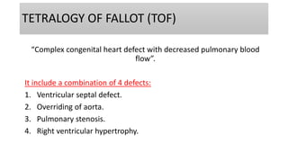
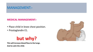
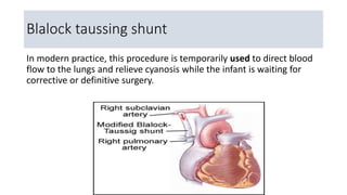
TETRALOGY OF FALLOT (TOF)
“Complex congenital heart defect with decreased pulmonary blood
flow”.
It include a combination of 4 defects:
1. Ventricular septal defect.
2. Overriding of aorta.
3. Pulmonary stenosis.
4. Right ventricular hypertrophy.
RIGHT VENTRICULAR HYPERTROPHY.
• Right ventricular hypertrophy is a heart disorder characterized by
thickening of the walls of the right ventricle.
• It can be caused by excessive stress on the right ventricle.
PULMONARY STENOSIS.
• Pulmonary stenosis is a condition characterized by obstruction to
blood flow from the right ventricle to the pulmonary artery.
• This obstruction is caused by narrowing (stenosis) at one or more
points from the right ventricle to the pulmonary artery.
OVERRIDING OF AORTA.
CLINICAL FEATURES
• Cyanosis.
• Clubbing of fingers.
• Dyspnea.
• Children are small and their nutritional status is poor.
TET’S SPELL
DIAGNOSTIC EVALUATION
• Cardiac examination
• Electrocardiogram
• Chest radiograph
• Echocardiogram
MANAGEMENT:-
MEDICAL MANAGEMENT:-
• Place child in knee chest position.
• Prostaglandin E1.
but why?
This will increase blood flow to the lungs.
And to calm the child.
SURGICALMANAGEMENT:-
Palliative surgery:
• Blalock taussing shunt.
• Pott’s procedure.
• Waterston shunt.
• Brock’s procedure.
Blalock taussing shunt
In modern practice, this procedure is temporarily used to direct blood
flow to the lungs and relieve cyanosis while the infant is waiting for
corrective or definitive surgery.
Corrective surgery:
• A patch closure of VSD.
• Pulmonary valvotomy for relief of pulmonary stenosis.
• Surgical pulmonary valvotomy is a procedure done to open up a
valve.
• It is done by cutting in to the valve leaflets which have become
sealed.
• This is an open-heart procedure
SURGICAL MANAGEMENT
POST OPERATIVE COMPLICATIONS:-
• Conduction abnormalities
• Residual ventricular septal defect
• Residual pulmonary stenosis
• Pulmonary valve regurgitation

TOF

  • 2.
    TETRALOGY OF FALLOT(TOF) “Complex congenital heart defect with decreased pulmonary blood flow”. It include a combination of 4 defects: 1. Ventricular septal defect. 2. Overriding of aorta. 3. Pulmonary stenosis. 4. Right ventricular hypertrophy.
  • 5.
    RIGHT VENTRICULAR HYPERTROPHY. •Right ventricular hypertrophy is a heart disorder characterized by thickening of the walls of the right ventricle. • It can be caused by excessive stress on the right ventricle.
  • 6.
    PULMONARY STENOSIS. • Pulmonarystenosis is a condition characterized by obstruction to blood flow from the right ventricle to the pulmonary artery. • This obstruction is caused by narrowing (stenosis) at one or more points from the right ventricle to the pulmonary artery.
  • 7.
  • 8.
    CLINICAL FEATURES • Cyanosis. •Clubbing of fingers. • Dyspnea. • Children are small and their nutritional status is poor.
  • 9.
  • 10.
    DIAGNOSTIC EVALUATION • Cardiacexamination • Electrocardiogram • Chest radiograph • Echocardiogram
  • 11.
    MANAGEMENT:- MEDICAL MANAGEMENT:- • Placechild in knee chest position. • Prostaglandin E1. but why? This will increase blood flow to the lungs. And to calm the child.
  • 12.
    SURGICALMANAGEMENT:- Palliative surgery: • Blalocktaussing shunt. • Pott’s procedure. • Waterston shunt. • Brock’s procedure.
  • 14.
    Blalock taussing shunt Inmodern practice, this procedure is temporarily used to direct blood flow to the lungs and relieve cyanosis while the infant is waiting for corrective or definitive surgery.
  • 15.
    Corrective surgery: • Apatch closure of VSD. • Pulmonary valvotomy for relief of pulmonary stenosis. • Surgical pulmonary valvotomy is a procedure done to open up a valve. • It is done by cutting in to the valve leaflets which have become sealed. • This is an open-heart procedure SURGICAL MANAGEMENT
  • 16.
    POST OPERATIVE COMPLICATIONS:- •Conduction abnormalities • Residual ventricular septal defect • Residual pulmonary stenosis • Pulmonary valve regurgitation