
Downloaded 120 times


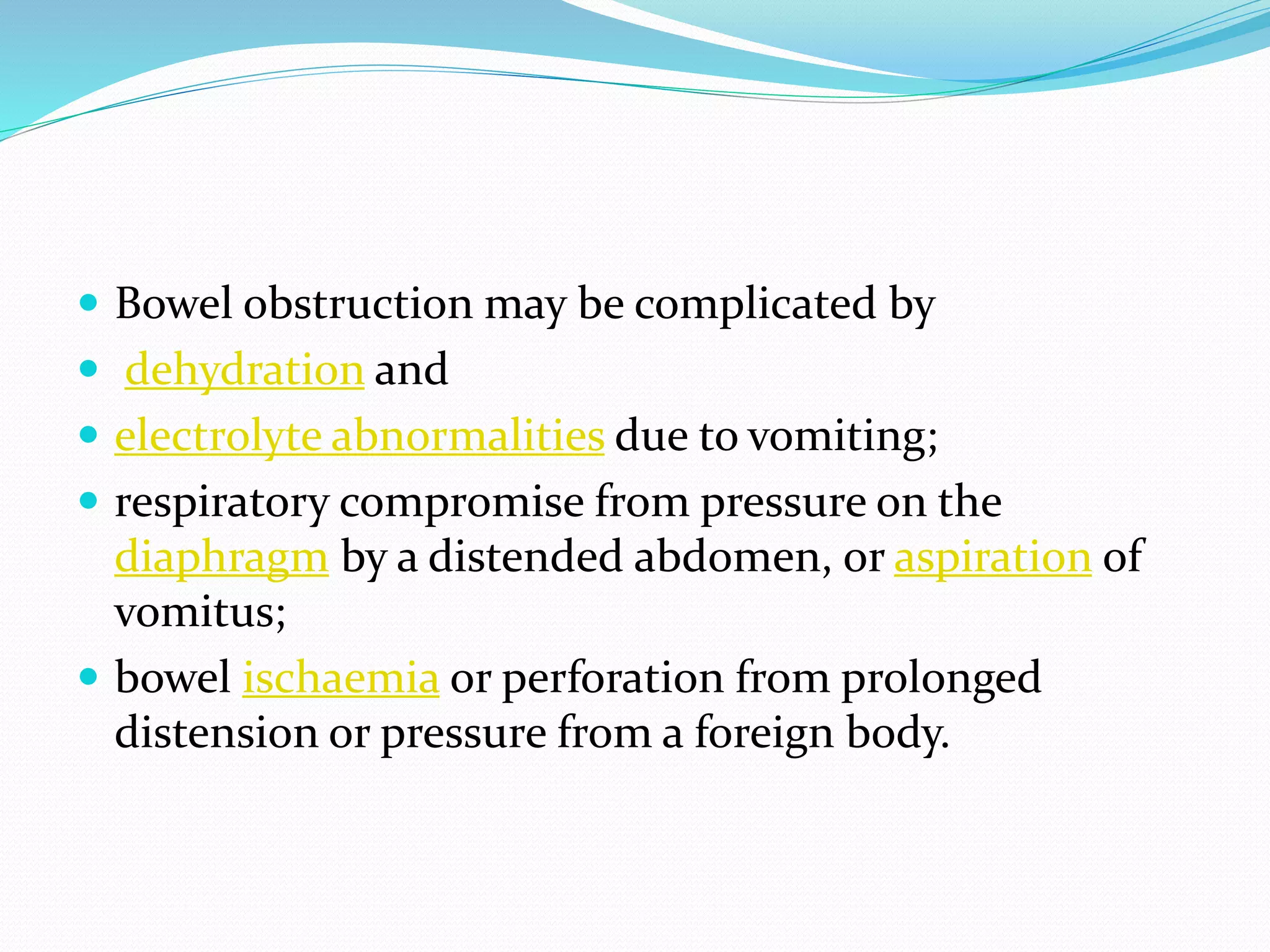
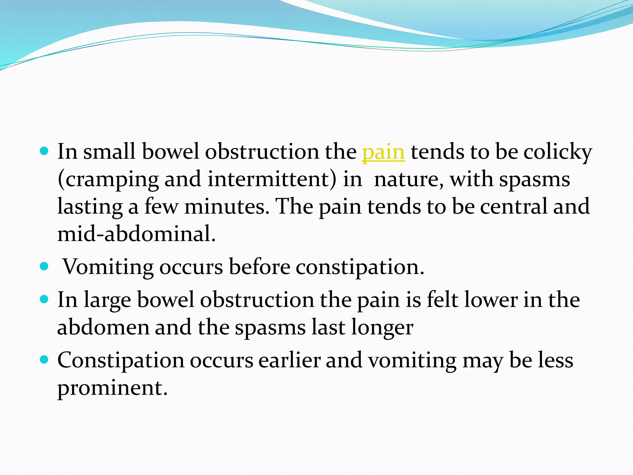

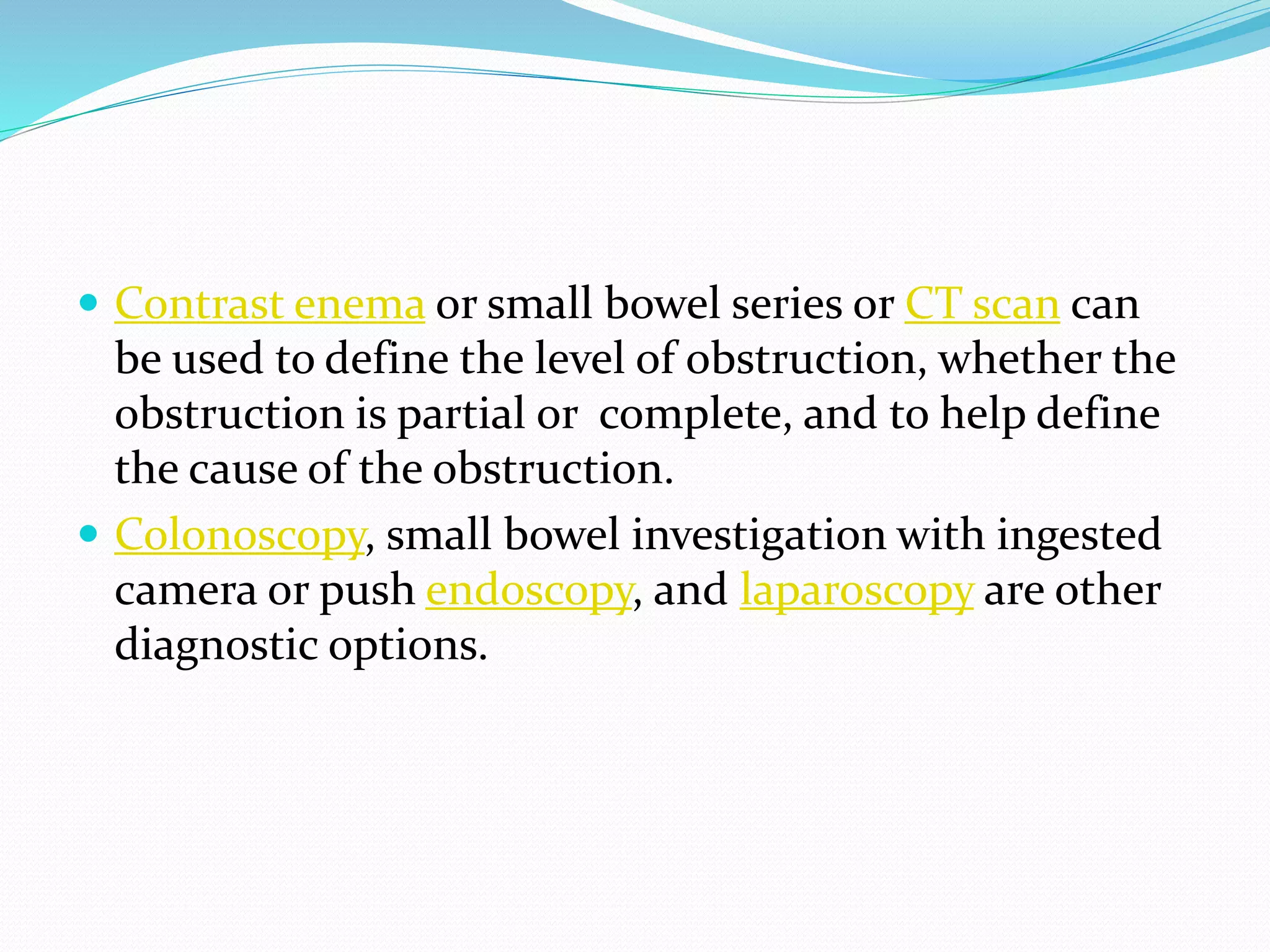

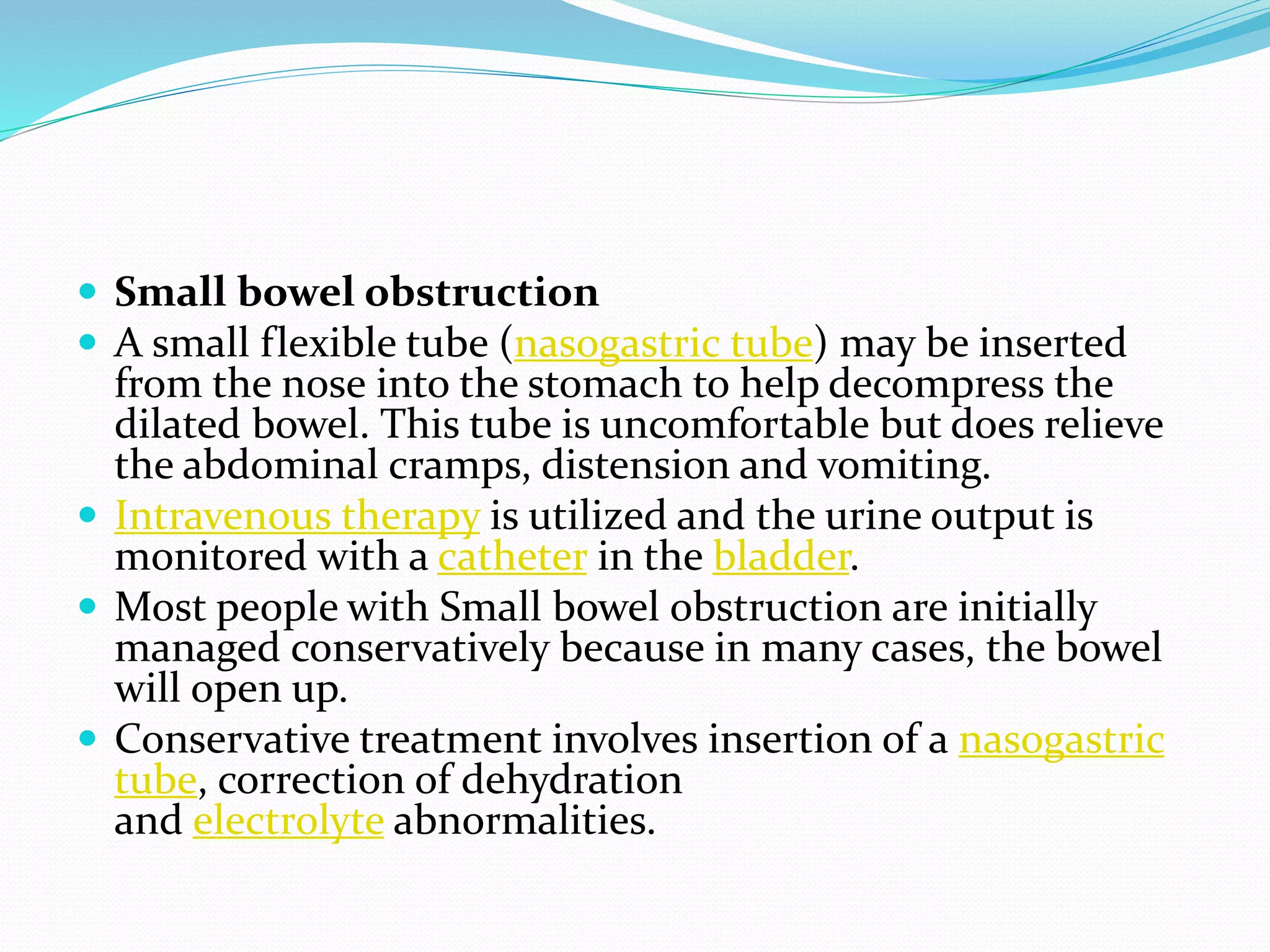


Bowel obstruction occurs when the intestines become blocked, preventing normal movement of digestive contents. There are several types, including small and large bowel obstruction. Causes include adhesions from prior surgery, hernias, tumors, inflammatory bowel disease, and foreign bodies. Symptoms depend on the location but commonly include abdominal pain, distension, vomiting, and constipation. Diagnosis involves medical history, physical exam, imaging tests like CT scans, and sometimes endoscopy or surgery to determine the specific cause. Treatment aims to resolve the blockage through conservative management with NG tubes and IV fluids or potentially surgery. Complications can include dehydration, electrolyte imbalances, infection, and bowel perforation if not properly treated


