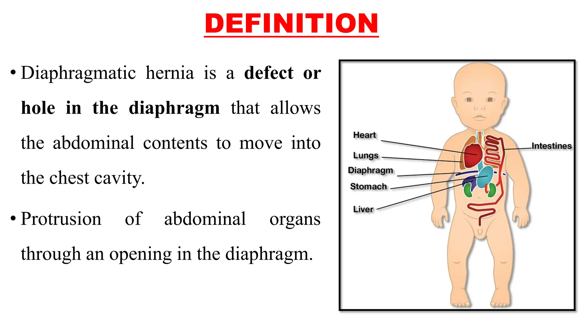
2. DEFINITION
• Diaphragmatic hernia is a defect or
hole in the diaphragm that allows
the abdominal contents to move into
the chest cavity.
• Protrusion of abdominal organs
through an opening in the diaphragm.
3.
4.
5. INCIDENCE
• CDH is a life-threatening condition affecting one in every 2,500-
4,000 births.
• The most serious complication of CDH is inadequate lung
development.
6. TYPES OF CONGENITAL
DIAPHRAGMATIC HERNIA
• There are two types of congenital diaphragmatic hernia (CDH) –
Bochdalek hernia. Morgagni hernia.
7. BOCHDALEK HERNIA
• A Bochdalek hernia is a hole in the diaphragm that can occur on the right or
left side.
• It is caused by either a malformation of the diaphragm or the intestines’
movement into the chest cavity as the diaphragm forms.
• Left-sided Bochdalek hernias are most common, accounting for 80% of cases.
• Bochdalek hernias that occur on the right side are far less common (10% of
cases), but can lead to worse outcomes due to the liver’s movement into the
diaphragm.
8. MORGAGNI HERNIA
• A Morgagni hernia is a hole in the middle of the diaphragm near the chest.
• It happens when the tendon (fibrous connective tissue that attaches muscle to
bone) in the middle of the diaphragm develops abnormally.
• Morgagni hernias are much less common than all Bochdalek hernias.
9. • There is no known cause for the development of congenital
diaphragmatic hernias.
• There is nothing someone can do to cause or prevent their baby from
developing a congenital diaphragmatic hernia.
CAUSES OF CONGENITAL
DIAPHRAGMATIC HERNIA
10. CLINICAL MANIFESTATIONS
• When CDH is not diagnosed prenatally, the symptoms of a congenital
diaphragmatic hernia are observable soon after birth. While they vary
between each child, the most common symptoms of CDH include:
1) Breathing difficulty
2) Rapid breathing (hyperventilating)
3) Rapid heart rate (tachycardia)
11. 4) Blue skin color (cyanosis)
5) One side of the chest is larger (Abnormal Chest Development)
6) Belly looks caved-in (concave/scaphoid)
The specific symptoms of CDH are dependent on the type of hernia,
Bochdalek vs. Morgagni hernias.
Babies with Morgagni hernias sometimes show no symptoms.
CLINICAL MANIFESTATIONS
12. DIAGNOSTIC EVALUATION
1) Fetal ultrasound: Diaphragmatic hernia can be diagnosed using fetal
ultrasound in the second and third trimesters of pregnancy.
• At the 20-week ultrasound, and is often first detected on a routine pre-natal
ultrasound.
2) A fetal echocardiogram: (ultrasound of the heart) may be done to check for
heart abnormalities before birth.
3) X-ray
4) Pulmonary Function Test
5) ABG analysis
13. TREATMENT
• Babies with a congenital diaphragmatic hernia
(CDH) can have different treatment plans
depending on the severity of the case.
• Most times, a baby is diagnosed with CDH in
utero, and during the pregnancy, a treatment
plan is developed prior to giving birth.
14. • Resuscitation with ventilatory support is of prime importance in
patients born with a CDH.
• ECMO has been shown to decrease the mortality of CDH.
• ECMO –(extracorporeal membrane oxygenation)
MEDICAL TREATMENT
15. • ECMO is a therapy used to treat people with life-threatening heart and
lung failure. ECMO involves the use a machine to replace some of the
functions of a patient's lungs or heart, or both simultaneously. While
ECMO therapy can be life-saving, it is not itself a treatment.
• The decision to utilize ECMO is made early in the disease process, usually
within 24 hours of birth.
• An ECMO machine circulates blood through a machine to remove carbon
dioxide and add oxygen.
MEDICAL TREATMENT
17. • Maternal antenatal corticosteroid (e.g. Betamethasone and
dexamethasone) administration has also been employed in an effort
to improve fetal lung growth and maturity, but the available evidence
is insufficient to support its routine use.
• Prostaglandin E1 (PGE1) has been used to treat severe pulmonary
hypertension in patients with CDH.
MEDICAL TREATMENT
19. Median sternotomy
• Median sternotomy is a type of surgical procedure in which a vertical inline
incision is made along the sternum, after which the sternum itself is divided
using a sternal saw (bone cutter).
• This procedure provides access to the heart and lungs for surgical
procedures such as heart transplant, lung transplant.
20. Thoracotomy
• A thoracotomy is a surgical procedure in which a cut is made
between the ribs to see and reach the lungs or other organs in the
chest or thorax.
• A thoracotomy is performed for diagnosis or treatment of a disease
and allows doctors to visualize, biopsy or remove tissue as needed.
22. • The surgery is done under general anesthesia.
• The surgeon usually makes a cut (incision) in the belly under the
lower ribs. This allows the organs in the area to be reached. The
surgeon gently pulls these organs down into place through the
opening in the diaphragm and into the abdominal cavity.
CONGENITAL DIAPHRAGMATIC HERNIA
REPAIR
23. • In less severe cases, the surgery can be done using smaller incisions in the
chest. A small video camera called a thoracoscope is placed through one of
the incisions. This allows the surgeon to view inside the chest.
• If the hole is small, it may be repaired with stitches. Otherwise, an
artificial patch is used to cover the hole.
CONGENITAL DIAPHRAGMATIC HERNIA
REPAIR
24. Fetoscopic Endoluminal Tracheal
Occlusion (FETO)
• Fetoscopic endoluminal tracheal occlusion, or FETO,
is a surgical procedure to treat the most severe cases
of congenital diaphragmatic hernia (CDH) that are
diagnosed in utero.
27. NURSING MANAGEMENT
• PRE-OPERATIVE CARE –
i. Reduce stimulation
ii. Resuscitation, if respiratory distress occurs
iii. Maintain oxygen saturation
iv. IV fluids are given
v. Keep the infant in semi-fowler’s position
vi. For pain relief ,after surgery administer analgesics
vii. Provide rest and comfort to the baby.