SlideShare a Scribd company logo
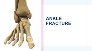
ANKLE
FRACTURE
ANKLE ANATOMY
 Ankle is a three bone joint composed of the
tibia , fibula and talus
 Talus articulates with the tibial plafond
superiorly , posterior malleolus of the tibia
posteriorly and medial malleolus medially
 Lateral articulation is with malleolus of fibula
Anatomy: Lateral Side
Medial view of fibula
Articular surface
Malleolar fossa
Lateral Ridge
Lateral Ligamentous Complex
Ankle Fracture
 Ankle fractures are among the most common injuries and management of
these fractures depends upon careful identification of the extent of bony
injury as well as soft tissue and ligamentous damage.
 Once defined, the key to successful outcome following rotational ankle
fractures is anatomic restoration and healing of ankle mortise.
 The male-to-female ratio for ankle fracture is 2:1
Fracture Stability
•Isolated medial or lateral malleolar fracture •Usually stable
•Posterior malleolus fracture (refers to
the posterior tibia)
•Usually unstable, as often associated with other
malleolar fractures
•Bimalleolar fracture (both medial and lateral
malleolus)
•Mostly unstable
•Trimalleolar fracture (medial, lateral and posterior mall
leolus)
•Always unstable
CLASSIFICATION
 2 Common Classifications
1. Weber Classification
2. Lauge Hansen Classification
The Weber classification categorizes ankle fractures according to the level of the
fibular fracture in relation to the ankle syndesmosis .
(a) Below syndesmosis (b) Level of syndesmosis
(c) Above level of syndesmosis
Weber A: fracture of the lateral malleolus
below the syndesmosis (intact syndesmosis,)
Usually stable.
Weber B: fracture of the lateral malleolus at
the level of the syndesmosis (possible
syndesmotic injury)Variable stability.
Weber C: fracture of the lateral malleolus
above the syndesmosis (ruptured syndesmosis,
torn interosseous membrane), Unstable.
Maisonneuve fracture: proximal subcapital
Weber C fracture; rupture of the syndesmosis
(definite lesions in red); medial malleolus
fracture and/or deltoid ligament tear (possible
lesions in blue) , Unstable
Lauge-Hansen(Depends
on the mechanism of
Injury)
Types:
Supination External Rotation
Supination Adduction
Pronation External Rotation
Pronation Abduction
•based on foot position and force of applied
stress/force
•has been shown to predict the observed (via MRI)
ligamentous injury in less than 50% of operatively
treated fracture
Supination-External Rotation
Stage 1 Anterior tibio- fibular
ligament
Stage 2 Fibula fracture
Stage 3 Posterior malleolus
fracture or posterior tibio-fibular
ligament
Stage 4 Deltoid ligament tear or
medial malleolus fracture
Supination-External
Rotation
Stage 2: Stable
Lateral Injury: classic posterosuperioranteroinferior fibula fracture
Medial Injury: Stability maintained
• Stage 1: fibula fracture is transverse
below mortise.
• Stage 2: medial malleolus fracture is
classic vertical pattern.
Supination Adduction
1
2
Supination Adduction: Stage 2
Lateral Injury: transverse fibular fracture at/below level of mortise
Medial injury: vertical shear type medial malleolar fracture
 Stage 1 Deltoid
ligament tear or
medial malleolus
fracture
 Stage 2 Anterior
tibio-fibular
ligament and
interosseous
membrane
 Stage 3 Spiral,
proximal fibula
fracture
 Stage 4 Posterior
malleolus fracture
or posterior tibio-
fibular ligament
Pronation-External Rotation
Pronation External Rotation: Stage 4
Medial injury: deltoid ligament tear &/or transverse medial malleolar fracture
Lateral Injury: spiral proximal lateral malleolar fracture
HIGHLY UNSTABLE…SYNDESMOTIC INJURY COMMON
 Stage 1 Transverse
medial malleolus fracture
distal to mortise
 Stage 2 Posterior
malleolus fracture or
posterior tibio-fibular
ligament
 Stage 3 Fibula fracture,
typically proximal to
mortise, often with a
butterfly fragment
Pronation-Abduction
Pronation-Abduction
Medial injury: tranverse to short oblique medial malleolar fracture
Lateral Injury: comminuted impaction type distal lateral malleolar fracture
CLINICAL FEATURES
• Local pain, swelling and hematoma
• Tenderness, especially in the area of the malleoli,
the syndesmosis and the posterior aspect of the ankle
joint
• Restricted range of movement
• Skin abnormalities (lacerations, discolorations, tenting,
or blistering)
• In some cases, accompanying injury (e.g., fracture of
the proximal fibula, knee, or foot)
Parts of the distal tibia can be seen protruding
from an 8 cm long wound above the medial
ankle. The foot is in a slightly pronated position.
X-ray findings show a distal fibula fracture at the
level of the syndesmosis (Weber B fracture).
MEHANISM OF INJURY
 Pattern of ankle fracture depends on many factors;
Position of foot and direction of force,
Chronicity or recurrent trauma leading to ligament injury
or laxity and distorted ankle biomechanics.
Patients age,
Bone quality
CLINICAL EVALUATION
 Variable presentation (limp to nonambulatory with severe pain,
swelling and deformity)
 Extent of soft tissue injury must be evaluated.
 Neurovascular status should be carefully documented.
 Entire length of fibula should be palpated for tenderness.
 A dislocated ankle should be reduced and splinted immediately.
• Ottawa ankle rules: used to indicate whether x-ray for ankle and
midfoot injuries is necessary X-ray (plain) of the ankle is indicated
when the patient experiences pain in the malleolar region and has one
one of the following features
• Tenderness at the posterior border or tip of the lateral malleolus
• Tenderness at the posterior border or tip of the medial malleolus
• Inability to bear weight
RADIOGRAPHIC EXAMINATION
 Plain X-ray Films:
•Anterio-posterior view of ankle
•Lateral view of ankle
•Mortise view of ankle
•Stress views when required
•Image the entire tibia, ankle to knee joint,
•Foot films when tender to palpation.
 On the anteroposterior view:-
 -The distal tibia and fibula, including the medial and
lateral malleoli, are well demonstrated.
 -Important note is that the fibular (lateral) malleolus is
longer than the tibial (medial) malleolus.
 -This anatomic feature, important for maintaining
ankle stability, is crucial for reconstruction of the
fractured ankle joint.
 -Even minimal displacement or shortening of the
lateral malleolus allows lateral talar shift to occur and
may cause incongruity in the ankle joint, possibly
leading to posttraumatic arthritis.
ANTEROPOSTERIOR VIEW
 Tibiofibular overlap
<10mm is abnormal – implies syndesmotic injury.
 Tibiofibular clear space
>5mm is abnormal – implies syndesmotic injury.
•Talar tilt
>2mm is considered abnormal.
LATERALVIEW
 Posterior malleolar fractures
can be identified.
 AP Talar Subluxation : Dome
of the talus should be centered
under the tibia and congruous
with the tibial plafond
 Associated injuries to
: Talus
: Calcaneum
MORTISE X-RAY
 The mortise view is about 15 degree of Internal
rotation.
 Useful in evaluation of articular surface between
talar dome and mortise.
 Medial Clear Space
- Between lateral border of medial malleolus and
medial talus
< = 4mm is Normal
>4mm suggests lateral shift of talus
Ankle fracture with displacement
Lateral radiograph of the
ankle:
Ankle fracture with
displacement of the talus (Ta)
in relation to the tibia (Ti).
The shaded area represents
the physiological, anatomical
position of the talus: talus and
tibia usually form the
talocrural joint. Because of
the displacement of the talus
(green line), the joint cannot
function normally.
OTHER IMAGING MODALITIES
 CT views
- Joint involvement
- Posterior malleolar fracture pattern
- Pre-operative planning
- Evaluate hindfoot and midfoot it needed
 MRI
-Ligament and tendon injury
- Syndesmosis injuries
TREATMENT
• Initial management: rest, ice, compression, and elevation
• Conservative treatment
• Indications: stable fractures (isolated/nondisplaced malleolar fractures)
• Short leg cast for 4–6 weeks
• Surgical treatment: to ensure normal alignment of bone and cartilage to prevent ankle arthritis
and to regain functionality
• Indications: unstable/displaced fractures, open ankle fractures, and cases of neurovascular damage
• Technique: reposition and internal or external fixation with metal plates and/or screws
• Aim of the operation is to ensure anatomical reduction of the ankle-mortise.This means
ensuring anatomical reduction of medial and lateral malleoli,and reduction of the talus
accurately within the mortise
 Unstable fractures require surgery, most often an open reduction and internal
fixation (ORIF), which is usually performed with permanently implanted metal hardware
that holds the bones in place while the natural healing process occurs.
A cast or splint will be required to immobilize the ankle following surgery
 Fractures with displacement
Aim of the operation is to ensure anatomical reduction of the ankle-
mortise.This means ensuring anatomical reduction of medial and lateral
malleoli,and reduction of the talus accurately within the mortise
WEBER B FRACTURE OF THE UPPER RIGHT ANKLE AFTER SURGERY
Posteroanterior X-ray of the right upper ankle: The fibula
fracture was treated by internal fixation by plate
osteosynthesis. An adjusting screw is used to fix the position
of the joint until healing of the syndesmotic injury is
achieved.
WEBER B FRACTURE OF THE UPPER RIGHT ANKLE POST-SURGERY
X-ray of the upper right anke (lateral view):
internal fixation using plate osteosynthesis and
an adjusting screw
COMPLICATIONS
 General complications of Fracture
• Damage to the peroneal nerve or saphenous nerve
• Post-traumatic osteoarthritis
REFERENCE
 http://rad.desk.nl/en/p420a20ca7196b/ankle-fracture-weber-and-lauge-
hansen-classification.html
 https://www.physio-pedia.com/Ankle_and_Foot_Fractures
 https://www.orthobullets.com/trauma/1047/ankle-fractures
 https://app.pulsenotes.com/surgery/orthopaedics/notes/ankle-fractures
 https://www.hss.edu/condition-list_ankle-fractures.asp
 https://orthoinfo.aaos.org/en/diseases--conditions/ankle-fractures-broken-
ankle/
Thank you

More Related Content

What's hot

Triple arthrodesis seminar by Dr Chirag Patel
Triple arthrodesis seminar by Dr Chirag PatelTriple arthrodesis seminar by Dr Chirag Patel
Triple arthrodesis seminar by Dr Chirag Patel
Chirag Patel
 
CORA (center of rotation of angulation)
CORA (center of rotation of angulation)CORA (center of rotation of angulation)
CORA (center of rotation of angulation)
Morshed Abir
 
Various ankle injuries and it’s management
Various ankle injuries and it’s managementVarious ankle injuries and it’s management
Various ankle injuries and it’s management
Amol Gaikwad
 
Tarsal coalition
Tarsal coalitionTarsal coalition
Tarsal coalition
Harsha Nandini
 
Radial head fracture
Radial head fractureRadial head fracture
Radial head fracture
Krunal Patel
 
Pelvic fracture classification
Pelvic fracture classificationPelvic fracture classification
Pelvic fracture classification
Zahid Askar
 
Knee dislocation
Knee dislocationKnee dislocation
Knee dislocation
Gajanan Pandit
 
Osteotomies around the hip
Osteotomies around the hipOsteotomies around the hip
Osteotomies around the hip
Santoshi Tanabuddi
 
Principles of deformity correction
Principles of deformity correctionPrinciples of deformity correction
Principles of deformity correction
Abdulla Kamal
 
Triple arthrodesis
Triple arthrodesisTriple arthrodesis
Triple arthrodesis
Ponnilavan Ponz
 
Primary total knee arthroplasty
Primary total knee arthroplastyPrimary total knee arthroplasty
Primary total knee arthroplastyjatinder12345
 
Superior Shoulder Suspensory Complex injuries (SSSC)
Superior Shoulder Suspensory Complex injuries (SSSC)Superior Shoulder Suspensory Complex injuries (SSSC)
Superior Shoulder Suspensory Complex injuries (SSSC)
Jaganmohan Sontyana
 
Protrusio acetabuli
Protrusio acetabuliProtrusio acetabuli
Protrusio acetabuli
Ghazwan Bayaty
 
Congenital vertical talus BY DR.NAVEEN RATHOR
Congenital vertical talus BY DR.NAVEEN RATHORCongenital vertical talus BY DR.NAVEEN RATHOR
Congenital vertical talus BY DR.NAVEEN RATHOR
DR.Naveen Rathor
 
Templating X-rays in THR
Templating X-rays in THR Templating X-rays in THR
Templating X-rays in THR
Dr. Bushu Harna
 
Fracture of talus ppt
Fracture of talus pptFracture of talus ppt
Fracture of talus ppt
sunil kumar daha
 
Mipo
Mipo Mipo
Non union neck of femur
Non union neck of femurNon union neck of femur
Non union neck of femur
ramachandra reddy
 
Fractures and dislocations of hand
Fractures and dislocations of handFractures and dislocations of hand
Fractures and dislocations of hand
Aftab Alam
 
Fracture of Distal End Humerus.
Fracture of Distal End Humerus.Fracture of Distal End Humerus.
Fracture of Distal End Humerus.
Dr.Anshu Sharma
 

What's hot (20)

Triple arthrodesis seminar by Dr Chirag Patel
Triple arthrodesis seminar by Dr Chirag PatelTriple arthrodesis seminar by Dr Chirag Patel
Triple arthrodesis seminar by Dr Chirag Patel
 
CORA (center of rotation of angulation)
CORA (center of rotation of angulation)CORA (center of rotation of angulation)
CORA (center of rotation of angulation)
 
Various ankle injuries and it’s management
Various ankle injuries and it’s managementVarious ankle injuries and it’s management
Various ankle injuries and it’s management
 
Tarsal coalition
Tarsal coalitionTarsal coalition
Tarsal coalition
 
Radial head fracture
Radial head fractureRadial head fracture
Radial head fracture
 
Pelvic fracture classification
Pelvic fracture classificationPelvic fracture classification
Pelvic fracture classification
 
Knee dislocation
Knee dislocationKnee dislocation
Knee dislocation
 
Osteotomies around the hip
Osteotomies around the hipOsteotomies around the hip
Osteotomies around the hip
 
Principles of deformity correction
Principles of deformity correctionPrinciples of deformity correction
Principles of deformity correction
 
Triple arthrodesis
Triple arthrodesisTriple arthrodesis
Triple arthrodesis
 
Primary total knee arthroplasty
Primary total knee arthroplastyPrimary total knee arthroplasty
Primary total knee arthroplasty
 
Superior Shoulder Suspensory Complex injuries (SSSC)
Superior Shoulder Suspensory Complex injuries (SSSC)Superior Shoulder Suspensory Complex injuries (SSSC)
Superior Shoulder Suspensory Complex injuries (SSSC)
 
Protrusio acetabuli
Protrusio acetabuliProtrusio acetabuli
Protrusio acetabuli
 
Congenital vertical talus BY DR.NAVEEN RATHOR
Congenital vertical talus BY DR.NAVEEN RATHORCongenital vertical talus BY DR.NAVEEN RATHOR
Congenital vertical talus BY DR.NAVEEN RATHOR
 
Templating X-rays in THR
Templating X-rays in THR Templating X-rays in THR
Templating X-rays in THR
 
Fracture of talus ppt
Fracture of talus pptFracture of talus ppt
Fracture of talus ppt
 
Mipo
Mipo Mipo
Mipo
 
Non union neck of femur
Non union neck of femurNon union neck of femur
Non union neck of femur
 
Fractures and dislocations of hand
Fractures and dislocations of handFractures and dislocations of hand
Fractures and dislocations of hand
 
Fracture of Distal End Humerus.
Fracture of Distal End Humerus.Fracture of Distal End Humerus.
Fracture of Distal End Humerus.
 

Similar to Ankle fracture

L12 ankle fxs
L12 ankle fxsL12 ankle fxs
L12 ankle fxs
Claudiu Cucu
 
Ankle fracture
Ankle fracture Ankle fracture
Ankle fracture
AashishYadav35
 
Anklefracturesfinal 120314092959-phpapp01
Anklefracturesfinal 120314092959-phpapp01Anklefracturesfinal 120314092959-phpapp01
Anklefracturesfinal 120314092959-phpapp01
Drpraveen Kumar
 
Ankle fracture
Ankle fractureAnkle fracture
Ankle fracture
mrudevgandhi1
 
Ankle seminar
Ankle seminarAnkle seminar
Ankle seminar
Arun Sivaram
 
Injuries around the ankle by Dr Bipul Borthakur ppt
Injuries around the ankle by Dr Bipul Borthakur pptInjuries around the ankle by Dr Bipul Borthakur ppt
Injuries around the ankle by Dr Bipul Borthakur ppt
BipulBorthakur
 
Ankle and foot injuries
Ankle and foot injuriesAnkle and foot injuries
Ankle and foot injuries
Amardeep kaur
 
Ankle injuries & Trimallelor fracture classification
Ankle injuries & Trimallelor  fracture classification Ankle injuries & Trimallelor  fracture classification
Ankle injuries & Trimallelor fracture classification
Ponnilavan Ponz
 
Closed ankle injuries
Closed ankle injuriesClosed ankle injuries
Closed ankle injuries
Asi-oqua Bassey
 
Ankle fractures management
Ankle fractures   managementAnkle fractures   management
Ankle fractures management
Sunil Santhosh
 
Ankle fractures
Ankle fracturesAnkle fractures
Ankle fractures
BipulBorthakur
 
SPORTS INJURIES OF ANKLE AND FOOT original.pptx
SPORTS INJURIES OF ANKLE AND FOOT original.pptxSPORTS INJURIES OF ANKLE AND FOOT original.pptx
SPORTS INJURIES OF ANKLE AND FOOT original.pptx
MeghaPrakash9
 
Orthopedics 5th year, 5th lecture (Dr. Ali A.Nabi)
Orthopedics 5th year, 5th lecture (Dr. Ali A.Nabi)Orthopedics 5th year, 5th lecture (Dr. Ali A.Nabi)
Orthopedics 5th year, 5th lecture (Dr. Ali A.Nabi)
College of Medicine, Sulaymaniyah
 
Ankle injuries - Copy.pptx
Ankle injuries - Copy.pptxAnkle injuries - Copy.pptx
Ankle injuries - Copy.pptx
SwetaUpadhyay18
 
Ankle joint final
Ankle   joint finalAnkle   joint final
Ankle joint final
Sahil Arora
 
ANKLE FRACTURES
ANKLE FRACTURESANKLE FRACTURES
ANKLE FRACTURES
Umesh Yadav
 
Orthopedics 5th year, 7th/part two & 8th lectures (Dr. Ali A.Nabi)
Orthopedics 5th year, 7th/part two & 8th lectures (Dr. Ali A.Nabi)Orthopedics 5th year, 7th/part two & 8th lectures (Dr. Ali A.Nabi)
Orthopedics 5th year, 7th/part two & 8th lectures (Dr. Ali A.Nabi)
College of Medicine, Sulaymaniyah
 
Ankle injury amanj
Ankle injury amanjAnkle injury amanj
Ankle injury amanjAmanj Gardi
 
Lower Limb Fractures.pptx
Lower Limb Fractures.pptxLower Limb Fractures.pptx
Lower Limb Fractures.pptx
Udit Biswal
 

Similar to Ankle fracture (20)

L12 ankle fxs
L12 ankle fxsL12 ankle fxs
L12 ankle fxs
 
Ankle fracture
Ankle fracture Ankle fracture
Ankle fracture
 
Anklefracturesfinal 120314092959-phpapp01
Anklefracturesfinal 120314092959-phpapp01Anklefracturesfinal 120314092959-phpapp01
Anklefracturesfinal 120314092959-phpapp01
 
Ankle fracture
Ankle fractureAnkle fracture
Ankle fracture
 
Ankle seminar
Ankle seminarAnkle seminar
Ankle seminar
 
Injuries around the ankle by Dr Bipul Borthakur ppt
Injuries around the ankle by Dr Bipul Borthakur pptInjuries around the ankle by Dr Bipul Borthakur ppt
Injuries around the ankle by Dr Bipul Borthakur ppt
 
Ankle and foot injuries
Ankle and foot injuriesAnkle and foot injuries
Ankle and foot injuries
 
Ankle injuries & Trimallelor fracture classification
Ankle injuries & Trimallelor  fracture classification Ankle injuries & Trimallelor  fracture classification
Ankle injuries & Trimallelor fracture classification
 
Closed ankle injuries
Closed ankle injuriesClosed ankle injuries
Closed ankle injuries
 
Ankle fractures management
Ankle fractures   managementAnkle fractures   management
Ankle fractures management
 
Ankle fractures
Ankle fracturesAnkle fractures
Ankle fractures
 
Ankle fractures
Ankle fracturesAnkle fractures
Ankle fractures
 
SPORTS INJURIES OF ANKLE AND FOOT original.pptx
SPORTS INJURIES OF ANKLE AND FOOT original.pptxSPORTS INJURIES OF ANKLE AND FOOT original.pptx
SPORTS INJURIES OF ANKLE AND FOOT original.pptx
 
Orthopedics 5th year, 5th lecture (Dr. Ali A.Nabi)
Orthopedics 5th year, 5th lecture (Dr. Ali A.Nabi)Orthopedics 5th year, 5th lecture (Dr. Ali A.Nabi)
Orthopedics 5th year, 5th lecture (Dr. Ali A.Nabi)
 
Ankle injuries - Copy.pptx
Ankle injuries - Copy.pptxAnkle injuries - Copy.pptx
Ankle injuries - Copy.pptx
 
Ankle joint final
Ankle   joint finalAnkle   joint final
Ankle joint final
 
ANKLE FRACTURES
ANKLE FRACTURESANKLE FRACTURES
ANKLE FRACTURES
 
Orthopedics 5th year, 7th/part two & 8th lectures (Dr. Ali A.Nabi)
Orthopedics 5th year, 7th/part two & 8th lectures (Dr. Ali A.Nabi)Orthopedics 5th year, 7th/part two & 8th lectures (Dr. Ali A.Nabi)
Orthopedics 5th year, 7th/part two & 8th lectures (Dr. Ali A.Nabi)
 
Ankle injury amanj
Ankle injury amanjAnkle injury amanj
Ankle injury amanj
 
Lower Limb Fractures.pptx
Lower Limb Fractures.pptxLower Limb Fractures.pptx
Lower Limb Fractures.pptx
 

Recently uploaded

ARTHROLOGY PPT NCISM SYLLABUS AYURVEDA STUDENTS
ARTHROLOGY PPT NCISM SYLLABUS AYURVEDA STUDENTSARTHROLOGY PPT NCISM SYLLABUS AYURVEDA STUDENTS
ARTHROLOGY PPT NCISM SYLLABUS AYURVEDA STUDENTS
Dr. Vinay Pareek
 
Prix Galien International 2024 Forum Program
Prix Galien International 2024 Forum ProgramPrix Galien International 2024 Forum Program
Prix Galien International 2024 Forum Program
Levi Shapiro
 
Pharynx and Clinical Correlations BY Dr.Rabia Inam Gandapore.pptx
Pharynx and Clinical Correlations BY Dr.Rabia Inam Gandapore.pptxPharynx and Clinical Correlations BY Dr.Rabia Inam Gandapore.pptx
Pharynx and Clinical Correlations BY Dr.Rabia Inam Gandapore.pptx
Dr. Rabia Inam Gandapore
 
BENIGN PROSTATIC HYPERPLASIA.BPH. BPHpdf
BENIGN PROSTATIC HYPERPLASIA.BPH. BPHpdfBENIGN PROSTATIC HYPERPLASIA.BPH. BPHpdf
BENIGN PROSTATIC HYPERPLASIA.BPH. BPHpdf
DR SETH JOTHAM
 
24 Upakrama.pptx class ppt useful in all
24 Upakrama.pptx class ppt useful in all24 Upakrama.pptx class ppt useful in all
24 Upakrama.pptx class ppt useful in all
DrSathishMS1
 
Charaka Samhita Sutra sthana Chapter 15 Upakalpaniyaadhyaya
Charaka Samhita Sutra sthana Chapter 15 UpakalpaniyaadhyayaCharaka Samhita Sutra sthana Chapter 15 Upakalpaniyaadhyaya
Charaka Samhita Sutra sthana Chapter 15 Upakalpaniyaadhyaya
Dr KHALID B.M
 
KDIGO 2024 guidelines for diabetologists
KDIGO 2024 guidelines for diabetologistsKDIGO 2024 guidelines for diabetologists
KDIGO 2024 guidelines for diabetologists
د.محمود نجيب
 
Cervical & Brachial Plexus By Dr. RIG.pptx
Cervical & Brachial Plexus By Dr. RIG.pptxCervical & Brachial Plexus By Dr. RIG.pptx
Cervical & Brachial Plexus By Dr. RIG.pptx
Dr. Rabia Inam Gandapore
 
Tom Selleck Health: A Comprehensive Look at the Iconic Actor’s Wellness Journey
Tom Selleck Health: A Comprehensive Look at the Iconic Actor’s Wellness JourneyTom Selleck Health: A Comprehensive Look at the Iconic Actor’s Wellness Journey
Tom Selleck Health: A Comprehensive Look at the Iconic Actor’s Wellness Journey
greendigital
 
Hemodialysis: Chapter 3, Dialysis Water Unit - Dr.Gawad
Hemodialysis: Chapter 3, Dialysis Water Unit - Dr.GawadHemodialysis: Chapter 3, Dialysis Water Unit - Dr.Gawad
Hemodialysis: Chapter 3, Dialysis Water Unit - Dr.Gawad
NephroTube - Dr.Gawad
 
Flu Vaccine Alert in Bangalore Karnataka
Flu Vaccine Alert in Bangalore KarnatakaFlu Vaccine Alert in Bangalore Karnataka
Flu Vaccine Alert in Bangalore Karnataka
addon Scans
 
New Drug Discovery and Development .....
New Drug Discovery and Development .....New Drug Discovery and Development .....
New Drug Discovery and Development .....
NEHA GUPTA
 
ARTIFICIAL INTELLIGENCE IN HEALTHCARE.pdf
ARTIFICIAL INTELLIGENCE IN  HEALTHCARE.pdfARTIFICIAL INTELLIGENCE IN  HEALTHCARE.pdf
ARTIFICIAL INTELLIGENCE IN HEALTHCARE.pdf
Anujkumaranit
 
Alcohol_Dr. Jeenal Mistry MD Pharmacology.pdf
Alcohol_Dr. Jeenal Mistry MD Pharmacology.pdfAlcohol_Dr. Jeenal Mistry MD Pharmacology.pdf
Alcohol_Dr. Jeenal Mistry MD Pharmacology.pdf
Dr Jeenal Mistry
 
The Normal Electrocardiogram - Part I of II
The Normal Electrocardiogram - Part I of IIThe Normal Electrocardiogram - Part I of II
The Normal Electrocardiogram - Part I of II
MedicoseAcademics
 
Physiology of Chemical Sensation of smell.pdf
Physiology of Chemical Sensation of smell.pdfPhysiology of Chemical Sensation of smell.pdf
Physiology of Chemical Sensation of smell.pdf
MedicoseAcademics
 
ACUTE SCROTUM.....pdf. ACUTE SCROTAL CONDITIOND
ACUTE SCROTUM.....pdf. ACUTE SCROTAL CONDITIONDACUTE SCROTUM.....pdf. ACUTE SCROTAL CONDITIOND
ACUTE SCROTUM.....pdf. ACUTE SCROTAL CONDITIOND
DR SETH JOTHAM
 
263778731218 Abortion Clinic /Pills In Harare ,
263778731218 Abortion Clinic /Pills In Harare ,263778731218 Abortion Clinic /Pills In Harare ,
263778731218 Abortion Clinic /Pills In Harare ,
sisternakatoto
 
Are There Any Natural Remedies To Treat Syphilis.pdf
Are There Any Natural Remedies To Treat Syphilis.pdfAre There Any Natural Remedies To Treat Syphilis.pdf
Are There Any Natural Remedies To Treat Syphilis.pdf
Little Cross Family Clinic
 
Lung Cancer: Artificial Intelligence, Synergetics, Complex System Analysis, S...
Lung Cancer: Artificial Intelligence, Synergetics, Complex System Analysis, S...Lung Cancer: Artificial Intelligence, Synergetics, Complex System Analysis, S...
Lung Cancer: Artificial Intelligence, Synergetics, Complex System Analysis, S...
Oleg Kshivets
 

Recently uploaded (20)

ARTHROLOGY PPT NCISM SYLLABUS AYURVEDA STUDENTS
ARTHROLOGY PPT NCISM SYLLABUS AYURVEDA STUDENTSARTHROLOGY PPT NCISM SYLLABUS AYURVEDA STUDENTS
ARTHROLOGY PPT NCISM SYLLABUS AYURVEDA STUDENTS
 
Prix Galien International 2024 Forum Program
Prix Galien International 2024 Forum ProgramPrix Galien International 2024 Forum Program
Prix Galien International 2024 Forum Program
 
Pharynx and Clinical Correlations BY Dr.Rabia Inam Gandapore.pptx
Pharynx and Clinical Correlations BY Dr.Rabia Inam Gandapore.pptxPharynx and Clinical Correlations BY Dr.Rabia Inam Gandapore.pptx
Pharynx and Clinical Correlations BY Dr.Rabia Inam Gandapore.pptx
 
BENIGN PROSTATIC HYPERPLASIA.BPH. BPHpdf
BENIGN PROSTATIC HYPERPLASIA.BPH. BPHpdfBENIGN PROSTATIC HYPERPLASIA.BPH. BPHpdf
BENIGN PROSTATIC HYPERPLASIA.BPH. BPHpdf
 
24 Upakrama.pptx class ppt useful in all
24 Upakrama.pptx class ppt useful in all24 Upakrama.pptx class ppt useful in all
24 Upakrama.pptx class ppt useful in all
 
Charaka Samhita Sutra sthana Chapter 15 Upakalpaniyaadhyaya
Charaka Samhita Sutra sthana Chapter 15 UpakalpaniyaadhyayaCharaka Samhita Sutra sthana Chapter 15 Upakalpaniyaadhyaya
Charaka Samhita Sutra sthana Chapter 15 Upakalpaniyaadhyaya
 
KDIGO 2024 guidelines for diabetologists
KDIGO 2024 guidelines for diabetologistsKDIGO 2024 guidelines for diabetologists
KDIGO 2024 guidelines for diabetologists
 
Cervical & Brachial Plexus By Dr. RIG.pptx
Cervical & Brachial Plexus By Dr. RIG.pptxCervical & Brachial Plexus By Dr. RIG.pptx
Cervical & Brachial Plexus By Dr. RIG.pptx
 
Tom Selleck Health: A Comprehensive Look at the Iconic Actor’s Wellness Journey
Tom Selleck Health: A Comprehensive Look at the Iconic Actor’s Wellness JourneyTom Selleck Health: A Comprehensive Look at the Iconic Actor’s Wellness Journey
Tom Selleck Health: A Comprehensive Look at the Iconic Actor’s Wellness Journey
 
Hemodialysis: Chapter 3, Dialysis Water Unit - Dr.Gawad
Hemodialysis: Chapter 3, Dialysis Water Unit - Dr.GawadHemodialysis: Chapter 3, Dialysis Water Unit - Dr.Gawad
Hemodialysis: Chapter 3, Dialysis Water Unit - Dr.Gawad
 
Flu Vaccine Alert in Bangalore Karnataka
Flu Vaccine Alert in Bangalore KarnatakaFlu Vaccine Alert in Bangalore Karnataka
Flu Vaccine Alert in Bangalore Karnataka
 
New Drug Discovery and Development .....
New Drug Discovery and Development .....New Drug Discovery and Development .....
New Drug Discovery and Development .....
 
ARTIFICIAL INTELLIGENCE IN HEALTHCARE.pdf
ARTIFICIAL INTELLIGENCE IN  HEALTHCARE.pdfARTIFICIAL INTELLIGENCE IN  HEALTHCARE.pdf
ARTIFICIAL INTELLIGENCE IN HEALTHCARE.pdf
 
Alcohol_Dr. Jeenal Mistry MD Pharmacology.pdf
Alcohol_Dr. Jeenal Mistry MD Pharmacology.pdfAlcohol_Dr. Jeenal Mistry MD Pharmacology.pdf
Alcohol_Dr. Jeenal Mistry MD Pharmacology.pdf
 
The Normal Electrocardiogram - Part I of II
The Normal Electrocardiogram - Part I of IIThe Normal Electrocardiogram - Part I of II
The Normal Electrocardiogram - Part I of II
 
Physiology of Chemical Sensation of smell.pdf
Physiology of Chemical Sensation of smell.pdfPhysiology of Chemical Sensation of smell.pdf
Physiology of Chemical Sensation of smell.pdf
 
ACUTE SCROTUM.....pdf. ACUTE SCROTAL CONDITIOND
ACUTE SCROTUM.....pdf. ACUTE SCROTAL CONDITIONDACUTE SCROTUM.....pdf. ACUTE SCROTAL CONDITIOND
ACUTE SCROTUM.....pdf. ACUTE SCROTAL CONDITIOND
 
263778731218 Abortion Clinic /Pills In Harare ,
263778731218 Abortion Clinic /Pills In Harare ,263778731218 Abortion Clinic /Pills In Harare ,
263778731218 Abortion Clinic /Pills In Harare ,
 
Are There Any Natural Remedies To Treat Syphilis.pdf
Are There Any Natural Remedies To Treat Syphilis.pdfAre There Any Natural Remedies To Treat Syphilis.pdf
Are There Any Natural Remedies To Treat Syphilis.pdf
 
Lung Cancer: Artificial Intelligence, Synergetics, Complex System Analysis, S...
Lung Cancer: Artificial Intelligence, Synergetics, Complex System Analysis, S...Lung Cancer: Artificial Intelligence, Synergetics, Complex System Analysis, S...
Lung Cancer: Artificial Intelligence, Synergetics, Complex System Analysis, S...
 

Ankle fracture

  • 2. ANKLE ANATOMY  Ankle is a three bone joint composed of the tibia , fibula and talus  Talus articulates with the tibial plafond superiorly , posterior malleolus of the tibia posteriorly and medial malleolus medially  Lateral articulation is with malleolus of fibula
  • 3.
  • 4. Anatomy: Lateral Side Medial view of fibula Articular surface Malleolar fossa Lateral Ridge Lateral Ligamentous Complex
  • 5. Ankle Fracture  Ankle fractures are among the most common injuries and management of these fractures depends upon careful identification of the extent of bony injury as well as soft tissue and ligamentous damage.  Once defined, the key to successful outcome following rotational ankle fractures is anatomic restoration and healing of ankle mortise.  The male-to-female ratio for ankle fracture is 2:1
  • 6. Fracture Stability •Isolated medial or lateral malleolar fracture •Usually stable •Posterior malleolus fracture (refers to the posterior tibia) •Usually unstable, as often associated with other malleolar fractures •Bimalleolar fracture (both medial and lateral malleolus) •Mostly unstable •Trimalleolar fracture (medial, lateral and posterior mall leolus) •Always unstable
  • 7. CLASSIFICATION  2 Common Classifications 1. Weber Classification 2. Lauge Hansen Classification The Weber classification categorizes ankle fractures according to the level of the fibular fracture in relation to the ankle syndesmosis . (a) Below syndesmosis (b) Level of syndesmosis (c) Above level of syndesmosis
  • 8. Weber A: fracture of the lateral malleolus below the syndesmosis (intact syndesmosis,) Usually stable. Weber B: fracture of the lateral malleolus at the level of the syndesmosis (possible syndesmotic injury)Variable stability. Weber C: fracture of the lateral malleolus above the syndesmosis (ruptured syndesmosis, torn interosseous membrane), Unstable. Maisonneuve fracture: proximal subcapital Weber C fracture; rupture of the syndesmosis (definite lesions in red); medial malleolus fracture and/or deltoid ligament tear (possible lesions in blue) , Unstable
  • 9. Lauge-Hansen(Depends on the mechanism of Injury) Types: Supination External Rotation Supination Adduction Pronation External Rotation Pronation Abduction •based on foot position and force of applied stress/force •has been shown to predict the observed (via MRI) ligamentous injury in less than 50% of operatively treated fracture
  • 10. Supination-External Rotation Stage 1 Anterior tibio- fibular ligament Stage 2 Fibula fracture Stage 3 Posterior malleolus fracture or posterior tibio-fibular ligament Stage 4 Deltoid ligament tear or medial malleolus fracture
  • 11. Supination-External Rotation Stage 2: Stable Lateral Injury: classic posterosuperioranteroinferior fibula fracture Medial Injury: Stability maintained
  • 12. • Stage 1: fibula fracture is transverse below mortise. • Stage 2: medial malleolus fracture is classic vertical pattern. Supination Adduction 1 2
  • 13. Supination Adduction: Stage 2 Lateral Injury: transverse fibular fracture at/below level of mortise Medial injury: vertical shear type medial malleolar fracture
  • 14.  Stage 1 Deltoid ligament tear or medial malleolus fracture  Stage 2 Anterior tibio-fibular ligament and interosseous membrane  Stage 3 Spiral, proximal fibula fracture  Stage 4 Posterior malleolus fracture or posterior tibio- fibular ligament Pronation-External Rotation
  • 15. Pronation External Rotation: Stage 4 Medial injury: deltoid ligament tear &/or transverse medial malleolar fracture Lateral Injury: spiral proximal lateral malleolar fracture HIGHLY UNSTABLE…SYNDESMOTIC INJURY COMMON
  • 16.  Stage 1 Transverse medial malleolus fracture distal to mortise  Stage 2 Posterior malleolus fracture or posterior tibio-fibular ligament  Stage 3 Fibula fracture, typically proximal to mortise, often with a butterfly fragment Pronation-Abduction
  • 17. Pronation-Abduction Medial injury: tranverse to short oblique medial malleolar fracture Lateral Injury: comminuted impaction type distal lateral malleolar fracture
  • 18. CLINICAL FEATURES • Local pain, swelling and hematoma • Tenderness, especially in the area of the malleoli, the syndesmosis and the posterior aspect of the ankle joint • Restricted range of movement • Skin abnormalities (lacerations, discolorations, tenting, or blistering) • In some cases, accompanying injury (e.g., fracture of the proximal fibula, knee, or foot) Parts of the distal tibia can be seen protruding from an 8 cm long wound above the medial ankle. The foot is in a slightly pronated position. X-ray findings show a distal fibula fracture at the level of the syndesmosis (Weber B fracture).
  • 19. MEHANISM OF INJURY  Pattern of ankle fracture depends on many factors; Position of foot and direction of force, Chronicity or recurrent trauma leading to ligament injury or laxity and distorted ankle biomechanics. Patients age, Bone quality
  • 20. CLINICAL EVALUATION  Variable presentation (limp to nonambulatory with severe pain, swelling and deformity)  Extent of soft tissue injury must be evaluated.  Neurovascular status should be carefully documented.  Entire length of fibula should be palpated for tenderness.  A dislocated ankle should be reduced and splinted immediately.
  • 21. • Ottawa ankle rules: used to indicate whether x-ray for ankle and midfoot injuries is necessary X-ray (plain) of the ankle is indicated when the patient experiences pain in the malleolar region and has one one of the following features • Tenderness at the posterior border or tip of the lateral malleolus • Tenderness at the posterior border or tip of the medial malleolus • Inability to bear weight
  • 22. RADIOGRAPHIC EXAMINATION  Plain X-ray Films: •Anterio-posterior view of ankle •Lateral view of ankle •Mortise view of ankle •Stress views when required •Image the entire tibia, ankle to knee joint, •Foot films when tender to palpation.
  • 23.  On the anteroposterior view:-  -The distal tibia and fibula, including the medial and lateral malleoli, are well demonstrated.  -Important note is that the fibular (lateral) malleolus is longer than the tibial (medial) malleolus.  -This anatomic feature, important for maintaining ankle stability, is crucial for reconstruction of the fractured ankle joint.  -Even minimal displacement or shortening of the lateral malleolus allows lateral talar shift to occur and may cause incongruity in the ankle joint, possibly leading to posttraumatic arthritis.
  • 24. ANTEROPOSTERIOR VIEW  Tibiofibular overlap <10mm is abnormal – implies syndesmotic injury.  Tibiofibular clear space >5mm is abnormal – implies syndesmotic injury. •Talar tilt >2mm is considered abnormal.
  • 25. LATERALVIEW  Posterior malleolar fractures can be identified.  AP Talar Subluxation : Dome of the talus should be centered under the tibia and congruous with the tibial plafond  Associated injuries to : Talus : Calcaneum
  • 26. MORTISE X-RAY  The mortise view is about 15 degree of Internal rotation.  Useful in evaluation of articular surface between talar dome and mortise.  Medial Clear Space - Between lateral border of medial malleolus and medial talus < = 4mm is Normal >4mm suggests lateral shift of talus
  • 27. Ankle fracture with displacement Lateral radiograph of the ankle: Ankle fracture with displacement of the talus (Ta) in relation to the tibia (Ti). The shaded area represents the physiological, anatomical position of the talus: talus and tibia usually form the talocrural joint. Because of the displacement of the talus (green line), the joint cannot function normally.
  • 28. OTHER IMAGING MODALITIES  CT views - Joint involvement - Posterior malleolar fracture pattern - Pre-operative planning - Evaluate hindfoot and midfoot it needed  MRI -Ligament and tendon injury - Syndesmosis injuries
  • 29. TREATMENT • Initial management: rest, ice, compression, and elevation • Conservative treatment • Indications: stable fractures (isolated/nondisplaced malleolar fractures) • Short leg cast for 4–6 weeks • Surgical treatment: to ensure normal alignment of bone and cartilage to prevent ankle arthritis and to regain functionality • Indications: unstable/displaced fractures, open ankle fractures, and cases of neurovascular damage • Technique: reposition and internal or external fixation with metal plates and/or screws • Aim of the operation is to ensure anatomical reduction of the ankle-mortise.This means ensuring anatomical reduction of medial and lateral malleoli,and reduction of the talus accurately within the mortise  Unstable fractures require surgery, most often an open reduction and internal fixation (ORIF), which is usually performed with permanently implanted metal hardware that holds the bones in place while the natural healing process occurs. A cast or splint will be required to immobilize the ankle following surgery
  • 30.  Fractures with displacement Aim of the operation is to ensure anatomical reduction of the ankle- mortise.This means ensuring anatomical reduction of medial and lateral malleoli,and reduction of the talus accurately within the mortise
  • 31. WEBER B FRACTURE OF THE UPPER RIGHT ANKLE AFTER SURGERY Posteroanterior X-ray of the right upper ankle: The fibula fracture was treated by internal fixation by plate osteosynthesis. An adjusting screw is used to fix the position of the joint until healing of the syndesmotic injury is achieved.
  • 32. WEBER B FRACTURE OF THE UPPER RIGHT ANKLE POST-SURGERY X-ray of the upper right anke (lateral view): internal fixation using plate osteosynthesis and an adjusting screw
  • 33. COMPLICATIONS  General complications of Fracture • Damage to the peroneal nerve or saphenous nerve • Post-traumatic osteoarthritis
  • 34. REFERENCE  http://rad.desk.nl/en/p420a20ca7196b/ankle-fracture-weber-and-lauge- hansen-classification.html  https://www.physio-pedia.com/Ankle_and_Foot_Fractures  https://www.orthobullets.com/trauma/1047/ankle-fractures  https://app.pulsenotes.com/surgery/orthopaedics/notes/ankle-fractures  https://www.hss.edu/condition-list_ankle-fractures.asp  https://orthoinfo.aaos.org/en/diseases--conditions/ankle-fractures-broken- ankle/