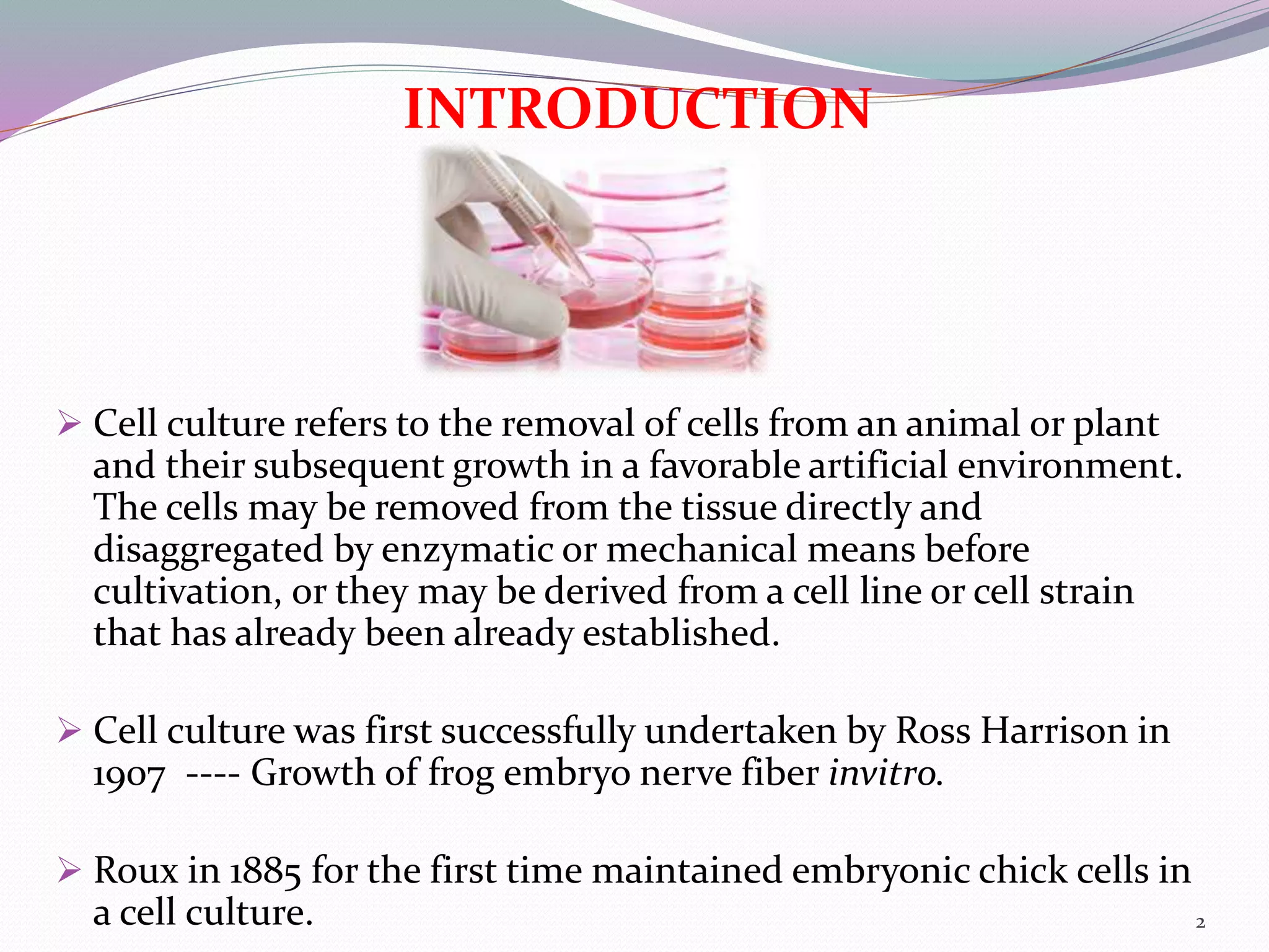
Presented by
S. Nivedhitha
M.Pharmacy 1st year
Pharmacology
Madras Medical College
INTRODUCTION
 Cell culture refers to the removal of cells from an animal or plant
and their subsequent growth in a favorable artificial environment.
The cells may be removed from the tissue directly and
disaggregated by enzymatic or mechanical means before
cultivation, or they may be derived from a cell line or cell strain
that has already been already established.
 Cell culture was first successfully undertaken by Ross Harrison in
1907 ---- Growth of frog embryo nerve fiber invitro.
 Roux in 1885 for the first time maintained embryonic chick cells in
a cell culture. 2
Major developments in cell culture
technology
First development was the use of antibiotics which
inhibits the growth of contaminants.
Second was the use of trypsin to remove adherent cells
to subculture further from the culture vessel.
Third was the use of chemically defined culture
medium.
3
Terminologies
Organ culture: Culture of native tissue (undisaggregated
tissue) that retains most of the in vivo histological
features. – whole organs/ intact organ fragments.
Cell culture: Culture of dispersed (disaggregated) cells
obtained from original tissue.
Histotypic culture: Culturing of cells for their
reaggregation to form a tissue-like structure.
Primary culture: Culture produced by the freshly isolated
cells taken from an organism. Cells- heterogeneous, slow
growing & represent the tissue of origin with regard to
their properties
4
Cell line: The subculturing of the primary culture gives
rise to cell lines.
Passage/subculture: The process of transfer of cells
from one culture vessel to another culture vessel (fresh
media).
Passage number: No. of times a culture has been
subcultured.
Finite cell lines: The cell lines with limited /finite
culture life span.
Continuous cell lines: Indefinite growth of cells in the
subsequent subcultures.
5
TISSUE CULTURE LABORATORY DESIGN
6
Equipments used in cell culture lab
The specific requirements of a cell culture lab depends
mainly on the type of research conducted.
All culture lab should be free from pathogenic
microbes (aseptic) and share some basic equipments.
Cell culture hood -biosafety cabinet/laminar flow hood
 Horizontal LAF : parallel to work surface, not
recirculating.
 Vertical LAF : From top of cabinet & drawn through
work surface, recirculated.
Fitted with HEPA filters and routine maintenance
checks –every 3-6 months. 7
US centers for disease control and prevention (CDC)
classifies BSC into 3 classes in two ways:
The level of – personnel & environmental protection
provided
- product protection provided
Class I
8
CLASS II
TYPES:
 A1, A2, B1, B2, C1 –based
on diff in minimum inflow velocities & exhaust systems.
 90% all BSC installed are type A2 cabinets.
Type A2 Type B2
9
Class III
10
Incubator: Temp 370C , CO2 2-5%, 95% Air at 99%
relative humidity.
Refrigerators: Liquid media at 40c, enzymes (trypsin) &
media components (glutamine, serum) at -200c,-800c
Microscopes: Inverted & dissecting microscope.
11
Centrifuge : Separate the particles
from a solution according to their
sizes, shapes, density, viscosity of
the medium by using the principle
of centrifugal force.
Liquid N2 container: long term storage
with low liq. N2 evaporation.
Vapour phase and liquid phase.
Haemocytometer : To estimate the
cell number and viability.
12
Water bath : Temp 5-1000 c. For thawing of cells taken
out from the freezer or liq. N2 tank to prevent any
mechanical damage to the cells .
Flow cytometry : Analyze the characteristics of the cells.
Culture vessels : Multiwell plates, petri dishes, flasks,
stirrer bottles. Anchorage dependent cells attach to the
surface of vessel & grow.
While, some cells undergo transformation and become
anchorage independent.
13
Culture media
 Provides the required nutrients and components for the cells to
grow and divide.
 Choice of media depends on– type of cells to be cultured and –
purpose of culture (growth, differentiation, production of
desired products).
Natural media- media components are from the natural origin
that provide sufficient nutritional support for proliferation of
animal cells.
E.g. clots, biological fluids, tissue extracts.
Artificial media: They are chemically synthesized.
 Serum containing media
 Serum free media
 Chemically defined media
 Protein free media
14
Types of culture media
15
16
17
Chemically defined media
These media contain contamination-free ultra pure
inorganic and organic ingredients, and may also
contain pure protein additives, like growth factors.
All the chemical components are known.
Protein free media
Do not contain any protein and only contain non-
protein constituents.
Formulations like MEM, RPMI-1640 are protein free
and protein supplement is provided when required.
18
Properties and requirements of media
19
• pH
•7.4
• indicator-phenol red.
20
• Buffers(Bicarbonate and HEPES)
•Bicarbonate buffered media requires CO2 atmosphere
• HEPES strong chemical buffer range pH 7.2 – 7.6
• Keto acids(oxalacetate and pyruvate)
• Intermediate in glycolysis/krebs cycle
• Keto acids added to the media as additional energy source
• Maintain maximum cell metabolism
• Carbohydrates
• Energy source
• Glucose and galactose
• Low (1g/L) and high(4.5 g/L) concentrations of sugars
used in basal media.
• Vitamins
• Precursors of numerous co-factors
• Vit B necessary for cell growth and proliferation
• Commonly found- riboflavin, thiamine, biotin
• Trace elements
• Zinc, copper, selenium & tricarboxylic acid intermediates.
• Supplements
• L-glutamine
• Non-essential amino acids(NEAA)
• Growth factors and hormones
21
Morphology of Cells in culture
 Fibroblastic(or fibroblast-like)
 Epithelial-like cells
 Lymphoblast-like cells
 Neuronal cells
22
Types of cell culture
Cell culture is classified into three:
• Primary cell culture
 Adherent cell culture
 Suspension cell culture
• Secondary cell culture
• Cell line
 Finite cell line
 Continuous cell line
23
Culturing cells in the laboratory
24
Primary cultures
25
26
27
28
SUB CULTURING or PASSAGING
CELLS
Check confluency of cells
Remove spent medium
Wash with PBS
Incubate with trypsin/EDTA
Resuspend in serum containing media
Transfer to culture flask
Why passage cells?
 To maintain cells in culture (i.e. don’t over grow)
 To increase cell number for experiments / storage
29
Standard growth curve of cells
30
 Adherent cell culture
• Those cell which attach to the surface of the culture
vessel/flask.
• Forms a monolayer
• They have to detached from the surface before they get
sub cultured.
• Growth limited to the surface area
 Suspension culture
• Those cell doesn’t get attached to the surface of culture
flask.
• They are free floating.
• Cells in blood stream.
• Growth is limited to concentration of cells
31
Subculture of monolayer
32
SECONDARY CELL CULTURE
 Derived from a primary cell culture.
 Isolated by selection or cloning.
 Becoming a more homogeneous cell population.
 Finite life span in vitro.
 Retain differentiated phenotype.
 Mainly anchorage dependant.
 Exhibit contact inhibition.
33
CELL LINE
Finite cell line
Limited life span
Go through a limited number
of cell generation.
Properties:
•Contact inhibition
• Density limitation
• Anchorage dependence
Less growth rate
Doubling time (24-92 hrs)
Continuous cell line
Grow indefinitely
Grows either in monolayer or
in suspension
Properties:
•Absence contact inhibition
•Absence anchorage
dependence
High growth rate
Doubling time(12-24hrs)
34
35
COMMON CELL LINES
Vero, Cos 7 – African green monkey kidney cells
CHO – Hamster
Sf 9, Sf 21 – Insect cells
CRYOPRESEVATION OF CELLS
Passage cells
Resuspend cells in serum containing media
Centrifuge & aspirate supernatant
Resuspend cells in 10% DMSO in FCS
Transfer to cryovial freeze at -800C
Transfer to liquid Nitrogen tank
Why cryopreserve cells?
 Reduced risk of microbial contamination
 Reduced risk of cross contamination with other cells
 Reduced risk of genetic drift and morphological changes
 Research conducted using cells at consistent low passage
36
37
Cryoprotectants
 Cell membrane permeating (eg: dimethyl sulfoxide(DMSO),
glycerol, 1,2 propanediol)
 Cell membrane non –permeating (eg: 2-metyl-2,4-pentanediol,
polymers –polyvinyl pyrrolidone, hydroxyethyl starch)
Freezing methods
 Slow freezing methods- cooling rate 10c/min,< 1.0M CPA
 Vitrification – Aq. State to glassy state in liqN2 ,high conc.
CPA(40-60% weight/volume)
Application of cryopreservation
 Cryopreservation of cell or organs
 Cryosurgery
 Biochemistry and molecular biology
 Food sciences
 Ecology and plant physiology
 Medical applications such as, blood transfusion, bone
marrow transplantation, artificial insemination and IVF
38
Advantages of cell culture
 Controlled physiological environment (pH, temp., osmotic
pressure, o2)
 Regulation of physiological conditions
 Homogeneity of cell types (achieved through serial passages)
 Easy to characterize cells for cytological and immunological
studies
 Storage of cultured cells for several years
 Legal, moral and ethical questions of animal experimentation
are avoided.
Limitations
 Expertise is needed
 Expensive – 10 times higher than direct use of animal tissue
 Unstable aneuploid chromosome constitution
 Control of environmental factors not easy.
39
APPLICATION OF CELL CULTURE
 Excellent model system for studying
• Normal physiology, cell biology and biochemistry of cells
• The effects of drugs, radiation and toxic compounds on the cell
• The interaction between disease causing agents and cells
(mutagenesis, carcinogenesis)
• Nutritional studies
• The process and triggers of aging.
 Toxicity testing
 Genetic engineering and gene therapy
Studies dealing with genetics (eg: gene transfer, genetic analysis,
transformation, immortalization, senescence)
 Cell-based manufacturing
Laboratory production of medical and pharmaceutical compounds
for wide range of applications. 40
 vaccines, insulin, interferon, hormones, other therapeutic
protein
 Genetic counselling (amniocentesis)
 Virology
•Cultivation of virus for vaccine production, also used to
study the infectious cycle.
 Cancer research
•Study the functions of various chemicals, virus and radiation
to convert normal cultured cells to cancerous cells.
 Drug screening and development
41
References
 Biotechnology by Satyanarayanan.
 Butler. M (2005). Animal cell culture: recent
achievements and perspectives in the production of
biopharmaceuticals. Applied microbiology and
biotechnology,68(3),283-291
 https://www.slideshare.net/mobile/meghabedekar/pri
nciple-of-cell-culture
 https://www.ncbi.nlm.nih.gov/pmc/articles
 www.biotechnologynotes.com/animals/animal-cell-
culture-history-types-and-applications/671
 Animal cell culture practical approach by John
R.W.Masters
42
THANK YOU
43

Cell culture techniques

  • 1.
    Presented by S. Nivedhitha M.Pharmacy1st year Pharmacology Madras Medical College
  • 2.
    INTRODUCTION  Cell culturerefers to the removal of cells from an animal or plant and their subsequent growth in a favorable artificial environment. The cells may be removed from the tissue directly and disaggregated by enzymatic or mechanical means before cultivation, or they may be derived from a cell line or cell strain that has already been already established.  Cell culture was first successfully undertaken by Ross Harrison in 1907 ---- Growth of frog embryo nerve fiber invitro.  Roux in 1885 for the first time maintained embryonic chick cells in a cell culture. 2
  • 3.
    Major developments incell culture technology First development was the use of antibiotics which inhibits the growth of contaminants. Second was the use of trypsin to remove adherent cells to subculture further from the culture vessel. Third was the use of chemically defined culture medium. 3
  • 4.
    Terminologies Organ culture: Cultureof native tissue (undisaggregated tissue) that retains most of the in vivo histological features. – whole organs/ intact organ fragments. Cell culture: Culture of dispersed (disaggregated) cells obtained from original tissue. Histotypic culture: Culturing of cells for their reaggregation to form a tissue-like structure. Primary culture: Culture produced by the freshly isolated cells taken from an organism. Cells- heterogeneous, slow growing & represent the tissue of origin with regard to their properties 4
  • 5.
    Cell line: Thesubculturing of the primary culture gives rise to cell lines. Passage/subculture: The process of transfer of cells from one culture vessel to another culture vessel (fresh media). Passage number: No. of times a culture has been subcultured. Finite cell lines: The cell lines with limited /finite culture life span. Continuous cell lines: Indefinite growth of cells in the subsequent subcultures. 5
  • 6.
  • 7.
    Equipments used incell culture lab The specific requirements of a cell culture lab depends mainly on the type of research conducted. All culture lab should be free from pathogenic microbes (aseptic) and share some basic equipments. Cell culture hood -biosafety cabinet/laminar flow hood  Horizontal LAF : parallel to work surface, not recirculating.  Vertical LAF : From top of cabinet & drawn through work surface, recirculated. Fitted with HEPA filters and routine maintenance checks –every 3-6 months. 7
  • 8.
    US centers fordisease control and prevention (CDC) classifies BSC into 3 classes in two ways: The level of – personnel & environmental protection provided - product protection provided Class I 8
  • 9.
    CLASS II TYPES:  A1,A2, B1, B2, C1 –based on diff in minimum inflow velocities & exhaust systems.  90% all BSC installed are type A2 cabinets. Type A2 Type B2 9
  • 10.
  • 11.
    Incubator: Temp 370C, CO2 2-5%, 95% Air at 99% relative humidity. Refrigerators: Liquid media at 40c, enzymes (trypsin) & media components (glutamine, serum) at -200c,-800c Microscopes: Inverted & dissecting microscope. 11
  • 12.
    Centrifuge : Separatethe particles from a solution according to their sizes, shapes, density, viscosity of the medium by using the principle of centrifugal force. Liquid N2 container: long term storage with low liq. N2 evaporation. Vapour phase and liquid phase. Haemocytometer : To estimate the cell number and viability. 12
  • 13.
    Water bath :Temp 5-1000 c. For thawing of cells taken out from the freezer or liq. N2 tank to prevent any mechanical damage to the cells . Flow cytometry : Analyze the characteristics of the cells. Culture vessels : Multiwell plates, petri dishes, flasks, stirrer bottles. Anchorage dependent cells attach to the surface of vessel & grow. While, some cells undergo transformation and become anchorage independent. 13
  • 14.
    Culture media  Providesthe required nutrients and components for the cells to grow and divide.  Choice of media depends on– type of cells to be cultured and – purpose of culture (growth, differentiation, production of desired products). Natural media- media components are from the natural origin that provide sufficient nutritional support for proliferation of animal cells. E.g. clots, biological fluids, tissue extracts. Artificial media: They are chemically synthesized.  Serum containing media  Serum free media  Chemically defined media  Protein free media 14
  • 15.
  • 16.
  • 17.
  • 18.
    Chemically defined media Thesemedia contain contamination-free ultra pure inorganic and organic ingredients, and may also contain pure protein additives, like growth factors. All the chemical components are known. Protein free media Do not contain any protein and only contain non- protein constituents. Formulations like MEM, RPMI-1640 are protein free and protein supplement is provided when required. 18
  • 19.
    Properties and requirementsof media 19 • pH •7.4 • indicator-phenol red.
  • 20.
    20 • Buffers(Bicarbonate andHEPES) •Bicarbonate buffered media requires CO2 atmosphere • HEPES strong chemical buffer range pH 7.2 – 7.6 • Keto acids(oxalacetate and pyruvate) • Intermediate in glycolysis/krebs cycle • Keto acids added to the media as additional energy source • Maintain maximum cell metabolism • Carbohydrates • Energy source • Glucose and galactose • Low (1g/L) and high(4.5 g/L) concentrations of sugars used in basal media.
  • 21.
    • Vitamins • Precursorsof numerous co-factors • Vit B necessary for cell growth and proliferation • Commonly found- riboflavin, thiamine, biotin • Trace elements • Zinc, copper, selenium & tricarboxylic acid intermediates. • Supplements • L-glutamine • Non-essential amino acids(NEAA) • Growth factors and hormones 21
  • 22.
    Morphology of Cellsin culture  Fibroblastic(or fibroblast-like)  Epithelial-like cells  Lymphoblast-like cells  Neuronal cells 22
  • 23.
    Types of cellculture Cell culture is classified into three: • Primary cell culture  Adherent cell culture  Suspension cell culture • Secondary cell culture • Cell line  Finite cell line  Continuous cell line 23
  • 24.
    Culturing cells inthe laboratory 24
  • 25.
  • 26.
  • 27.
  • 28.
  • 29.
    SUB CULTURING orPASSAGING CELLS Check confluency of cells Remove spent medium Wash with PBS Incubate with trypsin/EDTA Resuspend in serum containing media Transfer to culture flask Why passage cells?  To maintain cells in culture (i.e. don’t over grow)  To increase cell number for experiments / storage 29
  • 30.
  • 31.
     Adherent cellculture • Those cell which attach to the surface of the culture vessel/flask. • Forms a monolayer • They have to detached from the surface before they get sub cultured. • Growth limited to the surface area  Suspension culture • Those cell doesn’t get attached to the surface of culture flask. • They are free floating. • Cells in blood stream. • Growth is limited to concentration of cells 31
  • 32.
  • 33.
    SECONDARY CELL CULTURE Derived from a primary cell culture.  Isolated by selection or cloning.  Becoming a more homogeneous cell population.  Finite life span in vitro.  Retain differentiated phenotype.  Mainly anchorage dependant.  Exhibit contact inhibition. 33
  • 34.
    CELL LINE Finite cellline Limited life span Go through a limited number of cell generation. Properties: •Contact inhibition • Density limitation • Anchorage dependence Less growth rate Doubling time (24-92 hrs) Continuous cell line Grow indefinitely Grows either in monolayer or in suspension Properties: •Absence contact inhibition •Absence anchorage dependence High growth rate Doubling time(12-24hrs) 34
  • 35.
    35 COMMON CELL LINES Vero,Cos 7 – African green monkey kidney cells CHO – Hamster Sf 9, Sf 21 – Insect cells
  • 36.
    CRYOPRESEVATION OF CELLS Passagecells Resuspend cells in serum containing media Centrifuge & aspirate supernatant Resuspend cells in 10% DMSO in FCS Transfer to cryovial freeze at -800C Transfer to liquid Nitrogen tank Why cryopreserve cells?  Reduced risk of microbial contamination  Reduced risk of cross contamination with other cells  Reduced risk of genetic drift and morphological changes  Research conducted using cells at consistent low passage 36
  • 37.
    37 Cryoprotectants  Cell membranepermeating (eg: dimethyl sulfoxide(DMSO), glycerol, 1,2 propanediol)  Cell membrane non –permeating (eg: 2-metyl-2,4-pentanediol, polymers –polyvinyl pyrrolidone, hydroxyethyl starch)
  • 38.
    Freezing methods  Slowfreezing methods- cooling rate 10c/min,< 1.0M CPA  Vitrification – Aq. State to glassy state in liqN2 ,high conc. CPA(40-60% weight/volume) Application of cryopreservation  Cryopreservation of cell or organs  Cryosurgery  Biochemistry and molecular biology  Food sciences  Ecology and plant physiology  Medical applications such as, blood transfusion, bone marrow transplantation, artificial insemination and IVF 38
  • 39.
    Advantages of cellculture  Controlled physiological environment (pH, temp., osmotic pressure, o2)  Regulation of physiological conditions  Homogeneity of cell types (achieved through serial passages)  Easy to characterize cells for cytological and immunological studies  Storage of cultured cells for several years  Legal, moral and ethical questions of animal experimentation are avoided. Limitations  Expertise is needed  Expensive – 10 times higher than direct use of animal tissue  Unstable aneuploid chromosome constitution  Control of environmental factors not easy. 39
  • 40.
    APPLICATION OF CELLCULTURE  Excellent model system for studying • Normal physiology, cell biology and biochemistry of cells • The effects of drugs, radiation and toxic compounds on the cell • The interaction between disease causing agents and cells (mutagenesis, carcinogenesis) • Nutritional studies • The process and triggers of aging.  Toxicity testing  Genetic engineering and gene therapy Studies dealing with genetics (eg: gene transfer, genetic analysis, transformation, immortalization, senescence)  Cell-based manufacturing Laboratory production of medical and pharmaceutical compounds for wide range of applications. 40
  • 41.
     vaccines, insulin,interferon, hormones, other therapeutic protein  Genetic counselling (amniocentesis)  Virology •Cultivation of virus for vaccine production, also used to study the infectious cycle.  Cancer research •Study the functions of various chemicals, virus and radiation to convert normal cultured cells to cancerous cells.  Drug screening and development 41
  • 42.
    References  Biotechnology bySatyanarayanan.  Butler. M (2005). Animal cell culture: recent achievements and perspectives in the production of biopharmaceuticals. Applied microbiology and biotechnology,68(3),283-291  https://www.slideshare.net/mobile/meghabedekar/pri nciple-of-cell-culture  https://www.ncbi.nlm.nih.gov/pmc/articles  www.biotechnologynotes.com/animals/animal-cell- culture-history-types-and-applications/671  Animal cell culture practical approach by John R.W.Masters 42
  • 43.