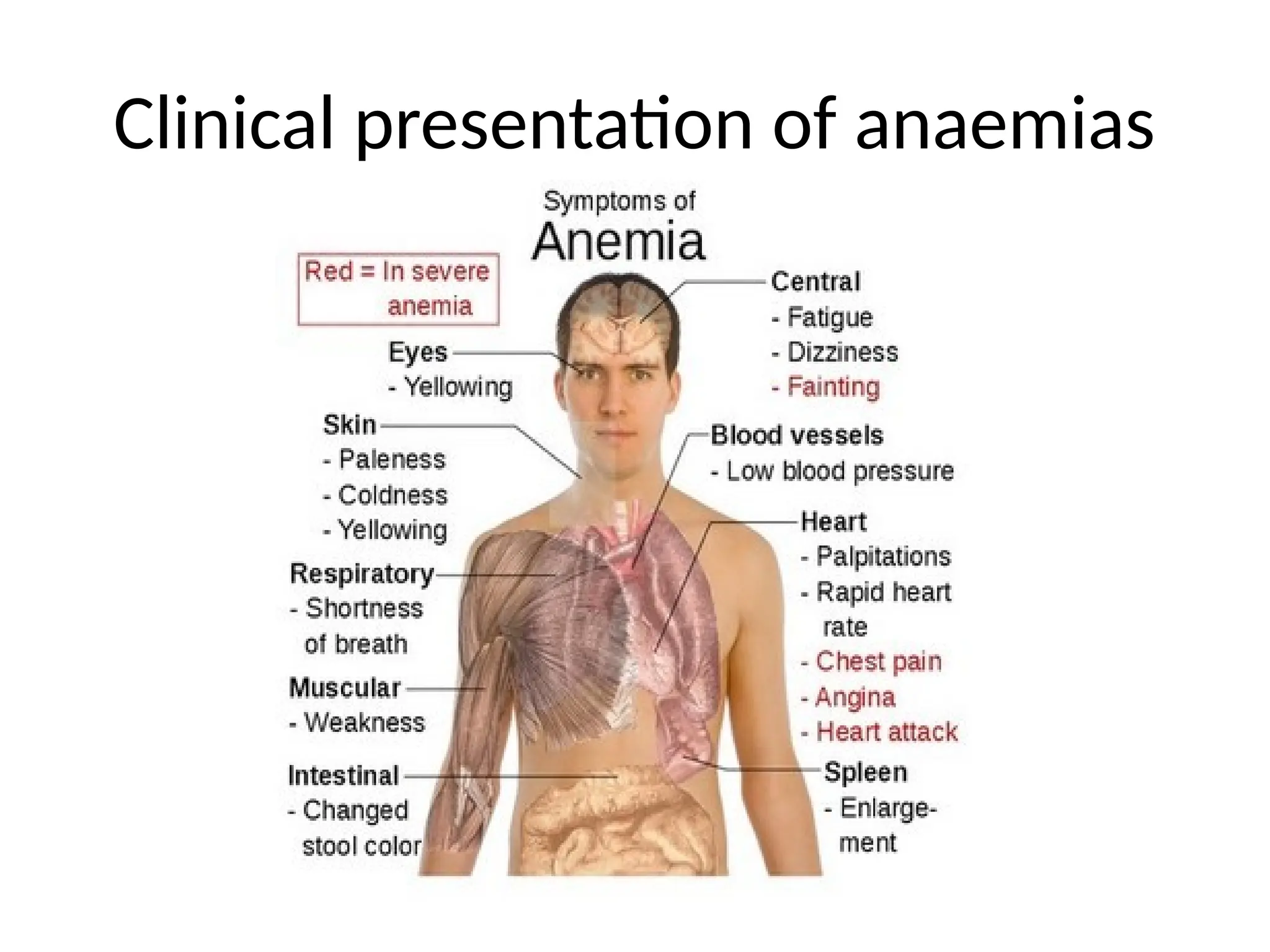
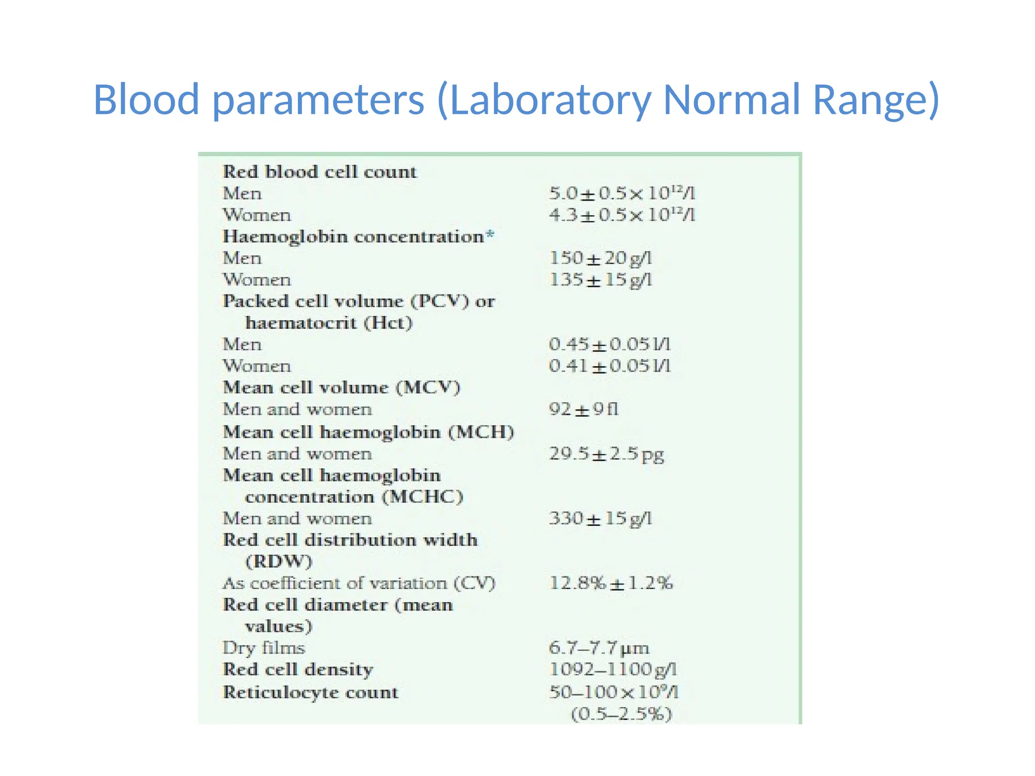
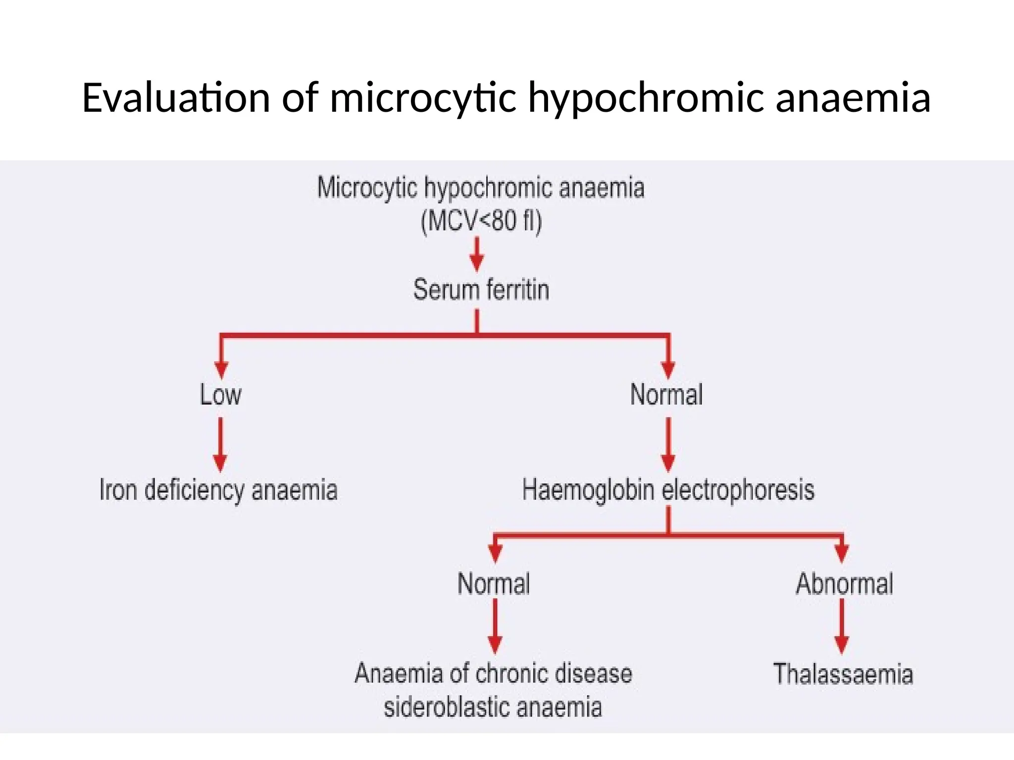
By Kawalya steven
Learning Objectives
At the end of this briefing, you should know
• Clinical presentation of anemia.
• Definition of anemia.
• Approach to diagnosis of anemia
• Classification of anaemia
• Distinct features of each type of anemia.
Clinical presentation of anemias
• Fatigue and weakness
• Headache
• Tinnitus
• Numbness and coldness
• Pallor
• Dyspnea and palpitations
• Angina pectoris
• Intermittent claudication
• Hemorrhages in the
fundus of eyes
• Menorrhagia
• Anorexia
• Flatulence
• Nausea
• Constipation
Clinical presentation of anaemias
Initial Laboratory Work-Up
• Hemoglobin concentration.
• Packed Cell Volume.
• Red cell indices.
• Peripheral Blood Smear.
6
Definition of Anaemia
• Anaemia is defined as a reduction in the concentration
of circulating haemoglobin below the level that is
expected for healthy personsof same age and sex in the
same environment.
Blood parameters (Laboratory Normal Range)
•In full term infants, hemoglobin is 18.0 ± 4.0 g/dl
•Children - 12.5 ± 1.5 g/dl
•Children(6-12 years) - 13.5 ± 2.0 g/dl
Grading of Anaemia
Classification of Anaemias
• Morphological Classification
• Etiological Classification
• Classification based on reticulocyte response.
Morphological Classification of Anemias
• Hematocrit: Proportion of the volume of red cells relative to the volume of blood
• Mean Corpuscular Volume: Dividing the total volume of red cells by the number of
red cells
• Index for average size of red cells
• Mean Corpuscular Hemoglobin: Average amount of haemoglobin in each red cell.
• Mean Corpuscular Hemoglobin ConcentrationThis represents the average
concentration of haemoglobin in a given volume of packed red cells.
• MCHC raised in hereditary spherocytosis.
• Decreased in hypochromic anaemia.
• Red cell distribution width: Variation in red cell size
• High in iron deficiency anaemia
• Normal in anaemia of chronic disease
:
13
1 Microcytic/hypochromic
3
1 2
2 Macrocytic/Normochromic
3 Normocytic/Normochromic
Morphologic Categories of Anemia
N.B. The nucleus of a small
lymphocyte (shown by the arrow)
is used as a reference to a normal
red cell size
Macrocytic anemia
• Low/normal reticulocyte
count, macrocytosis(oval and
round)
• Elevated MCV,MCHC
• Basophilic stippling
• Howell-jolly bodies
• Cabot rings
• Pancytopenia
• Hypersegmented neutrophils
• Bone marrow- megaloblastic
maturation,sieve like
chromatin,nuclear-
cytoplasmic
asynchrony,maturation arrest
Normocytic Normochromic anaemia with
effective erythropoiesis
Etiological Classification of Anaemias
Basic Approach to a diagnosis of anemia
Evaluation of microcytic hypochromic anaemia
Evaluation of macrocytic anaemia
Evaluation of normocytic anaemia
Evaluation of haemolytic anaemia
A simplified approach to diagnosis of haemolytic
anaemias
25
Reticulocyte Count
(In the Diagnosis of Anemia)
• Reticulocytes are non-
nucleated RBCs that still
contain RNA.
• Visualized by staining with
supravital dyes, including
new methylene blue or
brilliant cresyl blue.
• Useful in determining
response and potential of
bone marrow.
• Normal range is 0.5-2.5%
of all erythrocytes.
Fe++
deficiency anemia
• Low hemoglobin and low packed cell volume
• Low MCV,MCH and MCHC
• Microcytosis & hypochromia are hallmarks
• RDW is increased
• Serum ferritin is less than 15 micro gram/dl
• Serum iron is low,TIBC is increased and transferrin saturation is less
than 10 percent
• Free erythrocyte protoporphyrin is increased.
• Increased soluble transferrin receptor in serum
• Bone marrow-micronormoblastic,absence of stainable iron in bone
marrow on Perls Prussian blue reaction
27
Megaloblastic Anemia
Mild to severe anemia,
– Increased MCV & MCH, normal MCHC
– Low RBC, HGB, WBC and PLT counts (fragile cells) due
to ineffective hematopoiesis.
– Low reticulocyte count
– Macrocytic ovalocytes and teardrops;
– Marked anisocytosis and poikilocytosis
– Schistocytes/microcytes - due to RBC breakage upon
leaving the BM
– Erythroid hyperplasia - low M:E ratio (1:1)
– Iron stores increased.
28
Macrocytic Ovalocytes
Blood NRBC Blood
Howell-Jolly body
Teardrop
Schistocyte
Stippled RBC &
Cabot Ring
Giant Platelet
Pap bodies Hypersegmented Neutrophil >5
lobes
Megaloblastic anemia
Tests
• Folate and B12 levels
• Schilling test may be useful to establish
etiology of B12 deficiency
– Assesses radioactive B12 absorption with and
without exogenous IF
• Other tests if pernicious anemia is suspected
– Anti- parietal cell antibodies, anti-IF antibodies
– Secondary causes of poor absorption should be
sought (gastritis, ileal problems, etc.)
Anemia of chronic disease
• Normocytic anemia with ineffective erythropoiesis
(reduced reticulocyte count)
• Normochromic
• Results from
– Chronic inflammation (e.g. rheumatologic disease):
Cytokines released by inflammatory cells cause
macrophages to accumulate iron and not transfer it to
plasma or developing red cells (iron block anemia)
– Inflammation
– malignancy
• Bone marrow suppression (EPO is elevated)
Anemia of chronic disease
• Decreased serum iron,decreased total iron
binding capacity and normal or raised ferritin
• Increased marrow storage iron
• ESR is high
Normochromic, normocytic anemia with
effective erythropoiesis
INCREASED reticulocyte count
• Acute blood loss
– Very acutely, with hypovolemia,
may have normal blood counts,
will become anemic with volume
replenishment
• Hemolytic anemia
– Increased reticulocyte production
cannot keep pace with loss of
RBCs peripherally.
• Response to specific therapy in
nutritional anemias
Aplastic anemia
• Pancytopenia caused by bone marrow
failure…decreased production of all cell
lines and replacement of marrow with fat.
• Inherited- Fanconis anaemia, Dyskeratosis
congenita
• Acquired - Idiopathic,drugs like
NSAIDs,chloramphenicol,benzene,parvo
virus,hepatitis and EB virus.
Hemolytic anemia
• Abnormality intrinsic to red cells-
1. Hereditary spherocytosis
2. Thalassamia
3. Sickle cell anaemia
4. Glucose -6-phosphate dehydrogenase deficiency
• Abnormality extrinsic to red cells-
1. Immune
2. Mechanical etc
Evaluation of haemolytic anaemia
Hereditary spherocytosis
• Inherited defect in the red cell membrane
cytoskeleton (spectrin, ankyrin or band 3)
leading to the formation of spherocytic red
cells.
• Autosomal dominant
• Mild to moderate anaemia
• Intermittent jaundice
• Splenomegaly
• Pigment gall stones
• Peripheral smear-microspherocytes
• Screening test-osmotic fragility
Thalassemia
• Decreased or absent globin
chains
• Alpha and beta thalassemias
• Microcytic
hypochromic,target
cells,basophilic stippling
• Reticulocytosis
• Hb F elevated in
electrophoresis
Sickle cell anaemia
• Presence of Hb S
• Point mutation in 6th place of beta
chain
• Substitution of valine for glutamic
acid
• On deoxygenation, sickle cells are
formed
• Chronic hemolytic anaemia, vaso-
occlusive crisis
• Aplastic crisis
• Hemolytic crisis
• Infections
Sickle cell anaemia
• Sickling test is positive.
• Solubility test is positive.
• Electrophoresis shows HbS.
• In sickle cell trait, electrophoresis shows 60
percent of Hb A and 40 percent Hb S
Glucose-6-phosphate dehydrogenase
deficiency
• X linked disorder
• Reduced activity of G6PD
• Inability to remove H2O2
• Accumulated H2O2 leads to
oxidation of hemoglobin with
precipitation of globin chains
• Heinz bodies
• Red cells with heinz bodies
destroyed in
spleen(extravascular
hemolysis)
Glucose-6-phosphate dehydrogenase
deficiency
• Asymptomatic
• Neonatal jaundice
• Acute hemolytic anaemia
• Chronic hemolytic anaemia
• On peripheral smear-
polychromasia,fragmented red
cells,spherocytes,bite cells,half
ghost cells
• Biochemical-increased
bilirubin,hemoglobinemia and
hemoglobinuria
Glucose-6-phosphate dehydrogenase
deficiency
• Screening tests-fluorescent spot
test,methemoglobin reduction test and dye
decolorisation test
Immune hemolytic anaemia
• Warm antibody-persons over 50 years,mild
jaundice and splenomegaly,red cells coated
with IgG,spherocytes.Seen in autoimmune
disorders,lymphoma
• Cold antibody-acrocyanosis,IgM.
• Seen in cold agglutinin disease, Paroxysmal
cold hemoglobinuria (PCH)
Coomb’s test
• Detects presence of either antibody on RBC or
of antibody in serum
• Helpful in determining if a hemolytic anemia is
immune-mediated
SUMMARY
• Microcytic hypochromic anaemia-iron
deficiency
• Macrocytic hyperchromic-megaloblastic
anaemia
• Normochromic normocytic-hemolytic
anaemia
• Pancytopenia-megaloblastic and aplastic
anemias
Anemia update diagnosis  and management .pptx

Anemia update diagnosis and management .pptx

  • 1.
  • 2.
    Learning Objectives At theend of this briefing, you should know • Clinical presentation of anemia. • Definition of anemia. • Approach to diagnosis of anemia • Classification of anaemia • Distinct features of each type of anemia.
  • 3.
    Clinical presentation ofanemias • Fatigue and weakness • Headache • Tinnitus • Numbness and coldness • Pallor • Dyspnea and palpitations • Angina pectoris • Intermittent claudication • Hemorrhages in the fundus of eyes • Menorrhagia • Anorexia • Flatulence • Nausea • Constipation
  • 4.
  • 5.
    Initial Laboratory Work-Up •Hemoglobin concentration. • Packed Cell Volume. • Red cell indices. • Peripheral Blood Smear.
  • 6.
    6 Definition of Anaemia •Anaemia is defined as a reduction in the concentration of circulating haemoglobin below the level that is expected for healthy personsof same age and sex in the same environment.
  • 7.
  • 8.
    Blood parameters (LaboratoryNormal Range) •In full term infants, hemoglobin is 18.0 ± 4.0 g/dl •Children - 12.5 ± 1.5 g/dl •Children(6-12 years) - 13.5 ± 2.0 g/dl
  • 9.
  • 10.
    Classification of Anaemias •Morphological Classification • Etiological Classification • Classification based on reticulocyte response.
  • 11.
  • 12.
    • Hematocrit: Proportionof the volume of red cells relative to the volume of blood • Mean Corpuscular Volume: Dividing the total volume of red cells by the number of red cells • Index for average size of red cells • Mean Corpuscular Hemoglobin: Average amount of haemoglobin in each red cell. • Mean Corpuscular Hemoglobin ConcentrationThis represents the average concentration of haemoglobin in a given volume of packed red cells. • MCHC raised in hereditary spherocytosis. • Decreased in hypochromic anaemia. • Red cell distribution width: Variation in red cell size • High in iron deficiency anaemia • Normal in anaemia of chronic disease :
  • 13.
    13 1 Microcytic/hypochromic 3 1 2 2Macrocytic/Normochromic 3 Normocytic/Normochromic Morphologic Categories of Anemia N.B. The nucleus of a small lymphocyte (shown by the arrow) is used as a reference to a normal red cell size
  • 16.
    Macrocytic anemia • Low/normalreticulocyte count, macrocytosis(oval and round) • Elevated MCV,MCHC • Basophilic stippling • Howell-jolly bodies • Cabot rings • Pancytopenia • Hypersegmented neutrophils • Bone marrow- megaloblastic maturation,sieve like chromatin,nuclear- cytoplasmic asynchrony,maturation arrest
  • 17.
    Normocytic Normochromic anaemiawith effective erythropoiesis
  • 18.
  • 19.
    Basic Approach toa diagnosis of anemia
  • 20.
    Evaluation of microcytichypochromic anaemia
  • 21.
  • 22.
  • 23.
  • 24.
    A simplified approachto diagnosis of haemolytic anaemias
  • 25.
    25 Reticulocyte Count (In theDiagnosis of Anemia) • Reticulocytes are non- nucleated RBCs that still contain RNA. • Visualized by staining with supravital dyes, including new methylene blue or brilliant cresyl blue. • Useful in determining response and potential of bone marrow. • Normal range is 0.5-2.5% of all erythrocytes.
  • 26.
    Fe++ deficiency anemia • Lowhemoglobin and low packed cell volume • Low MCV,MCH and MCHC • Microcytosis & hypochromia are hallmarks • RDW is increased • Serum ferritin is less than 15 micro gram/dl • Serum iron is low,TIBC is increased and transferrin saturation is less than 10 percent • Free erythrocyte protoporphyrin is increased. • Increased soluble transferrin receptor in serum • Bone marrow-micronormoblastic,absence of stainable iron in bone marrow on Perls Prussian blue reaction
  • 27.
    27 Megaloblastic Anemia Mild tosevere anemia, – Increased MCV & MCH, normal MCHC – Low RBC, HGB, WBC and PLT counts (fragile cells) due to ineffective hematopoiesis. – Low reticulocyte count – Macrocytic ovalocytes and teardrops; – Marked anisocytosis and poikilocytosis – Schistocytes/microcytes - due to RBC breakage upon leaving the BM – Erythroid hyperplasia - low M:E ratio (1:1) – Iron stores increased.
  • 28.
    28 Macrocytic Ovalocytes Blood NRBCBlood Howell-Jolly body Teardrop Schistocyte Stippled RBC & Cabot Ring Giant Platelet Pap bodies Hypersegmented Neutrophil >5 lobes Megaloblastic anemia
  • 29.
    Tests • Folate andB12 levels • Schilling test may be useful to establish etiology of B12 deficiency – Assesses radioactive B12 absorption with and without exogenous IF • Other tests if pernicious anemia is suspected – Anti- parietal cell antibodies, anti-IF antibodies – Secondary causes of poor absorption should be sought (gastritis, ileal problems, etc.)
  • 30.
    Anemia of chronicdisease • Normocytic anemia with ineffective erythropoiesis (reduced reticulocyte count) • Normochromic • Results from – Chronic inflammation (e.g. rheumatologic disease): Cytokines released by inflammatory cells cause macrophages to accumulate iron and not transfer it to plasma or developing red cells (iron block anemia) – Inflammation – malignancy • Bone marrow suppression (EPO is elevated)
  • 31.
    Anemia of chronicdisease • Decreased serum iron,decreased total iron binding capacity and normal or raised ferritin • Increased marrow storage iron • ESR is high
  • 32.
    Normochromic, normocytic anemiawith effective erythropoiesis INCREASED reticulocyte count • Acute blood loss – Very acutely, with hypovolemia, may have normal blood counts, will become anemic with volume replenishment • Hemolytic anemia – Increased reticulocyte production cannot keep pace with loss of RBCs peripherally. • Response to specific therapy in nutritional anemias
  • 33.
    Aplastic anemia • Pancytopeniacaused by bone marrow failure…decreased production of all cell lines and replacement of marrow with fat. • Inherited- Fanconis anaemia, Dyskeratosis congenita • Acquired - Idiopathic,drugs like NSAIDs,chloramphenicol,benzene,parvo virus,hepatitis and EB virus.
  • 34.
    Hemolytic anemia • Abnormalityintrinsic to red cells- 1. Hereditary spherocytosis 2. Thalassamia 3. Sickle cell anaemia 4. Glucose -6-phosphate dehydrogenase deficiency • Abnormality extrinsic to red cells- 1. Immune 2. Mechanical etc
  • 35.
  • 36.
    Hereditary spherocytosis • Inheriteddefect in the red cell membrane cytoskeleton (spectrin, ankyrin or band 3) leading to the formation of spherocytic red cells. • Autosomal dominant • Mild to moderate anaemia • Intermittent jaundice • Splenomegaly • Pigment gall stones • Peripheral smear-microspherocytes • Screening test-osmotic fragility
  • 37.
    Thalassemia • Decreased orabsent globin chains • Alpha and beta thalassemias • Microcytic hypochromic,target cells,basophilic stippling • Reticulocytosis • Hb F elevated in electrophoresis
  • 38.
    Sickle cell anaemia •Presence of Hb S • Point mutation in 6th place of beta chain • Substitution of valine for glutamic acid • On deoxygenation, sickle cells are formed • Chronic hemolytic anaemia, vaso- occlusive crisis • Aplastic crisis • Hemolytic crisis • Infections
  • 39.
    Sickle cell anaemia •Sickling test is positive. • Solubility test is positive. • Electrophoresis shows HbS. • In sickle cell trait, electrophoresis shows 60 percent of Hb A and 40 percent Hb S
  • 40.
    Glucose-6-phosphate dehydrogenase deficiency • Xlinked disorder • Reduced activity of G6PD • Inability to remove H2O2 • Accumulated H2O2 leads to oxidation of hemoglobin with precipitation of globin chains • Heinz bodies • Red cells with heinz bodies destroyed in spleen(extravascular hemolysis)
  • 41.
    Glucose-6-phosphate dehydrogenase deficiency • Asymptomatic •Neonatal jaundice • Acute hemolytic anaemia • Chronic hemolytic anaemia • On peripheral smear- polychromasia,fragmented red cells,spherocytes,bite cells,half ghost cells • Biochemical-increased bilirubin,hemoglobinemia and hemoglobinuria
  • 42.
    Glucose-6-phosphate dehydrogenase deficiency • Screeningtests-fluorescent spot test,methemoglobin reduction test and dye decolorisation test
  • 43.
    Immune hemolytic anaemia •Warm antibody-persons over 50 years,mild jaundice and splenomegaly,red cells coated with IgG,spherocytes.Seen in autoimmune disorders,lymphoma • Cold antibody-acrocyanosis,IgM. • Seen in cold agglutinin disease, Paroxysmal cold hemoglobinuria (PCH)
  • 44.
    Coomb’s test • Detectspresence of either antibody on RBC or of antibody in serum • Helpful in determining if a hemolytic anemia is immune-mediated
  • 45.
    SUMMARY • Microcytic hypochromicanaemia-iron deficiency • Macrocytic hyperchromic-megaloblastic anaemia • Normochromic normocytic-hemolytic anaemia • Pancytopenia-megaloblastic and aplastic anemias

Editor's Notes

  • #27 Lab findings in vitamin B12 or folate deficiency: Mild to SEVERE anemia; MCV 100-160 fl; increased MCH, normal MCHC. Low RBC and HGB values with mildly decreased WBC and PLT counts (fragile cells) due to ineffective hematopoiesis. Low retic - due to high RBC death in bone marrow and blood. Macrocytic ovalocytes and teardrops; marked aniso and poik is typical. Schistocytes/microcytes - due to RBC breakage upon leaving the bone marrow. Advanced anemia: multiple Howell-Jolly bodies, nucRBCs, basophilic stippling, pappenheimer bodies, Cabot rings. Megaloblastic anemias are noted for markedly increased LD levels and high bilirubin & iron levels due to destruction of fragile red cells in the bone marrow and blood. Hypersegmented neutrophils (>5 lobes) - 1st change to appear and last to disappear. (Normal segmented neutrophils have 2-5 lobes; hyperseg neutrophils have 6 lobes). May also see giant band neutrophils as well as giant platelets.
  • #39 Sickling test : When red cells containing HbS are subjected to deoxygenation, the become sickle-shaped while cells that do not contain HbS remain normal. Certain reducing chemical agents such as 2% sodium metabisulphite or sodium dithionite can deprive red cells of oxygen. Solubility test: Small amount of blood is added to a solution that contains high-phosphate buffer, a reducing agent (sodium dithionite) and saponin. Red cells are haemolysed and HbS, if present, is reduced by dithionite. Reduced HbS forms insoluble polymers, which refract light, and solution becomes turbid. A reader scale is held at the back of the tube; in negative test lines will be clearly seen since HbA is soluble in phosphate buffer, while lines will not be seen in positive test due to formation of polymers of HbS (Fig. 4.10).