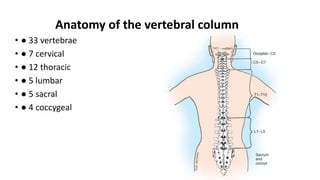
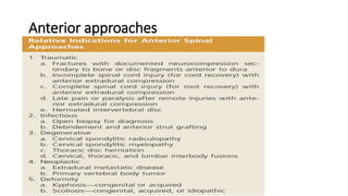
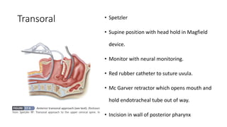
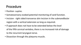
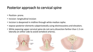
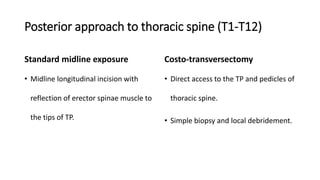
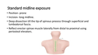
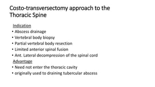
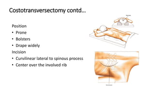
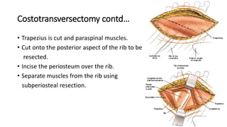
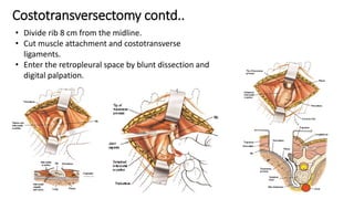
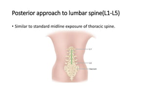
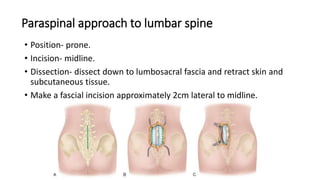
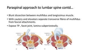
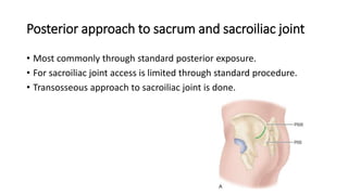
7. Transoral • Spetzler
• Supine position with head hold in Magfield
device.
• Monitor with neural monitoring.
• Red rubber catheter to suture uvula.
• Mc Garver retractor which opens mouth and
hold endotracheal tube out of way.
• Incision in wall of posterior pharynx
8. Anterior retropharyngeal approach
By McAfee et al.
Excellent for anterior debridement of the upper cervical spine
Allows placement of bone grafts for stabilization .
In contrast to the transoral approach, it is entirely extramucosal.
Has fewer complications of wound infection and neurologic deficit.
9. Procedure
• Position -supine.
• Somatosensory evoked potential monitoring of cord function.
• Incision- right sided transverse skin incision in the submandibular
region with a vertical extension as long as required.
• If approach does not have to be extended below the level
of the fifth cervical vertebra, there is no increased risk of damage
to the recurrent laryngeal nerve.
• Dissection through the platysma muscle.
10. Procedure contd..
• Need to protect carotid sheath.
• Feel for the pulsations of the carotid artery and protect the contents of
the carotid sheath.
• Resect the submandibular gland and ligate the duct to prevent formation
of a salivary fistula.
• Tag and divide tendon of digastric muscle.
• the hyoid bone and hypopharynx can be mobilized medially,
preventing exposure of the esophagus, hypopharynx and nasopharynx.
• Identify and mobilize the superior laryngeal nerve.
• Following adequate retraction of the carotid sheath laterally, Remove the
longus colli muscles subperiosteally.
13. Approach to C3-C7
Anterior approach Anterolateral approach
Most common complication of anterior approach – vocal cord palsy
due to recurrent laryngeal nerve injury.
Less common on left side since it is more vertical and protected in
esophago-tracheal groove.
Still right sided approach is preffered.
14. Anterior approach C3-C7
• By southwick and robinson.
• Supine position.
• Neuromonitoring.
transverse
• Incision (depending on surgeons preference)
longitudinal
For up to 2 levels- transverse incision.
For 3 or more levels- longitudinal incision.
15. Steps of anterior approach to C3-C7
From medial border of sternocleidomastoid center a transverse incision.
Incise platysma.
Longitudinally incise superficial layer of deep cervical fascia.
Carefully divide middle layer of deep cervical fascia enclosing omohyoid medial to carotid
sheath.
Now retract sternocleidomastoid and carotid sheath laterally.
16. • Retract trachea, esophagus, thyroid medially.
• Blunt dissection of deep layer of deep cervical fascia ,
consisting of pretracheal and prevertebral fascia
overlying longus colli muscle.
• Reflect longus colli muscle subperiosteally,
• Closure is always with drain to prevent hematoma
formation.
17. Anterolateral approach to C3-C7
• By Bruneau Et al and chibbaro Et al.
• Decompression of body and roots that are affected by unilateral
myelopathy / radiculopathy.
• A wedge of cervical vertebra can be taken without need for grafting.
• Direct exposure of vertebral artery and veins.
• Indication- elderly patients and smokers with unilateral anterior or lateral
bony compression without instability.
18. Anterolateral approach contd…
• Advantage- wide decompression with direct vision.
• Disadvantage- difficulty of dissection.
injury to vertebral artery and veins.
XI cranial nerve and sympathetic chain damage.
• Not a preferred approach.
19. Anterior approach to cervicothoracic junction
• Rapid transition from cervical lordosis to thoracic kyphosis.
• Abrupt change in depth of wound.
• Confluent area of vital structures that are not readily retracted.
low anterior cervical approach
• 3 approaches high transthoracic approach
transsternal approach
20. Low anterior cervical approach
(smith robinson approach)
• Position- supine
• Incision- transverse incision placed one finger breadth above clavicle
on left side.
• Extend across midline.
• Further steps similar to anterior approach.
• Prerequisite- lateral radiograph of extended neck.
21. Low anterior cervical approach contd…
• In lateral radiograph lowest instrumented vertebra should be visible.
• Line passing from planned skin incision site to this level of spine lies
cephalad to the manubrium.
22. High transthoracic approach
• In kyphotic deformity of thoracic spine cervical spine is forced into
chest.
• In such instances high transthoracic approach is logical.
• Position- lateral.
• Incision- periscapular incision to remove 2nd/3rd rib.
• Exposes interval between C6-T4.
23. Transsternal approach
• Position- supine.
• Incision- straight or Y-shaped incision from suprasternal notch to just
below xiphoid process.
• Extend proximal end diagonally to left/right.
• Provides limited access.
• High degree of surgical precision required.
24. Modified anterior approach to cervicothoracic junction
• Combination of median sternotomy and cervical incision.
• Excellent exposure from C3- T4.
• Incision- along anterior border of left sternocleidomastoid to sternal notch then
continue in midline to the level of 3rd costal cartilage.
25. Anterior approach to thoracic spine
• Transthoracic approach.
• Direct access to vertebral bodies (T2-T12).
• Left sided thoracotomy is preferred.
• On right side liver may present significant obstacle.
• Position- right lateral
• Incision- over rib corresponding to involved vertebra.
• Advantage- wide exposure.
• Disadvantage- respiratory problems.
26. Anterior approach to thoracic spine contd…
• Disarticulate rib from TP and hemifacets of vertebral body.
• Identify and preserve intercostal nerve.
• Incise parietal pleura and reflect it off the spine.
(one vertebra above and one vertebra below)
• Segmental vessels identified and ligated + divided.
• Reflect periosteum overlying spine.
27. Video assisted thoracic surgery (VATS)
• Used for anterior thoracic and thoracolumbar spine.
• scoliosis, kyphosis, tumor and fractures.
• Less morbidity in comparison to standard thoracotomy.
• But in case of procedure requiring extensive internal fixation open
thoracotomy preferred over VATS.
28. Anterior approach to thoracolumbar junction
• Technically difficult.
• Involves simultaneous exposure of thoracic cavity and retroperitoneal space.
• Diaphragm present at this level.
• Diaphragm incised around periphery to reduce interference with function of
diaphragm.
• Left sided approach preferred. (right side venacava and liver are there)
• Position- right lateral decubitus
29. Anterior approach to thoracolumbar junction contd..
• Incision- curvilinear incision.
• Resect 10th rib. (exposes from T10-L2)
• Incise diaphragm from lower ribs and crus
from side of spine.
• Incise prevertebral fascia.
• Rest steps similar to transthoracic approach.
30. Minimally invasive approach to thoracolumbar junction
• Extracelomic approach.
• Useful for centrally located pathologies.
e.g.- central thoracic disc herniation.
• Position- right lateral decubitus.
• Incision- 6 cm oblique incision in mid axillary line.
• Steps- 5cm of rib dissected.
Parietal pleura exposed.
Develop a plane between endothoracic fascia and pleura.
Blunt dissection to mobilise pleura anteriorly along with diaphragm.
31. Anterior approach to lumbar spine
Anterior retroperitoneal approach (L1-L5)
• Modification of anterolateral approach for sympathectomy by general
surgeons.
• Position- rt lateral decubitus(to avoid liver and inferior venacava)
• Incision- oblique incision over 12th rib from lateral border of quadratus
lumborum to lateral border of rectus abdominis.(L1-L2)
(several finger breadth below for L3-L5)
32. Anterior retroperitoneal approach (L1-L5) contd…
• Divide subcutaneous tissue, fascia and muscles (external oblique,
internal oblique, transversus abdominis, fascia transversalis) in line with
skin incision.
• Reflect peritoneum anteriorly by blunt dissection.
• Identify psoas muscle in retroperitoneal space.
• Allow ureter to fall anteriorly with retroperitoneal fat.
• Great vessels are protected with Deaver retractor.
• Psoas muscle retracted laterally with Richardson retractor.
33. Anterior transperitoneal approach to lumbosacral
junction(L5-S1)
• More extensive exposure.
• Disadvantage- damage to hypogastric plexus may lead to retrograde
ejaculation.
• Position- supine.
vertical
• Incision
transverse
Transverse incision more superior both cosmetically and for better
exposure.
34. Anterior transperitoneal approach to lumbosacral
junction(L5-S1) contd…
• Steps-
• Transection of rectus abdominis muscle.
• Open posterior rectus sheath and abdominal fascia to peritoneum.
• Peritoneum opened carefully.
• Pack off abdominal contents and identify posterior peritoneum over sacral
promontory.
• Make a longitudinal incision over posterior peritoneum midline along aortic
bifurcatrion.
• Extend incision distally to right along common iliac artery to its bifurcation.
• Use of electrocautery is avoided to prevent injury to hypogastric plexus.
35. Video assisted lumbar surgery
• Transperitoneal laparoscopic approaches
• Supine position
• Complication- vascular and peritoneal injury
36. Posterior approaches to spine
• Most direct access to spinous process, laminae, facets.
• Commonly used for degenerative and traumatic spinal disorders.
• Very commonly used.
37. Posterior approach to cervical spine
• Position- prone.
• Incision- longitudinal incision
• Incision is deepened in midline through white median raphe.
• expose posterior elements subperiosteally using electrocautery and elevators.
• While exposing upper cervical spine do not carry dissection farther than 1.5 cm
laterally on either side to avoid vertebral arteries.
38. Posterior approach to thoracic spine (T1-T12)
Standard midline exposure
• Midline longitudinal incision with
reflection of erector spinae muscle to
the tips of TP.
Costo-transversectomy
• Direct access to the TP and pedicles of
thoracic spine.
• Simple biopsy and local debridement.
39. Standard midline exposure
• Position- prone
• Incision- long midline.
• Deep dissection till the tip of spinous process through superficial and
lumbodorsal fascia.
• Reflect erector spinae muscle laterally from distal to proximal using
periosteal elevators.
40. Costo-transversectomy approach to the
Thoracic Spine
Indication
• Abscess drainage
• Vertebral body biopsy
• Partial vertebral body resection
• Limited anterior spinal fusion
• Ant. Lateral decompression of the spinal cord
Advantage
• Need not enter the thoracic cavity
• originally used to draining tubercular abscess
42. Costotransversectomy contd…
• Trapezius is cut and paraspinal muscles.
• Cut onto the posterior aspect of the rib to be
resected.
• Incise the periosteum over the rib.
• Separate muscles from the rib using
subperiosteal resection.
43. Costotransversectomy contd..
• Divide rib 8 cm from the midline.
• Cut muscle attachment and costotransverse
ligaments.
• Enter the retropleural space by blunt dissection and
digital palpation.
45. Posterior approach to lumbar spine(L1-L5)
• Similar to standard midline exposure of thoracic spine.
46. Paraspinal approach to lumbar spine
• Position- prone.
• Incision- midline.
• Dissection- dissect down to lumbosacral fascia and retract skin and
subcutaneous tissue.
• Make a fascial incision approximately 2cm lateral to midline.
47. Paraspinal approach to lumbar spine contd…
• Blunt dissection between multifidus and longissimus muscle.
• With cautery and elevators separate transverse fibres of multifidus
from fascial attachments.
• Expose TP , facet joint, lamina subperiosteally.
48. Posterior approach to sacrum and sacroiliac joint
• Most commonly through standard posterior exposure.
• For sacroiliac joint access is limited through standard procedure.
• Transosseous approach to sacroiliac joint is done.