SlideShare a Scribd company logo
Anatomy Of Ankle And Foot
Dr. Vaibhav Vira
Ankle
• A/k/a talocrural joint
• A diarthrodial articulation involving the
distal tibia and fibula and the body of the
talus
• the only example in the human body of a
true mortise joint
True Mortise
Foot
• The human foot is a complex structure
adapted to allow orthograde bipedal stance
and locomotion and is the only part of the
body that is in regular contact with the ground
• There are 28 separate bones in the human
foot, including the sesamoid bones of the first
metatarsophalangeal joint, and 31 joints,
including the ankle joint.
Foot (cont.)
• Functionally, the skeleton of the foot may be
divided into the tarsus, metatarsus and
phalanges.
• Anatomically it is divided into -
• The hindfoot comprises the calcaneus and talus
• The midfoot comprises the navicular, cuboid and
three cuneiforms
• The forefoot comprises five metatarsals, fourteen
phalanges and two sesamoid bones of the great
toe
Bones
Distal Tibia
• The distal end of the tibia has 5 surfaces namely
anterior, medial, posterior, lateral and distal
surfaces, and projects inferomedially as the
medial malleolus
• The distal surface, also called the tibial plafond,
articulates with the talus and is wider anteriorly
than posteriorly
• The medial malleolus is short and thick, and has a
smooth lateral surface with a crescentic or
comma-shaped facet that articulates with the
medial surface of the talar body
Distal Fibula
• The distal end of the fibula or lateral malleolus projects
distally and posteriorly relative to the medial malleolus
• Its lateral aspect is subcutaneous, the posterior surface
has a broad groove with a prominent lateral border,
and the anterior surface is rough and somewhat
rounded and articulates with the anteroinferior aspect
of the tibia.
• The medial surface has a triangular articular facet and
is vertically convex with its apex directed distally. It
articulates with the lateral talar surface. Behind the
facet is a rough malleolar fossa for ligamentous
attachment
Tarsus
• The seven tarsal bones occupy the proximal half of
the foot
• The tarsus and carpus are homologous, but the tarsal
elements are larger, reflecting their role in
supporting and distributing body weight.
• The proximal row is made up of the talus and
calcaneus
• The distal row contains, from medial to lateral, the
medial, intermediate and lateral cuneiforms and the
cuboid.
• Medially, there is an additional single
intermediate tarsal element, the navicular.
• Collectively, these bones display an arched
transverse alignment that is dorsally convex.
Medially, the navicular is interposed between the
head of the talus and the cuneiforms. Laterally,
the calcaneus articulates with the cuboid.
• The tarsus and metatarsus are arranged to form
intersecting longitudinal and transverse arches.
Hence, thrust and weight are not transmitted
from the tibia to the ground (or vice versa)
directly through the tarsus, but are distributed
through the tarsals and metatarsals to the ends
of the longitudinal arches.
Foot Bones
Talus
• The talus is an intercalated bone with no tendinous
attachments. It is the osseous link between the foot and leg
through the ankle joint. It is the second largest tarsal bone
• It Has Head, Neck And Body
• Head:- Directed distally and somewhat inferomedially, the
head has a distal surface, which is ovoid and convex; its
long axis is also inclined inferomedially to articulate with
the proximal navicular surface.
• The Neck is the narrow, medially inclined region between
the head and body.
• The body is cuboidal in shape, superior (trochlear) surface
articulates with tibia, inferior surface articulates with
calcaneum, medial surface articulates superiorly with
medial malleolus of tibia, lateral surface articulates with
lateral malleolus of fibula, and small posterior surface
Calcaneus
• In Latin Heel, forms prominence of heel
• The calcaneus is the largest of the tarsal bones
and projects posterior to the tibia and fibula
as a short lever for muscles of the calf
attached to its posterior surface.
• It is irregularly cuboidal, its long axis directed
forwards, upwards and laterally.
• It has 6 surfaces namely, anterior, posterior,
superior or dorsal, plantar, medial and lateral.
• Anterior surface is smallest surface and
articulates with cuboid.
• Posterior surface is where tendocalcaneus (tendo
achillis) and plantaris is attached.
• Superior surface is where it articulates with talus.
• The plantar surface is rough and has three
tubercles i.e. medial, lateral and anterior. Plantar
ligament is attached on this surface.
• Lateral surface is rough and flat
• Medial surface is concave and have process called
sustentaculum tali which assist in formation of
talocalcaneonavicular joint
Navicular
• Boat shaped
• The navicular articulates with the talar head
proximally and with the cuneiform bones
distally.
• 6 Surfaces namely anterior, posterior, superior
or dorsal, plantar, medial and lateral.
• Anterior surface articulates with three
cuneiforms
Cuneiform Bones
• Cuneiform = Wedge shaped
• 3 Bones:- medial, intermediate and lateral
• Medial is the largest and intermediate is smallest
cunieform bone
• Medial cuneiform has 6 surfaces
• Intermediate cuneiform has 4 surfaces
• Lateral cuneiform has 3 surfaces
• Articulate with the navicular proximally and with
the bases of the first to third metatarsals distally
Cuboid
• The cuboid, the most lateral bone in the distal
tarsal row, lies between the calcaneus
proximally and the fourth and fifth
metatarsals distally.
• As name suggest it is cuboid, it has 6 surfaces
namely anterior, posterior, superior (plantar)
or dorsal, medial and lateral.
Metatarsus
• Metatarsus is made up of 5 metatarsal bones
numbered from medial to lateral.
• Each metatarsal is miniature long bones
having shaft, base and head.
• Except for the first and fifth, the shafts are
long and slender, longitudinally convex
dorsally, and concave on their plantar aspects.
Phalanges
• 28 phalanges
• 14 in each foot
• 2 in great toe and 3 for each of other toes
Soft Tissues Around Ankle Foot
• Retinacula at the ankle:- Extensor Retinacula
(Superior and Inferior), Flexor Retinaculum
• Synovial Sheats at the Ankle
• Plantar Aponeurosis
• Fibular Retinacula (Superior And Inferior)
Extensor Retinacula
• Deep fascia is thickened to form bands called
retinaculum.
• So called because they retain tendons in the
place.
• On front of the ankle there are extensor
retinacula
• Superior Extensor Retinaculum:-
– Medially attached to the lower part of the anterior
border of tibia
– Laterally attached to the anterior boundary of the
elongated triangular area just above lateral malleolus.
Extensor Retinaculum (cont.)
• Inferior Extensor Retinaculum:-
– Y shaped band of deep fascia, situated in front of the ankle
joint and over posterior part of dorsum of the foot
– Stem of Y is laterally, and upper and lower bands medially
– Stem is attached to the anterior non articulating part of
the superior surface of the calcaneum, in front of the
sulcus calcanei
– Upper band passes upwards and medially, and attaches to
anterior border of medial malleolus
– Inferior band passes downward and medially and attaches
to the plantar aponeurosis
Extensor Retinaculum (cont.)
• Structures passing under Superior and Inferior
Extensor Retinaculum are (from Medial to
Lateral):-
– Tibialis Anterior
– Extensor Hallucis Longus
– Anterior Tibial Vessels
– Deep Peroneal Nerve
– Extensor Digitorum Longus
– The Peroneus Tertius
Extensor Retinaculum (cont.)
Flexor Retinaculum (Tarsal Tunnel)
• 2.5cm broad
• Attachments:-
– Anteriorly to the posterior border and tip of the
medial malleolus
– Posteriorly and laterally to the medial tubercle
• Structures passing deep to retinaculum are
(from medial to lateral):
– The tendon of the tibialis posterior
– The tendon of the flexor digitorum longus
– Posterior tibial artery and terminal branch with
accompanying veins.
– Tibial Nerve and its branches
– Tendon of Flexor hallucis longus
Lowest Part of
the deep surface
of retinaculum
give origin to
greater part of
abductor hallucis
muscle
Plantar Aponeurosis
• It is the thickened central part of deep fascia
• The plantar aponeurosis is composed of
densely compacted collagen fibres orientated
mainly longitudinally, but also transversely.
• It represents distal part of plantaris which was
seperated from rest of muscle during
evolution.
• It is triangular in shape with apex being
proximal.
• Apex is attached to medial tubercle of the
calcaneum, proximal to attachment of flexor
digitorum brevis
• Base is distal and divides into five processes near
the head of the metatarsal bones.
• Each process splits into superficial and deep slip.
• Superficial slip is attached to skin
• Deep slips embrace flexor tendons and blends
with fibrous flexor sheet and deep transverse
ligaments.
• It divides the sole into three compartments by
septas
• Function of Plantar Aponeurosis:
– Fixes the skin to the sole
– Protects the deeper structures
– Helps in maintaining the longitudinal arches of the
foot
– Gives origin to muscles of the first layer of the sole
Synovial Sheats at Ankle
• Anterior to the ankle, the synovial sheath for tibialis
anterior extends from the proximal margin of the superior
extensor retinaculum to the interval between the diverging
limbs of the inferior retinaculum.
• Posteromedial to the ankle, the sheath for tibialis posterior
starts approximately 4 cm above the medial malleolus and
ends just proximal to the attachment of the tendon to the
tuberosity of the navicular
• Posterolateral to the ankle, the tendons of fibularis longus
and brevis are enclosed in a single sheath deep to the
superior fibular retinaculum. This sheath splits into two
separate sheaths enclosing their respective tendons deep
to the inferior fibular retinaculum.
Fibular Retinacular
• The fibular retinacula are fibrous bands that retain the tendons
of fibularis longus and brevis in position as these tendons cross
the lateral aspect of the ankle region.
• The superior fibular retinaculum is a short band that extends
from the back of the lateral malleolus to the deep transverse
fascia of the leg and the lateral surface of the calcaneus. Damage
to the retinaculum can lead to instability of the tendons of
fibularis longus and brevis.
• The inferior fibular retinaculum is continuous in front with the
inferior extensor retinaculum, and is attached posteriorly to the
lateral surface of the calcaneus. Some of its fibres are fused with
the periosteum on the fibular trochlea (peroneal trochlea or
tubercle) of the calcaneus, forming a septum between the
tendons of fibularis longus and brevis.
Lateral Ligaments ("T" shaped)
• Anterior talofibular
(weakest and most
frequently injured)
• Calcaneofibular
(strongest of the
three ligaments)
• Posterior talofibular
Lateral
It consist of three bands namely
Specialised adipose tissue
• Heal and metatarsal pads.
• The heel is subject to repeated high impacts and is
anatomically adapted to withstand these pressures.
• The adult heel pad has an average thickness of 18 mm and
a mean epidermal thickness of 0.64 mm (dorsal epidermal
thickness averages 0.069 mm).
• The heel pad contains elastic adipose tissue organized as
spiral fibrous septa anchored to each other, to the
calcaneus and to the skin.
• The septa are U-shaped, fat-filled columns designed to
resist compressive loads and are reinforced internally with
elastic diagonal and transverse fibres, which separate the
fat into compartments.
• In the forefoot, the subcutaneous tissue consists of
fibrous lamellae arranged in a complex whorl
containing adipose tissue attached via vertical fibres to
the dermis superficially and the plantar aponeurosis
deeply.
• The fat is particularly thick in the region of the
metatarsophalangeal joints, which cushions the foot
during the toe-off phase of gait (see below).
• Like the heel pad, the metatarsal fat pad is designed to
withstand compressive and shearing forces.
• Atrophy of either may be a cause of persistent pain in
the distal plantar region.
JOINTS
• 33 joints and 26
bones.
• Two major joints
• The Ankle Joint
• The second major
joint is the Subtalar
Joint
T
MMLM
Ankle Joint
• The ankle joint is a synovial joint of hinge variety,
approximately uniaxial.
• The lower end of the tibia and its medial
malleolus, together with the lateral malleolus of
the fibula and inferior transverse tibiofibular
ligament, form a deep recess (‘mortise’) for the
body of the talus.
• Structurally it is a strong joint and stability is
ensured by close interlocking of articular surface,
strong collateral ligament and tendon that cross
the joint.
Ankle Joint
• Ligaments
– Fibrous Capsule
– The Deltoid or medial Ligament
– A lateral ligament
• Fibrous Capsule
– Surrounds the joint
– Weak anteriorly and posteriorly to allow hinge movement
– Attached all around the articular margins except
• Posterosuperiorly attached to inferior tibiofibular ligament
• Anteroinferiorly attached to dorsum of the neck of the talus at
some distance from trochlear surface
• Deltoid Or Medial Ligament
– It is a strong, triangular band, attached to the apex
and the anterior and posterior borders of the
medial malleolus. It consist of superficial and deep
part.
– Excessive tensile force results in avulsion fracture
rather than a tear of ligament
The Deltoid Ligament
• Deltoid ligament is
a combination of:
– Anterior talotibial
– Tibionavicular
– Tibiocalcaneal
– Posterior talotibial
Medial
SubTalar Joint
• 3 joints between talus and calcaneum namely
posterior, anterior and medial.
• The posterior joint is named talocalcanean or
subtalar joint.
• Anterior joints is part of talocalcaneonavicular
joint.
• Since the three joints form a single functional
unit, clinicians often include these joints
under the term subtalar joint.
• However, the sinus tarsi seperates posterior
articulation from the anterior and medial
articulations.
• The greater part of the talocalcaneonavicular
joint lies in front of the head of the talus and
not below it.
Talocalcanean Joint
• It is a plain synovial joint between the concave
facet on inferior surface of the body of talus
and convex facet on superior surface of the
calcaneum.
• The Bones are connected by:-
– A fibrous capsule
– The lateral and medial talocalcanean ligaments
– The interosseous talocalcanean ligament
– The cervical ligament
Interosseous talocalcanean ligament
• it is thick and very strong.
• It is the chief bond of union between talus and
calcaneum.
• Occupies sinus tarsi
• Seperates talocalcanean joint from
talocalcaneonavicular joint.
• Becomes taut in eversion and limits this
movement
• Cervical ligament:-
– It is placed lateral to sinus tarsi.
– It becomes taut in inversion and limits this
movement.
• Collateral ligaments of ankle joint provide
stability to the talocalcanean joint.
• Movement
– Inversion and Eversion
Talocalcaneonavicular Joint
• Some features of ball and socket joint
• Head of talus fits into a socket formed partly
by navicular bone and partly by calcaneum.
• Two ligaments also take part in forming the
socket
– Medially by the spring ligament
– Laterally by medial limb of bifurcate ligament
• Bones are connected by fibrous capsule
• Movements
– Inversion
– Eversion
Spring Ligament
• A/k/a Plantar calcaneonavicular ligament
• It is powerful
• Attached posteriorly to anterior margin of
substentaculum tali, and anteriorly to the plantar
surface of the navicular bone.
• Head of talus rest directly on the upper surface of
the ligament.
• Plantar surface supported by tendon of tibialis
posterior medially and by tendons of flexor
hallucis longus and flexor digitorum longus,
laterally.
• Most Important ligament for maintaining medial
longitudinal arch of foot.
Calcaneocuboid Joint
• Saddle Joint.
• Articular surface of calcaneum and cuboid is
concavoconvex.
• Bones are connected by
– A fibrous capsule
– The lateral limb of the bifurcate ligament
– The long plantar ligament
– The short plantar ligament
• Bifurcate Ligament
– Y shaped
– Stem attached to anterolateral part of sulcus
calcanei
– Medial limb/ calcaneonavicular ligament attached
to dorsolateral surface of navicular bone
– Lateral limb/ calcaneocuboid ligament attached to
dorsomedial surface of cuboid bone
• Long Plantar Ligament is a long and strong
ligament whose importance in maintaining
arches of foot.
• It is attached to plantar surface of calcaneum
posteriorly and cuboid bone anteriorly.
• Short plantar ligament lies deep to the long
plantar ligament.
• It is broad and strong ligament extending from
anterior tubercle of calcaneus to plantar surface
of cuboid bone.
Inversion And Eversion Of Foot
• Inversion is movement in which the medial
border of the foot is elevated, so that sole faces
medially.
• Eversion is a movement in which the lateral
border of foot is elevated, so that the sole faces
laterally.
• The movement can be performed voluntarily only
when foot is off the ground. When foot is on the
ground these movement help to adjust foot to
uneven ground.
• In these movements the entire part of the
foot below the talus moves together.
• It mainly takes place at the subtalar and
talocalcaneonavicular joints and partly at the
transverse tarsal joint.
• Inversion is accompanied by plantarflexion of
the foot and adduction of forefoot.
• Eversion is accompanied by dorsiflexion of the
foot and abduction of the forefoot.
• Joints Taking Part
– Main
• Subtalar (talocalcanean)
• Talocalcaneonavicular
– Accessory
• Transvese tarsal which includes calcaneocuboid and
talonavicular joints
• Muscle Producing movement
– Inversion: Tibialis Anterior and Tibialis Posterior
– Eversion: Peroneus Longus and Peroneus Brevis
• Limiting Factors
– Inversion
• Tension of peronei
• Tension of cervical ligament
– Eversion
• Tension of tibialis anterior
• Tension of tibialis posterior
• Tension of deltoid ligament
• Inversion and eversion greatly help the foot in
adjusting to uneven and slippery ground.
When feet are supporting weight, these
movement occur in a modified form called
supination and pronation, which are forced on
the foot by the body weight
Smaller Joints of forefoot
• These are plane joints between the navicular, the
cuneiforms, the cuboid and the metatarsal bones.
• They permit small gliding movements, which
allow elevation and depression of the heads of
the metatarsals as well as pronation and
supination of the foot.
• There are 6 joint cavities of the foot
(talocalcanean, talocalcaneonavicular,
calcaneocuboid, 1st cuneometatarsal,
cubometatarsal and calcaneonavicular with
extension i.e. navicular with three cuneiforms
and 2nd and 3rd cuneometatarsal)
TarsoMetatarsal Joint
• Tarsometatarsal articulations are approximately plane
synovial joints.
• The joints are approximately on an imaginary line
traced from the tubercle of the fifth metatarsal to the
tarsometatarsal joint of the great toe, except for that
between the second metatarsal and intermediate
cuneiform, which is 2–3 mm proximal to this line.
• Movements between the tarsals and metatarsals are
limited to flexion and extension, except in the first
tarsometatarsal joint, where some abduction and
rotation occur.
Metatarsophalangeal Joint
• Metatarsophalangeal articulations are ovoid
or ellipsoid joints between the rounded
metatarsal heads and shallow cavities on the
proximal phalangeal bases. They are usually
2.5 cm proximal to the web spaces of the toes.
• Flexion, extension, abduction and adduction
are the movements that occur in this joints
Interphalangeal Joint
• Interphalangeal articulations are almost pure
hinge joints, in which the trochlear surfaces
on the phalangeal heads articulate with
reciprocally curved surfaces on adjacent
phalangeal bases.
• Flexion and extension occurs in this joints
Muscles
• Extension of deep fascia form intermuscular
septa that divide leg into compartments.
• The anterior and posterior intermuscular
septa are attached to anterior and posterior
borders of fibula dividing leg into anterior,
posterior and lateral compartment.
• The posterior compartment is further
subdivided into superficialand deep parts by
transverse intermusclar septum
Muscle Compartments
• Anterior Compartment (Dorsal flexors)
– Tibialis anterior
– Peroneous tertius
– Extensor digitorum Longus
– Extensor digitorum brevis
– Extensor hallicus longus
• Lateral Compartment (Evertors)
– Peroneus longus
– Peroneus brevis
Muscle Compartments
• Deep Posterior Compartment (Plantar
Flexors)
– Flexor digitorum longus
– Flexor hallicus
– Tibialis Posterior
• Superficial Posterior Compartment(Plantar
Flexors)
– Gastrocnemius
– Soleus
– Plantaris
Ankle and Foot Muscles
• Superficial Posterior
Compartment
– Gastrocnemius
– Soleus
– Plantaris
• Deep Posterior
Compartment
– Flexor digitorum longus
– Flexor hallicus
– Tibialis Posterior
• Lateral Compartment
(Evertors)
– Peroneus longus
– Peroneus brevis
• Anterior Compartment
(Dorsal flexors)
– Tibialis anterior
– Peroneous tertius
– Ext. dig. Longus
– Ext. hallicus
Gastrocnemius
• Origin: posterior surface of the two
femur condyels
• Insertion: posterior surface of the
calcaneus via Achilles tendon
• Actions:
– plantar flexion of the foot
– flexion of the knee
• Stronger plantar flexion when the
knee is extended
• Superficial posterior compartment
Posterior
Soleus
• Located beneath the gastrocnemius
• Origin: upper 2/3 of the posterior
surfaces of the tibia and fibula
• Insertion: posterior surface of the
calcaneus via Achilles tendon
• Action:
– plantar flexion
• Superficial posterior compartment
Posterior
Gastrocnemius & Soleus
•Gastronemius and Soleus = “triceps surae” due to their three heads
Achilles Tendon
• Named after
Achilles
• Largest tendon
• 1000 pounds of
force
• Tendon of the
Gastrocnemius and
Soleus
Plantaris
• Absent in some humans
• Origin: lateral epicondyle
• Insertion: calcaneus
• Actions:
– plantar flexion
• Superficial posterior
compartment
Posterior
Tibialis posterior
• Origin: posterior surface of the upper
half of the adjacent surface of tibia &
fibula
• Insertion: navicular, cuneiforms, and
cuboid bones and bases of the 2nd-5th
metatarsal bones.
• Note: passes posterior to medial
malleolus.
• Actions:
– plantar flexion
– inversion of the foot
• Deep posterior compartment
Posterior
Flexor Digitorum Longus
• Origin: middle 1/3 of the posterior surface
of the tibia
• Insertion: base of the distal phalanges of
each of lateral four toes
• Note: passes posterior to medial malleolus.
• Actions:
– toe flexion
– plantar flexion,
– inversion of the foot
• Maintains the longitudinal arch
• Deep posterior compartment
Posterior
Flexor Hallicus Longus
• Origin: middle half of the posterior surface
of the fibula
• Insertion: distal phalanx of the large toe,
plantar surface
• Note: passes posterior to medial malleolus.
• Actions:
– Flexion of the great toe
– Inversion
– Plantar flexion
• Deep posterior compartment
Posterior
Tibialis anterior
• Origin: upper 2/3 of the anterior
surface of the tibia
• Insertion: medial cuneform and the
first metatarsal
• Note: passes anterior to medial
malleolus.
• Actions:
– Dorsal flexion
– Inversion.
• Anterior compartment
Anterior
Extensor hallicus longus
• Origin: middle 2/3 of the inner surface
of the front of the fibula
• Insertion: top of the distal phalanx of
the great toe
• Note: passes anterior
• Actions:
– Extension of big toe
– Dorsiflexion
– Weak inversion of the foot
• Anterior compartment
Anterior
Extensor digitorum longus
• Origin: lateral condyle of the tibia and
anterior surface of the fibula
• Insertion: middle and distal phalanges of
the four lateral toes.
• Note: passes anterior to lateral malleolus.
• Actions:
– Toe extension
– Dorsiflexion
– Eversion
• Anterior compartment
Anterior
Extensor Digitorum Brevis
• Origin: Anterior part of the superior
surface of the calcaneum
• Inserion: middle and distal phalanges
of the four medial toes.
• Note: Medial most part of muscle
which is distict is known as extensor
hallucis brevis
• Action:
– Toe Extension
• Anterior Compartment
Peroneous tertius
• Origin: lower fibula
• Insertion: dorsal surface of
the 5th metatarsal
• Note: passes anterior to
lateral malleolus.
• Action:
– Dorsiflexion
– Eversion
• Anterior compartment
Anterior
Peroneus longus muscle
• Origin: head and upper 2/3 of the outer
surface of the fibula
• Insertion: undersurfaces of the 1st
cuneiform and first metatarsal bones
• Note: passes posterior to lateral malleolus.
• Actions:
– Eversion
– Plantar flexion
• The tendon goes under the foot from the
lateral to the medial surface, thus aiding in
support for the transverse arch.
• Lateral compartment
Lateral
Peroneus brevis muscle
• Origin: lower 2/3 of the outer surface of
the fibula
• Insertion: dorsal surface of the 5th
metatarsal
• Note: passes posterior to lateral
malleolus.
• Actions:
– Plantar flexion
– Eversion
• Anterior compartment
Lateral
Lateral Compartment
Muscles and Tendons of the Sole
• Four Layers
• Muscles of the 1st layer
– Flexor digitorum brevis
– Abductor hallucis
– Abductor digiti minimi
• Muscles of the 2nd Layer
– Tendon of the flexor digitorum longus
– Flexor hallucis longus
– Flexor digitorum accessorius
– Lumbrical muscles
• Muscles of the 3rd layer
– Flexor haluucis brevis
– Flexor digiti minimi brevis
– Adductor hallucis
• Muscles of the 4th layer
– Interosseous muscles
– Tendon of tibialis posterior
– Tendon of peroneus longus
Arches of the Foot
• Arches of the foot help in walking, running and
jumping. In addition they help in weight bearing
and in providing upright posture.
• Arches are supported by intrinsic and extrinsic
muscles in the sole, ligaments, aponeurosis and
shape of the bones.
• The Foot has to act :-
– As a pliable platform to support weight in the upright
posture
– As a lever to propel the body forward
• Classification of Arches
– Longitudinal
• Medial
• Lateral
– Transverse
• Anterior
• Posterior
Medial Longitudinal Arch
• This arch is considerably higher, more mobile and
resilient than the lateral.
• It is considered as big arch of a small circle.
• Acts as a shock absorber.
• Anterior end:- Formed by head of 1st, 2nd, 3rd
metatarsals.
• Posterior End:- Medial tubercle of the calcaneum.
• Summit:- Superior articular surface of the body of
talus
• Pillars
– Anterior pillar is long and weak, formed by talus,
navicular, three cuneiform bones and first three
metatarsal bones.
– Posterior pillar is short and strong, formed by
medial part of the calcaneum
• Main Joint:- Talocalcaneonavicular joint
• Phalanges do not take part in formation of the
arches
Lateral Longitudinal Arch
• Characteristically low and limited mobility.
• Built to transmit weight and thrust to the ground.
• Considered as small arc of big circle
• Ends:-
– Anterior:- Heads of 4th and 5th metatarsal bones
– Posterior:- Lateral tubercle of the calcaneum
• Summit:- articular facets of the superior surface
of the calcaneum at the level of sub talar joint
• Pillars:-
– Anterior pillar:- long and weak, formed by cuboid
bone and by 4th and 5th metatarsal bones.
– Posterior pillar:- short and strong, formed by
lateral half of the calcaneum.
– Main Joint:- Calcaneocuboid Joint
Anterior Transverse Arch
• Formed by the heads of the five metatarsal
bones.
• Complete arch, as heads of 1st and 5th come in
contact with the ground and forms the two
end of the arch
Posterior Transverse Arch
• Formed by the greater part of tarsus and
metatarsus.
• Incomplete, as only the lateral end comes in
contact with the ground forming half dome,
which is completed by similar half dome of
the opposite foot.
Factors Responsible for Maintenance
of Arches
• Shape of the bones concerned
• Intersegmental ties/staples or ligaments (and
muscles)
• The beams or bowstrings that connect two
ends of the arch
• Slings that keep the summit of the arch pulled
up
• Suspension
Bony Factor
• Posterior transverse arch is formed and
maintained mainly because of the fact that
many of the tarsal bones involved (eg. the
cuneiform bones) and the heads of the
metatarsal bones, are wedge shaped, the apex
of wedge pointing downwards.
• The bony factor is not very important in the
case of other arches
Intersegmental Ties
• All arches are supported by ligaments uniting
the bones concerned.
• The most important of these are
– The spring ligament for the medial longitudinal
arch
– The long and short plantar ligaments for the
lateral longitudinal arch
– In the case of transvers arch, the metatarsal bones
are held together by the interosseous muscles.
The Beams
• The Longitudinal arches are prevented from
flattening by the plantar aponeurosis and by
the muscles of the first layer of the sole
(Flexor digitorum brevis, flexor digiti minimi
and abductor digiti minimi ).
• These structure keep the anterior and
posterior ends of the arches pulled together.
Slings
• The summit of the medial longitudinal arch is
pulled upwards by tendon passing from the
posterior compartment of the leg into the sole
i.e. tibialis posterior, flexor hallucis longus,
flexor digitorum longus.
• The summit of lateral longitudinal arch is
pulled upwards by peroneus longus and
peroneus brevis.
Slings (cont.)
• The tendons of tibialis anterior and peroneus
longus together form a sling (stirrup) which keeps
the middle of the foot pulled upwards, thus
supporting the longitudinal arches.
• Peroneus longus runs transversly across the sole,
it pulls the medial and lateral margins of the sole
closer together, thus maintaining the transverse
arch.
• The transverse arch is also supported by tibialis
posterior which grips many of the bones of the
sole through its slips
Suspension
• Medial Longitudinal Arch:- Tibialis Anterior
• Lateral Longitudinal Arch:- Peroneus Longus
Function of Arches
• Distribute body weight to the weight to the
weight bearing areas of the sole, mainly the heel
and the toes. Out of the later, weight is borne
mainly on 1st and 5th toe. The lateral border of
the foot bears some weight, but this is reduced
due to the presence of the lateral longitudinal
arch
• The arches acts as spring (chiefly the medial
longitudinal arch) which are of great help in
walking and running
Function of Arches (cont.)
• The act as shock absorbers in stepping and
particularly in jumping
• The concavity of the arches protects the soft
tissues of the sole against pressure
• The character of the medial longitudinal arch
is resiliency and that of lateral longitudinal
arch is rigidity.
Thank You

More Related Content

What's hot

Anatomy of knee joint
Anatomy of knee jointAnatomy of knee joint
Anatomy of knee joint
BipulBorthakur
 
The fibula
The fibulaThe fibula
The fibula
Idris Siddiqui
 
Ankle joint Anatomy
Ankle joint AnatomyAnkle joint Anatomy
Ankle joint Anatomy
dr.supriti verma bhatnagar
 
Shoulder anatomy
Shoulder anatomyShoulder anatomy
Shoulder anatomy
mrinal joshi
 
Anatomy of wrist joint
Anatomy of wrist jointAnatomy of wrist joint
Anatomy of wrist joint
BipulBorthakur
 
Wrist Joint
Wrist JointWrist Joint
Subtalar joint
Subtalar jointSubtalar joint
Subtalar joint
Idris Siddiqui
 
Ankle joint
Ankle jointAnkle joint
Ankle joint
Nosheen Almas
 
Ankle and foot complex
Ankle and foot complexAnkle and foot complex
Ankle and foot complex
Dr.Rajal Sukhiyaji
 
9. anatomy of the foot
9. anatomy of the foot9. anatomy of the foot
9. anatomy of the foot
Dr. Mohammad Mahmoud
 
The wrist joint
The wrist jointThe wrist joint
The wrist joint
Idris Siddiqui
 
Radial nerve - Course & Relations / Applied Anatomy
Radial nerve - Course & Relations / Applied Anatomy Radial nerve - Course & Relations / Applied Anatomy
Radial nerve - Course & Relations / Applied Anatomy
Uthamalingam Murali
 
The foot
The footThe foot
The foot
Idris Siddiqui
 
Muscles of foot
Muscles of footMuscles of foot
Muscles of foot
Idris Siddiqui
 
The patella
The patellaThe patella
The patella
Idris Siddiqui
 
Anatomy of Hip joint
Anatomy of Hip joint Anatomy of Hip joint
Anatomy of Hip joint
Ammedicine Medicine
 
Extensor retinaculum & dorsal digital expansion Dr.N.Mugunthan
Extensor retinaculum & dorsal digital expansion Dr.N.MugunthanExtensor retinaculum & dorsal digital expansion Dr.N.Mugunthan
Extensor retinaculum & dorsal digital expansion Dr.N.Mugunthan
MUGUNTHAN Dr.Mugunthan
 
2. front of the thigh ii
2. front of the thigh ii2. front of the thigh ii
2. front of the thigh ii
Dr. Mohammad Mahmoud
 

What's hot (20)

Anatomy of knee joint
Anatomy of knee jointAnatomy of knee joint
Anatomy of knee joint
 
The fibula
The fibulaThe fibula
The fibula
 
Ankle joint Anatomy
Ankle joint AnatomyAnkle joint Anatomy
Ankle joint Anatomy
 
Shoulder anatomy
Shoulder anatomyShoulder anatomy
Shoulder anatomy
 
Anatomy of wrist joint
Anatomy of wrist jointAnatomy of wrist joint
Anatomy of wrist joint
 
Median nerve
Median nerveMedian nerve
Median nerve
 
Wrist Joint
Wrist JointWrist Joint
Wrist Joint
 
Subtalar joint
Subtalar jointSubtalar joint
Subtalar joint
 
Ankle joint
Ankle jointAnkle joint
Ankle joint
 
Ankle and foot complex
Ankle and foot complexAnkle and foot complex
Ankle and foot complex
 
9. anatomy of the foot
9. anatomy of the foot9. anatomy of the foot
9. anatomy of the foot
 
Anatomy of Hand
Anatomy of HandAnatomy of Hand
Anatomy of Hand
 
The wrist joint
The wrist jointThe wrist joint
The wrist joint
 
Radial nerve - Course & Relations / Applied Anatomy
Radial nerve - Course & Relations / Applied Anatomy Radial nerve - Course & Relations / Applied Anatomy
Radial nerve - Course & Relations / Applied Anatomy
 
The foot
The footThe foot
The foot
 
Muscles of foot
Muscles of footMuscles of foot
Muscles of foot
 
The patella
The patellaThe patella
The patella
 
Anatomy of Hip joint
Anatomy of Hip joint Anatomy of Hip joint
Anatomy of Hip joint
 
Extensor retinaculum & dorsal digital expansion Dr.N.Mugunthan
Extensor retinaculum & dorsal digital expansion Dr.N.MugunthanExtensor retinaculum & dorsal digital expansion Dr.N.Mugunthan
Extensor retinaculum & dorsal digital expansion Dr.N.Mugunthan
 
2. front of the thigh ii
2. front of the thigh ii2. front of the thigh ii
2. front of the thigh ii
 

Similar to Anatomy of ankle and foot

Osseous System Part II.pptx
Osseous System Part II.pptxOsseous System Part II.pptx
Osseous System Part II.pptx
Swatiingle7
 
lower limb.pptx
lower limb.pptxlower limb.pptx
lower limb.pptx
ssuser31c469
 
medicolegal aspects of anatomy of lower limb bones
medicolegal aspects of anatomy of lower limb bonesmedicolegal aspects of anatomy of lower limb bones
medicolegal aspects of anatomy of lower limb bones
tsokos
 
group4autosaved-180113160013.pdf
group4autosaved-180113160013.pdfgroup4autosaved-180113160013.pdf
group4autosaved-180113160013.pdf
ssuserbf4af22
 
Anatomy of lower extremities
Anatomy of lower extremities Anatomy of lower extremities
Anatomy of lower extremities
Khaled Abdiaziz
 
Lecture 1 -Bones of Lower Limb (2).pptx
Lecture 1 -Bones of Lower Limb (2).pptxLecture 1 -Bones of Lower Limb (2).pptx
Lecture 1 -Bones of Lower Limb (2).pptx
IbrahimAdelzaid
 
Lecture 1
Lecture 1Lecture 1
Lecture 1
almawali10
 
02 osteologyLL.ppt
02 osteologyLL.ppt02 osteologyLL.ppt
02 osteologyLL.ppt
ELLYROTSPA
 
Lecture 7
Lecture 7Lecture 7
Lecture 7
Eimaan Ktk
 
Muscles Bones of Lower Limb Lower Limb Bones of LL HUMAN
Muscles  Bones of Lower Limb Lower Limb Bones of LL HUMANMuscles  Bones of Lower Limb Lower Limb Bones of LL HUMAN
Muscles Bones of Lower Limb Lower Limb Bones of LL HUMAN
nidhi sharma
 
Vertebral Colmn and Thorex.pptx
Vertebral Colmn and Thorex.pptxVertebral Colmn and Thorex.pptx
Vertebral Colmn and Thorex.pptx
ssusere3aa49
 
Vertebral Colmn and Thorex.pptx
Vertebral Colmn and Thorex.pptxVertebral Colmn and Thorex.pptx
Vertebral Colmn and Thorex.pptx
RCGaur1
 
Lecture 8 calcaneum
Lecture 8 calcaneumLecture 8 calcaneum
Lecture 8 calcaneum
Eimaan Ktk
 
The LEG.pptx
The LEG.pptxThe LEG.pptx
The LEG.pptx
AsHakourAaden
 
The ankle joint
The  ankle jointThe  ankle joint
The ankle joint
Idris Siddiqui
 
Ankle joint & bones of foot
Ankle joint & bones of footAnkle joint & bones of foot
Ankle joint & bones of foot
M S
 
lower limb.ppt
lower limb.pptlower limb.ppt
lower limb.ppt
Jemal64
 
Foot Anatomy.pptx
Foot Anatomy.pptxFoot Anatomy.pptx
Foot Anatomy.pptx
NelJohnFailagao
 
upper limb.pptx
upper limb.pptxupper limb.pptx
upper limb.pptx
Folafunmi
 
Ankle joint
Ankle jointAnkle joint
Ankle joint
Farhan Ali
 

Similar to Anatomy of ankle and foot (20)

Osseous System Part II.pptx
Osseous System Part II.pptxOsseous System Part II.pptx
Osseous System Part II.pptx
 
lower limb.pptx
lower limb.pptxlower limb.pptx
lower limb.pptx
 
medicolegal aspects of anatomy of lower limb bones
medicolegal aspects of anatomy of lower limb bonesmedicolegal aspects of anatomy of lower limb bones
medicolegal aspects of anatomy of lower limb bones
 
group4autosaved-180113160013.pdf
group4autosaved-180113160013.pdfgroup4autosaved-180113160013.pdf
group4autosaved-180113160013.pdf
 
Anatomy of lower extremities
Anatomy of lower extremities Anatomy of lower extremities
Anatomy of lower extremities
 
Lecture 1 -Bones of Lower Limb (2).pptx
Lecture 1 -Bones of Lower Limb (2).pptxLecture 1 -Bones of Lower Limb (2).pptx
Lecture 1 -Bones of Lower Limb (2).pptx
 
Lecture 1
Lecture 1Lecture 1
Lecture 1
 
02 osteologyLL.ppt
02 osteologyLL.ppt02 osteologyLL.ppt
02 osteologyLL.ppt
 
Lecture 7
Lecture 7Lecture 7
Lecture 7
 
Muscles Bones of Lower Limb Lower Limb Bones of LL HUMAN
Muscles  Bones of Lower Limb Lower Limb Bones of LL HUMANMuscles  Bones of Lower Limb Lower Limb Bones of LL HUMAN
Muscles Bones of Lower Limb Lower Limb Bones of LL HUMAN
 
Vertebral Colmn and Thorex.pptx
Vertebral Colmn and Thorex.pptxVertebral Colmn and Thorex.pptx
Vertebral Colmn and Thorex.pptx
 
Vertebral Colmn and Thorex.pptx
Vertebral Colmn and Thorex.pptxVertebral Colmn and Thorex.pptx
Vertebral Colmn and Thorex.pptx
 
Lecture 8 calcaneum
Lecture 8 calcaneumLecture 8 calcaneum
Lecture 8 calcaneum
 
The LEG.pptx
The LEG.pptxThe LEG.pptx
The LEG.pptx
 
The ankle joint
The  ankle jointThe  ankle joint
The ankle joint
 
Ankle joint & bones of foot
Ankle joint & bones of footAnkle joint & bones of foot
Ankle joint & bones of foot
 
lower limb.ppt
lower limb.pptlower limb.ppt
lower limb.ppt
 
Foot Anatomy.pptx
Foot Anatomy.pptxFoot Anatomy.pptx
Foot Anatomy.pptx
 
upper limb.pptx
upper limb.pptxupper limb.pptx
upper limb.pptx
 
Ankle joint
Ankle jointAnkle joint
Ankle joint
 

Recently uploaded

HOT NEW PRODUCT! BIG SALES FAST SHIPPING NOW FROM CHINA!! EU KU DB BK substit...
HOT NEW PRODUCT! BIG SALES FAST SHIPPING NOW FROM CHINA!! EU KU DB BK substit...HOT NEW PRODUCT! BIG SALES FAST SHIPPING NOW FROM CHINA!! EU KU DB BK substit...
HOT NEW PRODUCT! BIG SALES FAST SHIPPING NOW FROM CHINA!! EU KU DB BK substit...
GL Anaacs
 
heat stroke and heat exhaustion in children
heat stroke and heat exhaustion in childrenheat stroke and heat exhaustion in children
heat stroke and heat exhaustion in children
SumeraAhmad5
 
How STIs Influence the Development of Pelvic Inflammatory Disease.pptx
How STIs Influence the Development of Pelvic Inflammatory Disease.pptxHow STIs Influence the Development of Pelvic Inflammatory Disease.pptx
How STIs Influence the Development of Pelvic Inflammatory Disease.pptx
FFragrant
 
ACUTE SCROTUM.....pdf. ACUTE SCROTAL CONDITIOND
ACUTE SCROTUM.....pdf. ACUTE SCROTAL CONDITIONDACUTE SCROTUM.....pdf. ACUTE SCROTAL CONDITIOND
ACUTE SCROTUM.....pdf. ACUTE SCROTAL CONDITIOND
DR SETH JOTHAM
 
New Directions in Targeted Therapeutic Approaches for Older Adults With Mantl...
New Directions in Targeted Therapeutic Approaches for Older Adults With Mantl...New Directions in Targeted Therapeutic Approaches for Older Adults With Mantl...
New Directions in Targeted Therapeutic Approaches for Older Adults With Mantl...
i3 Health
 
Ozempic: Preoperative Management of Patients on GLP-1 Receptor Agonists
Ozempic: Preoperative Management of Patients on GLP-1 Receptor Agonists  Ozempic: Preoperative Management of Patients on GLP-1 Receptor Agonists
Ozempic: Preoperative Management of Patients on GLP-1 Receptor Agonists
Saeid Safari
 
ARTHROLOGY PPT NCISM SYLLABUS AYURVEDA STUDENTS
ARTHROLOGY PPT NCISM SYLLABUS AYURVEDA STUDENTSARTHROLOGY PPT NCISM SYLLABUS AYURVEDA STUDENTS
ARTHROLOGY PPT NCISM SYLLABUS AYURVEDA STUDENTS
Dr. Vinay Pareek
 
Surat @ℂall @Girls ꧁❤8527049040❤꧂@ℂall @Girls Service Vip Top Model Safe
Surat @ℂall @Girls ꧁❤8527049040❤꧂@ℂall @Girls Service Vip Top Model SafeSurat @ℂall @Girls ꧁❤8527049040❤꧂@ℂall @Girls Service Vip Top Model Safe
Surat @ℂall @Girls ꧁❤8527049040❤꧂@ℂall @Girls Service Vip Top Model Safe
Savita Shen $i11
 
Non-respiratory Functions of the Lungs.pdf
Non-respiratory Functions of the Lungs.pdfNon-respiratory Functions of the Lungs.pdf
Non-respiratory Functions of the Lungs.pdf
MedicoseAcademics
 
BENIGN PROSTATIC HYPERPLASIA.BPH. BPHpdf
BENIGN PROSTATIC HYPERPLASIA.BPH. BPHpdfBENIGN PROSTATIC HYPERPLASIA.BPH. BPHpdf
BENIGN PROSTATIC HYPERPLASIA.BPH. BPHpdf
DR SETH JOTHAM
 
Tom Selleck Health: A Comprehensive Look at the Iconic Actor’s Wellness Journey
Tom Selleck Health: A Comprehensive Look at the Iconic Actor’s Wellness JourneyTom Selleck Health: A Comprehensive Look at the Iconic Actor’s Wellness Journey
Tom Selleck Health: A Comprehensive Look at the Iconic Actor’s Wellness Journey
greendigital
 
24 Upakrama.pptx class ppt useful in all
24 Upakrama.pptx class ppt useful in all24 Upakrama.pptx class ppt useful in all
24 Upakrama.pptx class ppt useful in all
DrSathishMS1
 
Physiology of Special Chemical Sensation of Taste
Physiology of Special Chemical Sensation of TastePhysiology of Special Chemical Sensation of Taste
Physiology of Special Chemical Sensation of Taste
MedicoseAcademics
 
Prix Galien International 2024 Forum Program
Prix Galien International 2024 Forum ProgramPrix Galien International 2024 Forum Program
Prix Galien International 2024 Forum Program
Levi Shapiro
 
Maxilla, Mandible & Hyoid Bone & Clinical Correlations by Dr. RIG.pptx
Maxilla, Mandible & Hyoid Bone & Clinical Correlations by Dr. RIG.pptxMaxilla, Mandible & Hyoid Bone & Clinical Correlations by Dr. RIG.pptx
Maxilla, Mandible & Hyoid Bone & Clinical Correlations by Dr. RIG.pptx
Dr. Rabia Inam Gandapore
 
263778731218 Abortion Clinic /Pills In Harare ,
263778731218 Abortion Clinic /Pills In Harare ,263778731218 Abortion Clinic /Pills In Harare ,
263778731218 Abortion Clinic /Pills In Harare ,
sisternakatoto
 
Ophthalmology Clinical Tests for OSCE exam
Ophthalmology Clinical Tests for OSCE examOphthalmology Clinical Tests for OSCE exam
Ophthalmology Clinical Tests for OSCE exam
KafrELShiekh University
 
Hemodialysis: Chapter 3, Dialysis Water Unit - Dr.Gawad
Hemodialysis: Chapter 3, Dialysis Water Unit - Dr.GawadHemodialysis: Chapter 3, Dialysis Water Unit - Dr.Gawad
Hemodialysis: Chapter 3, Dialysis Water Unit - Dr.Gawad
NephroTube - Dr.Gawad
 
Lung Cancer: Artificial Intelligence, Synergetics, Complex System Analysis, S...
Lung Cancer: Artificial Intelligence, Synergetics, Complex System Analysis, S...Lung Cancer: Artificial Intelligence, Synergetics, Complex System Analysis, S...
Lung Cancer: Artificial Intelligence, Synergetics, Complex System Analysis, S...
Oleg Kshivets
 
Ocular injury ppt Upendra pal optometrist upums saifai etawah
Ocular injury  ppt  Upendra pal  optometrist upums saifai etawahOcular injury  ppt  Upendra pal  optometrist upums saifai etawah
Ocular injury ppt Upendra pal optometrist upums saifai etawah
pal078100
 

Recently uploaded (20)

HOT NEW PRODUCT! BIG SALES FAST SHIPPING NOW FROM CHINA!! EU KU DB BK substit...
HOT NEW PRODUCT! BIG SALES FAST SHIPPING NOW FROM CHINA!! EU KU DB BK substit...HOT NEW PRODUCT! BIG SALES FAST SHIPPING NOW FROM CHINA!! EU KU DB BK substit...
HOT NEW PRODUCT! BIG SALES FAST SHIPPING NOW FROM CHINA!! EU KU DB BK substit...
 
heat stroke and heat exhaustion in children
heat stroke and heat exhaustion in childrenheat stroke and heat exhaustion in children
heat stroke and heat exhaustion in children
 
How STIs Influence the Development of Pelvic Inflammatory Disease.pptx
How STIs Influence the Development of Pelvic Inflammatory Disease.pptxHow STIs Influence the Development of Pelvic Inflammatory Disease.pptx
How STIs Influence the Development of Pelvic Inflammatory Disease.pptx
 
ACUTE SCROTUM.....pdf. ACUTE SCROTAL CONDITIOND
ACUTE SCROTUM.....pdf. ACUTE SCROTAL CONDITIONDACUTE SCROTUM.....pdf. ACUTE SCROTAL CONDITIOND
ACUTE SCROTUM.....pdf. ACUTE SCROTAL CONDITIOND
 
New Directions in Targeted Therapeutic Approaches for Older Adults With Mantl...
New Directions in Targeted Therapeutic Approaches for Older Adults With Mantl...New Directions in Targeted Therapeutic Approaches for Older Adults With Mantl...
New Directions in Targeted Therapeutic Approaches for Older Adults With Mantl...
 
Ozempic: Preoperative Management of Patients on GLP-1 Receptor Agonists
Ozempic: Preoperative Management of Patients on GLP-1 Receptor Agonists  Ozempic: Preoperative Management of Patients on GLP-1 Receptor Agonists
Ozempic: Preoperative Management of Patients on GLP-1 Receptor Agonists
 
ARTHROLOGY PPT NCISM SYLLABUS AYURVEDA STUDENTS
ARTHROLOGY PPT NCISM SYLLABUS AYURVEDA STUDENTSARTHROLOGY PPT NCISM SYLLABUS AYURVEDA STUDENTS
ARTHROLOGY PPT NCISM SYLLABUS AYURVEDA STUDENTS
 
Surat @ℂall @Girls ꧁❤8527049040❤꧂@ℂall @Girls Service Vip Top Model Safe
Surat @ℂall @Girls ꧁❤8527049040❤꧂@ℂall @Girls Service Vip Top Model SafeSurat @ℂall @Girls ꧁❤8527049040❤꧂@ℂall @Girls Service Vip Top Model Safe
Surat @ℂall @Girls ꧁❤8527049040❤꧂@ℂall @Girls Service Vip Top Model Safe
 
Non-respiratory Functions of the Lungs.pdf
Non-respiratory Functions of the Lungs.pdfNon-respiratory Functions of the Lungs.pdf
Non-respiratory Functions of the Lungs.pdf
 
BENIGN PROSTATIC HYPERPLASIA.BPH. BPHpdf
BENIGN PROSTATIC HYPERPLASIA.BPH. BPHpdfBENIGN PROSTATIC HYPERPLASIA.BPH. BPHpdf
BENIGN PROSTATIC HYPERPLASIA.BPH. BPHpdf
 
Tom Selleck Health: A Comprehensive Look at the Iconic Actor’s Wellness Journey
Tom Selleck Health: A Comprehensive Look at the Iconic Actor’s Wellness JourneyTom Selleck Health: A Comprehensive Look at the Iconic Actor’s Wellness Journey
Tom Selleck Health: A Comprehensive Look at the Iconic Actor’s Wellness Journey
 
24 Upakrama.pptx class ppt useful in all
24 Upakrama.pptx class ppt useful in all24 Upakrama.pptx class ppt useful in all
24 Upakrama.pptx class ppt useful in all
 
Physiology of Special Chemical Sensation of Taste
Physiology of Special Chemical Sensation of TastePhysiology of Special Chemical Sensation of Taste
Physiology of Special Chemical Sensation of Taste
 
Prix Galien International 2024 Forum Program
Prix Galien International 2024 Forum ProgramPrix Galien International 2024 Forum Program
Prix Galien International 2024 Forum Program
 
Maxilla, Mandible & Hyoid Bone & Clinical Correlations by Dr. RIG.pptx
Maxilla, Mandible & Hyoid Bone & Clinical Correlations by Dr. RIG.pptxMaxilla, Mandible & Hyoid Bone & Clinical Correlations by Dr. RIG.pptx
Maxilla, Mandible & Hyoid Bone & Clinical Correlations by Dr. RIG.pptx
 
263778731218 Abortion Clinic /Pills In Harare ,
263778731218 Abortion Clinic /Pills In Harare ,263778731218 Abortion Clinic /Pills In Harare ,
263778731218 Abortion Clinic /Pills In Harare ,
 
Ophthalmology Clinical Tests for OSCE exam
Ophthalmology Clinical Tests for OSCE examOphthalmology Clinical Tests for OSCE exam
Ophthalmology Clinical Tests for OSCE exam
 
Hemodialysis: Chapter 3, Dialysis Water Unit - Dr.Gawad
Hemodialysis: Chapter 3, Dialysis Water Unit - Dr.GawadHemodialysis: Chapter 3, Dialysis Water Unit - Dr.Gawad
Hemodialysis: Chapter 3, Dialysis Water Unit - Dr.Gawad
 
Lung Cancer: Artificial Intelligence, Synergetics, Complex System Analysis, S...
Lung Cancer: Artificial Intelligence, Synergetics, Complex System Analysis, S...Lung Cancer: Artificial Intelligence, Synergetics, Complex System Analysis, S...
Lung Cancer: Artificial Intelligence, Synergetics, Complex System Analysis, S...
 
Ocular injury ppt Upendra pal optometrist upums saifai etawah
Ocular injury  ppt  Upendra pal  optometrist upums saifai etawahOcular injury  ppt  Upendra pal  optometrist upums saifai etawah
Ocular injury ppt Upendra pal optometrist upums saifai etawah
 

Anatomy of ankle and foot

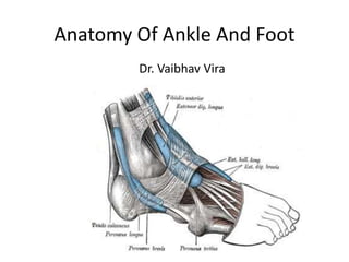
  • 1. Anatomy Of Ankle And Foot Dr. Vaibhav Vira
  • 2. Ankle • A/k/a talocrural joint • A diarthrodial articulation involving the distal tibia and fibula and the body of the talus • the only example in the human body of a true mortise joint
  • 4. Foot • The human foot is a complex structure adapted to allow orthograde bipedal stance and locomotion and is the only part of the body that is in regular contact with the ground • There are 28 separate bones in the human foot, including the sesamoid bones of the first metatarsophalangeal joint, and 31 joints, including the ankle joint.
  • 5. Foot (cont.) • Functionally, the skeleton of the foot may be divided into the tarsus, metatarsus and phalanges. • Anatomically it is divided into - • The hindfoot comprises the calcaneus and talus • The midfoot comprises the navicular, cuboid and three cuneiforms • The forefoot comprises five metatarsals, fourteen phalanges and two sesamoid bones of the great toe
  • 7. Distal Tibia • The distal end of the tibia has 5 surfaces namely anterior, medial, posterior, lateral and distal surfaces, and projects inferomedially as the medial malleolus • The distal surface, also called the tibial plafond, articulates with the talus and is wider anteriorly than posteriorly • The medial malleolus is short and thick, and has a smooth lateral surface with a crescentic or comma-shaped facet that articulates with the medial surface of the talar body
  • 8. Distal Fibula • The distal end of the fibula or lateral malleolus projects distally and posteriorly relative to the medial malleolus • Its lateral aspect is subcutaneous, the posterior surface has a broad groove with a prominent lateral border, and the anterior surface is rough and somewhat rounded and articulates with the anteroinferior aspect of the tibia. • The medial surface has a triangular articular facet and is vertically convex with its apex directed distally. It articulates with the lateral talar surface. Behind the facet is a rough malleolar fossa for ligamentous attachment
  • 9.
  • 10.
  • 11. Tarsus • The seven tarsal bones occupy the proximal half of the foot • The tarsus and carpus are homologous, but the tarsal elements are larger, reflecting their role in supporting and distributing body weight. • The proximal row is made up of the talus and calcaneus • The distal row contains, from medial to lateral, the medial, intermediate and lateral cuneiforms and the cuboid.
  • 12. • Medially, there is an additional single intermediate tarsal element, the navicular. • Collectively, these bones display an arched transverse alignment that is dorsally convex. Medially, the navicular is interposed between the head of the talus and the cuneiforms. Laterally, the calcaneus articulates with the cuboid. • The tarsus and metatarsus are arranged to form intersecting longitudinal and transverse arches. Hence, thrust and weight are not transmitted from the tibia to the ground (or vice versa) directly through the tarsus, but are distributed through the tarsals and metatarsals to the ends of the longitudinal arches.
  • 14. Talus • The talus is an intercalated bone with no tendinous attachments. It is the osseous link between the foot and leg through the ankle joint. It is the second largest tarsal bone • It Has Head, Neck And Body • Head:- Directed distally and somewhat inferomedially, the head has a distal surface, which is ovoid and convex; its long axis is also inclined inferomedially to articulate with the proximal navicular surface. • The Neck is the narrow, medially inclined region between the head and body. • The body is cuboidal in shape, superior (trochlear) surface articulates with tibia, inferior surface articulates with calcaneum, medial surface articulates superiorly with medial malleolus of tibia, lateral surface articulates with lateral malleolus of fibula, and small posterior surface
  • 15.
  • 16. Calcaneus • In Latin Heel, forms prominence of heel • The calcaneus is the largest of the tarsal bones and projects posterior to the tibia and fibula as a short lever for muscles of the calf attached to its posterior surface. • It is irregularly cuboidal, its long axis directed forwards, upwards and laterally. • It has 6 surfaces namely, anterior, posterior, superior or dorsal, plantar, medial and lateral.
  • 17. • Anterior surface is smallest surface and articulates with cuboid. • Posterior surface is where tendocalcaneus (tendo achillis) and plantaris is attached. • Superior surface is where it articulates with talus. • The plantar surface is rough and has three tubercles i.e. medial, lateral and anterior. Plantar ligament is attached on this surface. • Lateral surface is rough and flat • Medial surface is concave and have process called sustentaculum tali which assist in formation of talocalcaneonavicular joint
  • 18.
  • 19. Navicular • Boat shaped • The navicular articulates with the talar head proximally and with the cuneiform bones distally. • 6 Surfaces namely anterior, posterior, superior or dorsal, plantar, medial and lateral. • Anterior surface articulates with three cuneiforms
  • 20.
  • 21. Cuneiform Bones • Cuneiform = Wedge shaped • 3 Bones:- medial, intermediate and lateral • Medial is the largest and intermediate is smallest cunieform bone • Medial cuneiform has 6 surfaces • Intermediate cuneiform has 4 surfaces • Lateral cuneiform has 3 surfaces • Articulate with the navicular proximally and with the bases of the first to third metatarsals distally
  • 22. Cuboid • The cuboid, the most lateral bone in the distal tarsal row, lies between the calcaneus proximally and the fourth and fifth metatarsals distally. • As name suggest it is cuboid, it has 6 surfaces namely anterior, posterior, superior (plantar) or dorsal, medial and lateral.
  • 23.
  • 24. Metatarsus • Metatarsus is made up of 5 metatarsal bones numbered from medial to lateral. • Each metatarsal is miniature long bones having shaft, base and head. • Except for the first and fifth, the shafts are long and slender, longitudinally convex dorsally, and concave on their plantar aspects.
  • 25.
  • 26. Phalanges • 28 phalanges • 14 in each foot • 2 in great toe and 3 for each of other toes
  • 27. Soft Tissues Around Ankle Foot • Retinacula at the ankle:- Extensor Retinacula (Superior and Inferior), Flexor Retinaculum • Synovial Sheats at the Ankle • Plantar Aponeurosis • Fibular Retinacula (Superior And Inferior)
  • 28. Extensor Retinacula • Deep fascia is thickened to form bands called retinaculum. • So called because they retain tendons in the place. • On front of the ankle there are extensor retinacula • Superior Extensor Retinaculum:- – Medially attached to the lower part of the anterior border of tibia – Laterally attached to the anterior boundary of the elongated triangular area just above lateral malleolus.
  • 29. Extensor Retinaculum (cont.) • Inferior Extensor Retinaculum:- – Y shaped band of deep fascia, situated in front of the ankle joint and over posterior part of dorsum of the foot – Stem of Y is laterally, and upper and lower bands medially – Stem is attached to the anterior non articulating part of the superior surface of the calcaneum, in front of the sulcus calcanei – Upper band passes upwards and medially, and attaches to anterior border of medial malleolus – Inferior band passes downward and medially and attaches to the plantar aponeurosis
  • 30. Extensor Retinaculum (cont.) • Structures passing under Superior and Inferior Extensor Retinaculum are (from Medial to Lateral):- – Tibialis Anterior – Extensor Hallucis Longus – Anterior Tibial Vessels – Deep Peroneal Nerve – Extensor Digitorum Longus – The Peroneus Tertius
  • 32. Flexor Retinaculum (Tarsal Tunnel) • 2.5cm broad • Attachments:- – Anteriorly to the posterior border and tip of the medial malleolus – Posteriorly and laterally to the medial tubercle • Structures passing deep to retinaculum are (from medial to lateral): – The tendon of the tibialis posterior – The tendon of the flexor digitorum longus
  • 33. – Posterior tibial artery and terminal branch with accompanying veins. – Tibial Nerve and its branches – Tendon of Flexor hallucis longus Lowest Part of the deep surface of retinaculum give origin to greater part of abductor hallucis muscle
  • 34. Plantar Aponeurosis • It is the thickened central part of deep fascia • The plantar aponeurosis is composed of densely compacted collagen fibres orientated mainly longitudinally, but also transversely. • It represents distal part of plantaris which was seperated from rest of muscle during evolution. • It is triangular in shape with apex being proximal.
  • 35. • Apex is attached to medial tubercle of the calcaneum, proximal to attachment of flexor digitorum brevis • Base is distal and divides into five processes near the head of the metatarsal bones. • Each process splits into superficial and deep slip. • Superficial slip is attached to skin • Deep slips embrace flexor tendons and blends with fibrous flexor sheet and deep transverse ligaments.
  • 36. • It divides the sole into three compartments by septas • Function of Plantar Aponeurosis: – Fixes the skin to the sole – Protects the deeper structures – Helps in maintaining the longitudinal arches of the foot – Gives origin to muscles of the first layer of the sole
  • 37.
  • 38. Synovial Sheats at Ankle • Anterior to the ankle, the synovial sheath for tibialis anterior extends from the proximal margin of the superior extensor retinaculum to the interval between the diverging limbs of the inferior retinaculum. • Posteromedial to the ankle, the sheath for tibialis posterior starts approximately 4 cm above the medial malleolus and ends just proximal to the attachment of the tendon to the tuberosity of the navicular • Posterolateral to the ankle, the tendons of fibularis longus and brevis are enclosed in a single sheath deep to the superior fibular retinaculum. This sheath splits into two separate sheaths enclosing their respective tendons deep to the inferior fibular retinaculum.
  • 39. Fibular Retinacular • The fibular retinacula are fibrous bands that retain the tendons of fibularis longus and brevis in position as these tendons cross the lateral aspect of the ankle region. • The superior fibular retinaculum is a short band that extends from the back of the lateral malleolus to the deep transverse fascia of the leg and the lateral surface of the calcaneus. Damage to the retinaculum can lead to instability of the tendons of fibularis longus and brevis. • The inferior fibular retinaculum is continuous in front with the inferior extensor retinaculum, and is attached posteriorly to the lateral surface of the calcaneus. Some of its fibres are fused with the periosteum on the fibular trochlea (peroneal trochlea or tubercle) of the calcaneus, forming a septum between the tendons of fibularis longus and brevis.
  • 40.
  • 41. Lateral Ligaments ("T" shaped) • Anterior talofibular (weakest and most frequently injured) • Calcaneofibular (strongest of the three ligaments) • Posterior talofibular Lateral It consist of three bands namely
  • 42. Specialised adipose tissue • Heal and metatarsal pads. • The heel is subject to repeated high impacts and is anatomically adapted to withstand these pressures. • The adult heel pad has an average thickness of 18 mm and a mean epidermal thickness of 0.64 mm (dorsal epidermal thickness averages 0.069 mm). • The heel pad contains elastic adipose tissue organized as spiral fibrous septa anchored to each other, to the calcaneus and to the skin. • The septa are U-shaped, fat-filled columns designed to resist compressive loads and are reinforced internally with elastic diagonal and transverse fibres, which separate the fat into compartments.
  • 43. • In the forefoot, the subcutaneous tissue consists of fibrous lamellae arranged in a complex whorl containing adipose tissue attached via vertical fibres to the dermis superficially and the plantar aponeurosis deeply. • The fat is particularly thick in the region of the metatarsophalangeal joints, which cushions the foot during the toe-off phase of gait (see below). • Like the heel pad, the metatarsal fat pad is designed to withstand compressive and shearing forces. • Atrophy of either may be a cause of persistent pain in the distal plantar region.
  • 44. JOINTS • 33 joints and 26 bones. • Two major joints • The Ankle Joint • The second major joint is the Subtalar Joint T MMLM
  • 45. Ankle Joint • The ankle joint is a synovial joint of hinge variety, approximately uniaxial. • The lower end of the tibia and its medial malleolus, together with the lateral malleolus of the fibula and inferior transverse tibiofibular ligament, form a deep recess (‘mortise’) for the body of the talus. • Structurally it is a strong joint and stability is ensured by close interlocking of articular surface, strong collateral ligament and tendon that cross the joint.
  • 46.
  • 47. Ankle Joint • Ligaments – Fibrous Capsule – The Deltoid or medial Ligament – A lateral ligament • Fibrous Capsule – Surrounds the joint – Weak anteriorly and posteriorly to allow hinge movement – Attached all around the articular margins except • Posterosuperiorly attached to inferior tibiofibular ligament • Anteroinferiorly attached to dorsum of the neck of the talus at some distance from trochlear surface
  • 48. • Deltoid Or Medial Ligament – It is a strong, triangular band, attached to the apex and the anterior and posterior borders of the medial malleolus. It consist of superficial and deep part. – Excessive tensile force results in avulsion fracture rather than a tear of ligament
  • 49. The Deltoid Ligament • Deltoid ligament is a combination of: – Anterior talotibial – Tibionavicular – Tibiocalcaneal – Posterior talotibial Medial
  • 50.
  • 51.
  • 52. SubTalar Joint • 3 joints between talus and calcaneum namely posterior, anterior and medial. • The posterior joint is named talocalcanean or subtalar joint. • Anterior joints is part of talocalcaneonavicular joint. • Since the three joints form a single functional unit, clinicians often include these joints under the term subtalar joint.
  • 53. • However, the sinus tarsi seperates posterior articulation from the anterior and medial articulations. • The greater part of the talocalcaneonavicular joint lies in front of the head of the talus and not below it.
  • 54.
  • 55. Talocalcanean Joint • It is a plain synovial joint between the concave facet on inferior surface of the body of talus and convex facet on superior surface of the calcaneum. • The Bones are connected by:- – A fibrous capsule – The lateral and medial talocalcanean ligaments – The interosseous talocalcanean ligament – The cervical ligament
  • 56. Interosseous talocalcanean ligament • it is thick and very strong. • It is the chief bond of union between talus and calcaneum. • Occupies sinus tarsi • Seperates talocalcanean joint from talocalcaneonavicular joint. • Becomes taut in eversion and limits this movement
  • 57.
  • 58. • Cervical ligament:- – It is placed lateral to sinus tarsi. – It becomes taut in inversion and limits this movement. • Collateral ligaments of ankle joint provide stability to the talocalcanean joint. • Movement – Inversion and Eversion
  • 59.
  • 60. Talocalcaneonavicular Joint • Some features of ball and socket joint • Head of talus fits into a socket formed partly by navicular bone and partly by calcaneum. • Two ligaments also take part in forming the socket – Medially by the spring ligament – Laterally by medial limb of bifurcate ligament
  • 61. • Bones are connected by fibrous capsule • Movements – Inversion – Eversion
  • 62. Spring Ligament • A/k/a Plantar calcaneonavicular ligament • It is powerful • Attached posteriorly to anterior margin of substentaculum tali, and anteriorly to the plantar surface of the navicular bone. • Head of talus rest directly on the upper surface of the ligament. • Plantar surface supported by tendon of tibialis posterior medially and by tendons of flexor hallucis longus and flexor digitorum longus, laterally. • Most Important ligament for maintaining medial longitudinal arch of foot.
  • 63.
  • 64. Calcaneocuboid Joint • Saddle Joint. • Articular surface of calcaneum and cuboid is concavoconvex. • Bones are connected by – A fibrous capsule – The lateral limb of the bifurcate ligament – The long plantar ligament – The short plantar ligament
  • 65.
  • 66. • Bifurcate Ligament – Y shaped – Stem attached to anterolateral part of sulcus calcanei – Medial limb/ calcaneonavicular ligament attached to dorsolateral surface of navicular bone – Lateral limb/ calcaneocuboid ligament attached to dorsomedial surface of cuboid bone
  • 67.
  • 68. • Long Plantar Ligament is a long and strong ligament whose importance in maintaining arches of foot. • It is attached to plantar surface of calcaneum posteriorly and cuboid bone anteriorly. • Short plantar ligament lies deep to the long plantar ligament. • It is broad and strong ligament extending from anterior tubercle of calcaneus to plantar surface of cuboid bone.
  • 69.
  • 70. Inversion And Eversion Of Foot • Inversion is movement in which the medial border of the foot is elevated, so that sole faces medially. • Eversion is a movement in which the lateral border of foot is elevated, so that the sole faces laterally. • The movement can be performed voluntarily only when foot is off the ground. When foot is on the ground these movement help to adjust foot to uneven ground.
  • 71. • In these movements the entire part of the foot below the talus moves together. • It mainly takes place at the subtalar and talocalcaneonavicular joints and partly at the transverse tarsal joint. • Inversion is accompanied by plantarflexion of the foot and adduction of forefoot. • Eversion is accompanied by dorsiflexion of the foot and abduction of the forefoot.
  • 72.
  • 73. • Joints Taking Part – Main • Subtalar (talocalcanean) • Talocalcaneonavicular – Accessory • Transvese tarsal which includes calcaneocuboid and talonavicular joints • Muscle Producing movement – Inversion: Tibialis Anterior and Tibialis Posterior – Eversion: Peroneus Longus and Peroneus Brevis
  • 74. • Limiting Factors – Inversion • Tension of peronei • Tension of cervical ligament – Eversion • Tension of tibialis anterior • Tension of tibialis posterior • Tension of deltoid ligament
  • 75. • Inversion and eversion greatly help the foot in adjusting to uneven and slippery ground. When feet are supporting weight, these movement occur in a modified form called supination and pronation, which are forced on the foot by the body weight
  • 76. Smaller Joints of forefoot • These are plane joints between the navicular, the cuneiforms, the cuboid and the metatarsal bones. • They permit small gliding movements, which allow elevation and depression of the heads of the metatarsals as well as pronation and supination of the foot. • There are 6 joint cavities of the foot (talocalcanean, talocalcaneonavicular, calcaneocuboid, 1st cuneometatarsal, cubometatarsal and calcaneonavicular with extension i.e. navicular with three cuneiforms and 2nd and 3rd cuneometatarsal)
  • 77.
  • 78. TarsoMetatarsal Joint • Tarsometatarsal articulations are approximately plane synovial joints. • The joints are approximately on an imaginary line traced from the tubercle of the fifth metatarsal to the tarsometatarsal joint of the great toe, except for that between the second metatarsal and intermediate cuneiform, which is 2–3 mm proximal to this line. • Movements between the tarsals and metatarsals are limited to flexion and extension, except in the first tarsometatarsal joint, where some abduction and rotation occur.
  • 79. Metatarsophalangeal Joint • Metatarsophalangeal articulations are ovoid or ellipsoid joints between the rounded metatarsal heads and shallow cavities on the proximal phalangeal bases. They are usually 2.5 cm proximal to the web spaces of the toes. • Flexion, extension, abduction and adduction are the movements that occur in this joints
  • 80. Interphalangeal Joint • Interphalangeal articulations are almost pure hinge joints, in which the trochlear surfaces on the phalangeal heads articulate with reciprocally curved surfaces on adjacent phalangeal bases. • Flexion and extension occurs in this joints
  • 81. Muscles • Extension of deep fascia form intermuscular septa that divide leg into compartments. • The anterior and posterior intermuscular septa are attached to anterior and posterior borders of fibula dividing leg into anterior, posterior and lateral compartment. • The posterior compartment is further subdivided into superficialand deep parts by transverse intermusclar septum
  • 82. Muscle Compartments • Anterior Compartment (Dorsal flexors) – Tibialis anterior – Peroneous tertius – Extensor digitorum Longus – Extensor digitorum brevis – Extensor hallicus longus • Lateral Compartment (Evertors) – Peroneus longus – Peroneus brevis
  • 83. Muscle Compartments • Deep Posterior Compartment (Plantar Flexors) – Flexor digitorum longus – Flexor hallicus – Tibialis Posterior • Superficial Posterior Compartment(Plantar Flexors) – Gastrocnemius – Soleus – Plantaris
  • 84. Ankle and Foot Muscles • Superficial Posterior Compartment – Gastrocnemius – Soleus – Plantaris • Deep Posterior Compartment – Flexor digitorum longus – Flexor hallicus – Tibialis Posterior • Lateral Compartment (Evertors) – Peroneus longus – Peroneus brevis • Anterior Compartment (Dorsal flexors) – Tibialis anterior – Peroneous tertius – Ext. dig. Longus – Ext. hallicus
  • 85. Gastrocnemius • Origin: posterior surface of the two femur condyels • Insertion: posterior surface of the calcaneus via Achilles tendon • Actions: – plantar flexion of the foot – flexion of the knee • Stronger plantar flexion when the knee is extended • Superficial posterior compartment Posterior
  • 86. Soleus • Located beneath the gastrocnemius • Origin: upper 2/3 of the posterior surfaces of the tibia and fibula • Insertion: posterior surface of the calcaneus via Achilles tendon • Action: – plantar flexion • Superficial posterior compartment Posterior
  • 87. Gastrocnemius & Soleus •Gastronemius and Soleus = “triceps surae” due to their three heads
  • 88.
  • 89. Achilles Tendon • Named after Achilles • Largest tendon • 1000 pounds of force • Tendon of the Gastrocnemius and Soleus
  • 90. Plantaris • Absent in some humans • Origin: lateral epicondyle • Insertion: calcaneus • Actions: – plantar flexion • Superficial posterior compartment Posterior
  • 91.
  • 92. Tibialis posterior • Origin: posterior surface of the upper half of the adjacent surface of tibia & fibula • Insertion: navicular, cuneiforms, and cuboid bones and bases of the 2nd-5th metatarsal bones. • Note: passes posterior to medial malleolus. • Actions: – plantar flexion – inversion of the foot • Deep posterior compartment Posterior
  • 93. Flexor Digitorum Longus • Origin: middle 1/3 of the posterior surface of the tibia • Insertion: base of the distal phalanges of each of lateral four toes • Note: passes posterior to medial malleolus. • Actions: – toe flexion – plantar flexion, – inversion of the foot • Maintains the longitudinal arch • Deep posterior compartment Posterior
  • 94. Flexor Hallicus Longus • Origin: middle half of the posterior surface of the fibula • Insertion: distal phalanx of the large toe, plantar surface • Note: passes posterior to medial malleolus. • Actions: – Flexion of the great toe – Inversion – Plantar flexion • Deep posterior compartment Posterior
  • 95.
  • 96. Tibialis anterior • Origin: upper 2/3 of the anterior surface of the tibia • Insertion: medial cuneform and the first metatarsal • Note: passes anterior to medial malleolus. • Actions: – Dorsal flexion – Inversion. • Anterior compartment Anterior
  • 97. Extensor hallicus longus • Origin: middle 2/3 of the inner surface of the front of the fibula • Insertion: top of the distal phalanx of the great toe • Note: passes anterior • Actions: – Extension of big toe – Dorsiflexion – Weak inversion of the foot • Anterior compartment Anterior
  • 98. Extensor digitorum longus • Origin: lateral condyle of the tibia and anterior surface of the fibula • Insertion: middle and distal phalanges of the four lateral toes. • Note: passes anterior to lateral malleolus. • Actions: – Toe extension – Dorsiflexion – Eversion • Anterior compartment Anterior
  • 99. Extensor Digitorum Brevis • Origin: Anterior part of the superior surface of the calcaneum • Inserion: middle and distal phalanges of the four medial toes. • Note: Medial most part of muscle which is distict is known as extensor hallucis brevis • Action: – Toe Extension • Anterior Compartment
  • 100. Peroneous tertius • Origin: lower fibula • Insertion: dorsal surface of the 5th metatarsal • Note: passes anterior to lateral malleolus. • Action: – Dorsiflexion – Eversion • Anterior compartment Anterior
  • 101.
  • 102. Peroneus longus muscle • Origin: head and upper 2/3 of the outer surface of the fibula • Insertion: undersurfaces of the 1st cuneiform and first metatarsal bones • Note: passes posterior to lateral malleolus. • Actions: – Eversion – Plantar flexion • The tendon goes under the foot from the lateral to the medial surface, thus aiding in support for the transverse arch. • Lateral compartment Lateral
  • 103. Peroneus brevis muscle • Origin: lower 2/3 of the outer surface of the fibula • Insertion: dorsal surface of the 5th metatarsal • Note: passes posterior to lateral malleolus. • Actions: – Plantar flexion – Eversion • Anterior compartment Lateral
  • 105. Muscles and Tendons of the Sole • Four Layers • Muscles of the 1st layer – Flexor digitorum brevis – Abductor hallucis – Abductor digiti minimi • Muscles of the 2nd Layer – Tendon of the flexor digitorum longus – Flexor hallucis longus – Flexor digitorum accessorius – Lumbrical muscles
  • 106. • Muscles of the 3rd layer – Flexor haluucis brevis – Flexor digiti minimi brevis – Adductor hallucis • Muscles of the 4th layer – Interosseous muscles – Tendon of tibialis posterior – Tendon of peroneus longus
  • 107.
  • 108.
  • 109.
  • 110.
  • 111.
  • 112.
  • 113.
  • 114.
  • 115.
  • 116. Arches of the Foot • Arches of the foot help in walking, running and jumping. In addition they help in weight bearing and in providing upright posture. • Arches are supported by intrinsic and extrinsic muscles in the sole, ligaments, aponeurosis and shape of the bones. • The Foot has to act :- – As a pliable platform to support weight in the upright posture – As a lever to propel the body forward
  • 117. • Classification of Arches – Longitudinal • Medial • Lateral – Transverse • Anterior • Posterior
  • 118.
  • 119. Medial Longitudinal Arch • This arch is considerably higher, more mobile and resilient than the lateral. • It is considered as big arch of a small circle. • Acts as a shock absorber. • Anterior end:- Formed by head of 1st, 2nd, 3rd metatarsals. • Posterior End:- Medial tubercle of the calcaneum. • Summit:- Superior articular surface of the body of talus
  • 120. • Pillars – Anterior pillar is long and weak, formed by talus, navicular, three cuneiform bones and first three metatarsal bones. – Posterior pillar is short and strong, formed by medial part of the calcaneum • Main Joint:- Talocalcaneonavicular joint • Phalanges do not take part in formation of the arches
  • 121.
  • 122. Lateral Longitudinal Arch • Characteristically low and limited mobility. • Built to transmit weight and thrust to the ground. • Considered as small arc of big circle • Ends:- – Anterior:- Heads of 4th and 5th metatarsal bones – Posterior:- Lateral tubercle of the calcaneum • Summit:- articular facets of the superior surface of the calcaneum at the level of sub talar joint
  • 123. • Pillars:- – Anterior pillar:- long and weak, formed by cuboid bone and by 4th and 5th metatarsal bones. – Posterior pillar:- short and strong, formed by lateral half of the calcaneum. – Main Joint:- Calcaneocuboid Joint
  • 124.
  • 125. Anterior Transverse Arch • Formed by the heads of the five metatarsal bones. • Complete arch, as heads of 1st and 5th come in contact with the ground and forms the two end of the arch
  • 126. Posterior Transverse Arch • Formed by the greater part of tarsus and metatarsus. • Incomplete, as only the lateral end comes in contact with the ground forming half dome, which is completed by similar half dome of the opposite foot.
  • 127. Factors Responsible for Maintenance of Arches • Shape of the bones concerned • Intersegmental ties/staples or ligaments (and muscles) • The beams or bowstrings that connect two ends of the arch • Slings that keep the summit of the arch pulled up • Suspension
  • 128. Bony Factor • Posterior transverse arch is formed and maintained mainly because of the fact that many of the tarsal bones involved (eg. the cuneiform bones) and the heads of the metatarsal bones, are wedge shaped, the apex of wedge pointing downwards. • The bony factor is not very important in the case of other arches
  • 129. Intersegmental Ties • All arches are supported by ligaments uniting the bones concerned. • The most important of these are – The spring ligament for the medial longitudinal arch – The long and short plantar ligaments for the lateral longitudinal arch – In the case of transvers arch, the metatarsal bones are held together by the interosseous muscles.
  • 130. The Beams • The Longitudinal arches are prevented from flattening by the plantar aponeurosis and by the muscles of the first layer of the sole (Flexor digitorum brevis, flexor digiti minimi and abductor digiti minimi ). • These structure keep the anterior and posterior ends of the arches pulled together.
  • 131. Slings • The summit of the medial longitudinal arch is pulled upwards by tendon passing from the posterior compartment of the leg into the sole i.e. tibialis posterior, flexor hallucis longus, flexor digitorum longus. • The summit of lateral longitudinal arch is pulled upwards by peroneus longus and peroneus brevis.
  • 132. Slings (cont.) • The tendons of tibialis anterior and peroneus longus together form a sling (stirrup) which keeps the middle of the foot pulled upwards, thus supporting the longitudinal arches. • Peroneus longus runs transversly across the sole, it pulls the medial and lateral margins of the sole closer together, thus maintaining the transverse arch. • The transverse arch is also supported by tibialis posterior which grips many of the bones of the sole through its slips
  • 133. Suspension • Medial Longitudinal Arch:- Tibialis Anterior • Lateral Longitudinal Arch:- Peroneus Longus
  • 134. Function of Arches • Distribute body weight to the weight to the weight bearing areas of the sole, mainly the heel and the toes. Out of the later, weight is borne mainly on 1st and 5th toe. The lateral border of the foot bears some weight, but this is reduced due to the presence of the lateral longitudinal arch • The arches acts as spring (chiefly the medial longitudinal arch) which are of great help in walking and running
  • 135. Function of Arches (cont.) • The act as shock absorbers in stepping and particularly in jumping • The concavity of the arches protects the soft tissues of the sole against pressure • The character of the medial longitudinal arch is resiliency and that of lateral longitudinal arch is rigidity.
  • 136.