
Recommended
More Related Content
What's hot
What's hot (20)
Viewers also liked
Viewers also liked (20)
Similar to Anatomy and PhysiologySkeletal system II
Similar to Anatomy and PhysiologySkeletal system II (20)
More from mrhunterspage
More from mrhunterspage (20)
Anatomy and PhysiologySkeletal system II
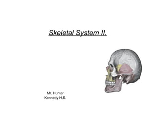
- 1. Skeletal System II. Mr. Hunter Kennedy H.S.
- 2. Mr. Hunter Anatomy and Physiology 11.15.2012 • Objective(s) • SWBAT • List the 5 regions of the spine • Describe the structure of vertebrae • Distinguish true ribs from false ribs and their attachment sites • Describe articulations of the upper extremities of axial and appendicular skeleton Bell Ringer: What is the name of the location marked X on the skeleton model?__________
- 3. Vertebral Column • Consists of a series of vertebrae connected so that they form a flexible curved rod. • Different regions of the spine: cervical, thoracic, lumbar, sacrum and coccyx • (7) cervical, (12) thoracic, (5) lumbar, (1) sacrum and (1) coccyx. • Sacrum = 5 bones in child • Coccyx = 3 to 5 in child
- 4. Vertebral Column • Cervical, thoracic and lumbar vertebrae consists of various parts: • The hole in the center is the vertebral foramen in which the spinal cord travels. • Transverse and articular processes are for controlled movement and rib attachments.
- 5. Vertebral Column Curves • Cervical and lumbar curves of the spine are concave. Thoracic and sacral are convex. • Curves of the spine provide strength to balance and support the weight of the body.
- 6. Thorax and Ribs • Twelve pairs of ribs, the sternum and the thoracic vertebrae form the thorax. • Each of the 12 pairs of ribs are attached posteriorly to a vertebra. • The first 7 pairs of ribs are attached to the sternum by costal cartilage. • The 8th, 9th and 10th pairs are attached to the cartilage of the 7th ribs = false ribs. • The last two pairs of ribs are not attached to any cartilage and appear to float.
- 7. Appendicular Skeleton • The scapula and the clavicle compose the shoulder / pectoral girdle • This connects the upper extremity to the axial skeleton. • This occurs at the sternoclavicular joint – The point of attachment between the clavicle and the sternum. • Fractures of the clavicle are common due to small size of the joint and the wide range of motion by the upper extremitiy.
- 8. Appendicular Skeleton • The humerus is the long bone of the arm and the second longest bone in the body. • Attached at the proximal end to the glenoid cavity of the scapula via a group of muscles called the rotator cuff • The distal end of the humerus articulates with the ulna and the radius. • The olecranon process of the ulna fits into the olecranon fossa of the humerus.
- 9. Appendicular Skeleton – Wrist and Hand • The wrist and the hand have more bones for their size than any other part of the body. • 8 carpal – wrist bones, 5 metacarpals – bones of the palm, 14 phalanges – fingers • There are 27 bones total.
- 10. Appendicular Skeleton – Lower Extremity • The pelvic girdle – hip connects the legs to the trunk. • Consists of two large coxal bones located on each side of the pelvis, attached inferiorly to the sacrum of the vertebral canal. • In the infant, the coxal bone consists of the ilium, ischium and the pubis.
- 11. Appendicular Skeleton – Lower Extremity • The femur is the longest bone in the body. It articulates with the coxal bone in a cup- shaped socket called the acetabulum. • Distally, the femur articulates with the patella - knee cap at the medial condyle of the tibia – shinbone. • The fibula lies along the outer lateral border of the leg.
- 12. Appendicular Skeleton – Foot and Ankle • Toe bones have the same as finger bones – phalanges. • Metatarsals and tarsals are located in the foot • Each foot has (5) metatarsal bones, and (7) tarsal bones and (14) phalanges • The largest tarsal bone is the calcaneous – heel bone
- 13. Appendicular Skeleton – Foot and Ankle • Strong ligaments and tendons hold the foot bones in a normal arched position for adequate support of body weight. • When the tendons or ligaments become damaged the arches can collapse – flat feet. • Two arches of the foot: Transverse and Longitudinal
- 14. Differences Between Male and Female Skeleton • Most male skeletons have larger bones due to skeletal muscle attachments. • The greater the tension on the bone via muscle attachments, the larger and denser the bone becomes at the point of attachment. • The coxal bones are also different. • The pubic angle in the female is wider than that of the male for the purpose of childbearing.
- 15. Skeletal Joints • Every bone in the body connects to at least one other bone via joints with the exception of the hyoid bone. • Joints allow movement to occur between certain bones. • They also hold our bones securely together.
- 16. Skeletal Joints • Synarthroses – no movement. Fibrous connective tissue joins the articulating bones and holds them close together. Ex. Cranial bones – sutures. • Amphiarthroses – slight movement. A joint in which cartilage joins the articulating bones. Ex. Joint between the two pubic bones and the joints between the body of vertebrae • Diarthroses – free movement.
- 17. Types of Skeletal Joints • There are several types of diarthroses. • Ball and socket joint • Hinge joint • Saddle joint • Pivot joint • Gliding joint • Condyloid joint • Different joint structures provide different ranges of motion. • Flexion, Extension,Rotation • Circumduction, Abduction and Adduction
- 18. Types of Skeletal Joints • Different joint structures provide different ranges of motion. • Flexion, Extension,Rotation • Circumduction, Abduction and Adduction • Pg. -131-132 • Read pgs. 141-145 • Answer Quick Check pg. 145 # 1-3