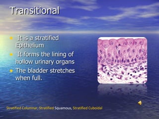
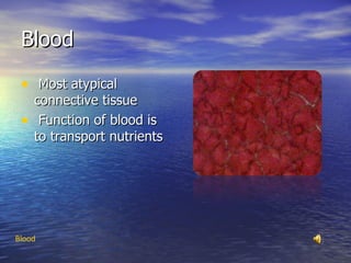
Tissues Epithelial Tissue Connective Tissue Muscle Tissue Nerve Tissue Tissue-  Epithelial ,  Connective ,  Muscle,  Nerve,  Credits
Epithelial Tissue Widespread throughout the whole body. Epithelial cells may be; Squamous, Cuboidal, or Columnar in shape.  Functions of Epithelial Tissue Simple Epithelial ,  Stratified Epithelial
Stratified Epithelial Two or more cell layers The Basal Cells Durability  Stratified Columnar ,  Transitional,  Stratified  Squamous ,  Stratified  Cuboidal
Stratified Columnar Limited Distribution in the body Transitional,   Stratified  Squamous ,  Stratified  Cuboidal
Transitional It is a stratified Epithelium It forms the lining of hollow urinary organs The bladder stretches when full.  Stratified Columnar,  Stratified  Squamous ,  Stratified  Cuboidal
Stratified Squamous Most widespread of the stratified epithelia Forms the external part of the skin The epidermis is keratinized Stratified Columnar ,  Transitional,  Stratified  Cuboidal
Stratified Cuboidal It’s rare Mostly found in the largest ducts of sweat glands Stratified Columnar,   Transitional  ,  Stratified  Squamous
Simple Epithelial Is composed of a single layer Very good at protection Found where absorption and filtration occur  Simple Columnar,  Pseudostratified  Columnar ,  Simple  Cuboidal ,  Simple  Squamous
Simple Columnar Tall closely packed cells Mostly associated with absorption  Pseudostratified  Columnar ,  Simple  Cuboidal ,  Simple  Squamous
Pseudostratified Columnar Vary in height Gives a false impression It is similar to Simple Columnar  Simple Columnar,  Simple  Cuboidal ,  Simple  Squamous
Simple Cuboidal Single layered cells String of Bead Located  in the smallest ducts of  sweat glands Simple Columnar ,  Pseudostratified  Columnar ,  Simple  Squamous
Simple Squamous Thin and shaped laterally flat Mesothelium  Located in the kidneys  Simple Columnar,  Pseudostratified  Columnar ,  Simple  Cuboidal
Connective Tissue It is found everywhere in the body Characteristics of connective tissue The four main classes are; Connective Tissue Proper, Cartilage, Bone, and Blood  Connective Tissue Proper,  Cartilage,  Bones , Blood
Connective Tissue- Proper Connective Tissue proper has two subclasses: Loose connective tissue, and Dense connective tissue Different from Epithelial Tissue Spread out in intervals Loose Connective,  Dense Connective
Loose Connective Found beneath the dermis of the skin Specialized Connective Tissue in the body Adipose ,  Reticular,  Areolar
Dense Connective Subclasses are; Elastic, Dense Connective, and Dense Regular Consists mainly of collage Elastic,  Dense Irregular ,  Dense Regular
Adipose Connective  Accounts for 90% of the tissue’s mass Packed closely together Without the fat stores in our Adipose tissue we could not live for more than a few days Reticular ,  Areolar
Reticular Connective Tissue Resembles Areolar Connective tissue Network of reticular fibers Adipose,  Areolar
Areolar Connective The functions of Areolar tissue Very loose Most widely distributed Connective tissue Adipose ,  Reticular
Elastic Connective Forms a fibrous membrane Dense Irregular ,  Dense Regular
Dense Irregular Connective Forms sheets in areas of the body Forms fibrous joint capsules Elastic ,  Dense Irregular
Dense Regular Connective Contains closely packed bundles of collagen fibers Great resistance to tension  Few cells  Elastic ,  Dense Irregular
Cartilage Withstands both tension and compression  Cartilage is tough  Receive nutrients from blood cells  Hyaline Cartilage ,  Fibrocartilage ,  Elastic Cartilage
Hyaline Cartilage Abundant cartilage in the body Firm support Fibrocartilage ,  Elastic Cartilage
Fibrocartilage Perfect intermediate between hyaline cartilage and dense regular connective tissue  Compressible and resists tension well Located in the intervertebral Hyaline Cartilage ,  Elastic Cartilage
Elastic Cartilage Nearly identical to hyaline cartilage Forms the external ear and epiglottis Hyaline Cartilage,  Fibrocartilage
Bone Rocklike hardness  Known as the osseous tissue Bones are what make up the skeleton Bone
Blood Most atypical connective tissue Function of blood is to transport nutrients  Blood
Muscle Tissue Highly vascularized tissue  Responsible for most types of body movement Myofilaments  Skeletal Muscle,  Cardiac Muscle , Smooth Muscle
Cardiac Muscle Found in the walls of the heart Similar to skeletal muscle cells Skeletal Muscle ,  Smooth Muscle
Skeletal Muscle Muscle forms the flesh of the body Muscle fibers Cardiac Muscle ,  Smooth Muscle
Smooth Muscle Found in the walls of the hollow organs Spindle Shaped  Cardiac Muscle ,  Skeletal Muscle
Nerve Main component of the nervous system  Neuron  Nerve
Credits All sounds on slides 1-33 were spoken by Angela DiPasquale  All slides were written by Angela DiPasquale Picture of tissue on slide one was taken by Zerlina Picture of Epithelial tissue on slide two was taken by Angela, Brittany, and Andie Picture of Stratified Epithelial on slide three was take by Akay Picture of Stratified Columnar on slide four was taken by Angela Andie, and Brittany Picture of Transitional tissue on slide five was taken by Akay Picture of Stratified Squamous on slide six was taken by Greenflame09 Picture of Stratified Cuboidal on slide seven was take by Akay Picture of Simple Epithelial on slide eight was taken by Nina Picture of Simple Columnar on slide nine was taken by  Greenflame09 Picture of Pseudostratified Columnar on slide ten was taken by Akay Picture of Simple Cuboidal on slide eleven was taken by Seancloud2511 Picture of Simple Squamous on slide twelve was taken by Akay Credits
Credits Picture of Connective tissue on slide thirteen was taken by Angela, Andie, and Brittany Picture of Connective tissue proper on slide fourteen was taken by Akay Picture of Loose connective tissue on slide fifteen was taken by Christi Picture of Dense connective tissue on slide sixteen was taken by Akay Picture of Adipose tissue on slide seventeen was taken by Angela, Brittany, and Andie Picture of Reticular Connective tissue on slide eighteen was take by Akay Picture of Areolar Connective tissue on slide nineteen was taken by Akay Picture of Elastic tissue on slide twenty was taken by wright.arron Picture of Dense Irregular tissue on slide twenty-one was taken by Christi Picture of Cartilage on slide twenty-three was taken by Akay Picture of Hyaline Cartilage on slide twenty-four was taken by Angela, Brittany, and Andie Picture of Fibrocartilage on slide twenty-five was taken by Kilpschy Picture of Elastic Cartilage on slide twenty-six was taken by Akay Picture of Bone on slide twenty-seven was taken by Curious expeditions Picture of Blood on slide twenty-eight was taken by Angela, Brittany, and Andie Picture of Muscle tissue on slide twenty-nine was taken by Sciencestuff Picture of Cardiac muscle on slide thirty was taken by Angela, Brittany, and Andie Picture of Skeletal Muscle on slide thirty-one was taken by Angela, Brittany, and Andie Picture of Smooth Muscle on slide thirty-two was taken by Christi Picture of Nerve tissue on slide thirty-three was taken by Akay Picture of Flowers on Credits page was taken from Ragazza1983 Song on credits page was sung by Beetoveen

Anatomy Power Point

  • 1.
    Tissues Epithelial TissueConnective Tissue Muscle Tissue Nerve Tissue Tissue- Epithelial , Connective , Muscle, Nerve, Credits
  • 2.
    Epithelial Tissue Widespreadthroughout the whole body. Epithelial cells may be; Squamous, Cuboidal, or Columnar in shape. Functions of Epithelial Tissue Simple Epithelial , Stratified Epithelial
  • 3.
    Stratified Epithelial Twoor more cell layers The Basal Cells Durability Stratified Columnar , Transitional, Stratified Squamous , Stratified Cuboidal
  • 4.
    Stratified Columnar LimitedDistribution in the body Transitional, Stratified Squamous , Stratified Cuboidal
  • 5.
    Transitional It isa stratified Epithelium It forms the lining of hollow urinary organs The bladder stretches when full. Stratified Columnar, Stratified Squamous , Stratified Cuboidal
  • 6.
    Stratified Squamous Mostwidespread of the stratified epithelia Forms the external part of the skin The epidermis is keratinized Stratified Columnar , Transitional, Stratified Cuboidal
  • 7.
    Stratified Cuboidal It’srare Mostly found in the largest ducts of sweat glands Stratified Columnar, Transitional , Stratified Squamous
  • 8.
    Simple Epithelial Iscomposed of a single layer Very good at protection Found where absorption and filtration occur Simple Columnar, Pseudostratified Columnar , Simple Cuboidal , Simple Squamous
  • 9.
    Simple Columnar Tallclosely packed cells Mostly associated with absorption Pseudostratified Columnar , Simple Cuboidal , Simple Squamous
  • 10.
    Pseudostratified Columnar Varyin height Gives a false impression It is similar to Simple Columnar Simple Columnar, Simple Cuboidal , Simple Squamous
  • 11.
    Simple Cuboidal Singlelayered cells String of Bead Located in the smallest ducts of sweat glands Simple Columnar , Pseudostratified Columnar , Simple Squamous
  • 12.
    Simple Squamous Thinand shaped laterally flat Mesothelium Located in the kidneys Simple Columnar, Pseudostratified Columnar , Simple Cuboidal
  • 13.
    Connective Tissue Itis found everywhere in the body Characteristics of connective tissue The four main classes are; Connective Tissue Proper, Cartilage, Bone, and Blood Connective Tissue Proper, Cartilage, Bones , Blood
  • 14.
    Connective Tissue- ProperConnective Tissue proper has two subclasses: Loose connective tissue, and Dense connective tissue Different from Epithelial Tissue Spread out in intervals Loose Connective, Dense Connective
  • 15.
    Loose Connective Foundbeneath the dermis of the skin Specialized Connective Tissue in the body Adipose , Reticular, Areolar
  • 16.
    Dense Connective Subclassesare; Elastic, Dense Connective, and Dense Regular Consists mainly of collage Elastic, Dense Irregular , Dense Regular
  • 17.
    Adipose Connective Accounts for 90% of the tissue’s mass Packed closely together Without the fat stores in our Adipose tissue we could not live for more than a few days Reticular , Areolar
  • 18.
    Reticular Connective TissueResembles Areolar Connective tissue Network of reticular fibers Adipose, Areolar
  • 19.
    Areolar Connective Thefunctions of Areolar tissue Very loose Most widely distributed Connective tissue Adipose , Reticular
  • 20.
    Elastic Connective Formsa fibrous membrane Dense Irregular , Dense Regular
  • 21.
    Dense Irregular ConnectiveForms sheets in areas of the body Forms fibrous joint capsules Elastic , Dense Irregular
  • 22.
    Dense Regular ConnectiveContains closely packed bundles of collagen fibers Great resistance to tension Few cells Elastic , Dense Irregular
  • 23.
    Cartilage Withstands bothtension and compression Cartilage is tough Receive nutrients from blood cells Hyaline Cartilage , Fibrocartilage , Elastic Cartilage
  • 24.
    Hyaline Cartilage Abundantcartilage in the body Firm support Fibrocartilage , Elastic Cartilage
  • 25.
    Fibrocartilage Perfect intermediatebetween hyaline cartilage and dense regular connective tissue Compressible and resists tension well Located in the intervertebral Hyaline Cartilage , Elastic Cartilage
  • 26.
    Elastic Cartilage Nearlyidentical to hyaline cartilage Forms the external ear and epiglottis Hyaline Cartilage, Fibrocartilage
  • 27.
    Bone Rocklike hardness Known as the osseous tissue Bones are what make up the skeleton Bone
  • 28.
    Blood Most atypicalconnective tissue Function of blood is to transport nutrients Blood
  • 29.
    Muscle Tissue Highlyvascularized tissue Responsible for most types of body movement Myofilaments Skeletal Muscle, Cardiac Muscle , Smooth Muscle
  • 30.
    Cardiac Muscle Foundin the walls of the heart Similar to skeletal muscle cells Skeletal Muscle , Smooth Muscle
  • 31.
    Skeletal Muscle Muscleforms the flesh of the body Muscle fibers Cardiac Muscle , Smooth Muscle
  • 32.
    Smooth Muscle Foundin the walls of the hollow organs Spindle Shaped Cardiac Muscle , Skeletal Muscle
  • 33.
    Nerve Main componentof the nervous system Neuron Nerve
  • 34.
    Credits All soundson slides 1-33 were spoken by Angela DiPasquale All slides were written by Angela DiPasquale Picture of tissue on slide one was taken by Zerlina Picture of Epithelial tissue on slide two was taken by Angela, Brittany, and Andie Picture of Stratified Epithelial on slide three was take by Akay Picture of Stratified Columnar on slide four was taken by Angela Andie, and Brittany Picture of Transitional tissue on slide five was taken by Akay Picture of Stratified Squamous on slide six was taken by Greenflame09 Picture of Stratified Cuboidal on slide seven was take by Akay Picture of Simple Epithelial on slide eight was taken by Nina Picture of Simple Columnar on slide nine was taken by Greenflame09 Picture of Pseudostratified Columnar on slide ten was taken by Akay Picture of Simple Cuboidal on slide eleven was taken by Seancloud2511 Picture of Simple Squamous on slide twelve was taken by Akay Credits
  • 35.
    Credits Picture ofConnective tissue on slide thirteen was taken by Angela, Andie, and Brittany Picture of Connective tissue proper on slide fourteen was taken by Akay Picture of Loose connective tissue on slide fifteen was taken by Christi Picture of Dense connective tissue on slide sixteen was taken by Akay Picture of Adipose tissue on slide seventeen was taken by Angela, Brittany, and Andie Picture of Reticular Connective tissue on slide eighteen was take by Akay Picture of Areolar Connective tissue on slide nineteen was taken by Akay Picture of Elastic tissue on slide twenty was taken by wright.arron Picture of Dense Irregular tissue on slide twenty-one was taken by Christi Picture of Cartilage on slide twenty-three was taken by Akay Picture of Hyaline Cartilage on slide twenty-four was taken by Angela, Brittany, and Andie Picture of Fibrocartilage on slide twenty-five was taken by Kilpschy Picture of Elastic Cartilage on slide twenty-six was taken by Akay Picture of Bone on slide twenty-seven was taken by Curious expeditions Picture of Blood on slide twenty-eight was taken by Angela, Brittany, and Andie Picture of Muscle tissue on slide twenty-nine was taken by Sciencestuff Picture of Cardiac muscle on slide thirty was taken by Angela, Brittany, and Andie Picture of Skeletal Muscle on slide thirty-one was taken by Angela, Brittany, and Andie Picture of Smooth Muscle on slide thirty-two was taken by Christi Picture of Nerve tissue on slide thirty-three was taken by Akay Picture of Flowers on Credits page was taken from Ragazza1983 Song on credits page was sung by Beetoveen