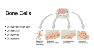
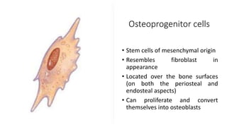
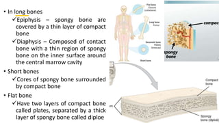
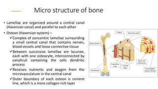
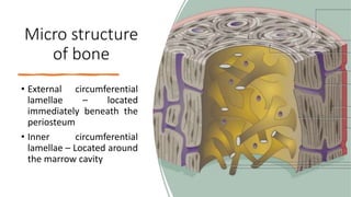
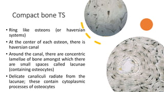
Histology of bone
Dr. P. Christabel Divya
Introduction
• Solid support for the body
• Protects vital organs such as those in
the cranial and thoracic cavities
• Harbors cavities containing bone
marrow where blood cells are
formed
• Reservoir of calcium, phosphorous
and other ions that can be released
or stored in a controlled fashion to
maintain constant concentration in
body fluids
Constituents
• Specialized connective tissue
• Cells
• Osteoprogenitor cells
• Osteoblasts
• Osteocytes
• Osteoclasts
• Calcified extracellular matrix
• Organic matter
• Fibres – Type 1 collagen
• Ground substances – Proteoglycans (aggregcan)
and Glycoproteins (Osteonectin) and water
• Inorganic material – 50% of dry weight
• Calcium hydroxyapatite
• Other ions - Bicarbonate, citrate, magnesium,
potassium, sodium ions
Bone membranes
• Bones are lined on both internal and
external surfaces by layer of connective
tissue containing osteoprogenitor cells
Endosteum – On the internal surface
surrounding the marrow cavity
Periosteum – On the external surface
Bone Cells
• Osteoprogenitor cells
• Osteoblasts
• Osteocytes
• Osteoclasts
Osteoprogenitor cells
• Stem cells of mesenchymal origin
• Resembles fibroblast in
appearance
• Located over the bone surfaces
(on both the periosteal and
endosteal aspects)
• Can proliferate and convert
themselves into osteoblasts
Osteoblast
• Bone forming cell
• Cuboidal to columnar shape with basophilic cytoplasm
• Located at the surfaces of bone matrix, usually side by
side in a layer
• Synthesize and secrete the organic components of
bone matrix, including type I collagen fibres,
proteoglycans and glycoproteins (osteonectin)
Osteoblast
contd
• Secrete matrix components at the cell surface in contact
with the existing bone matrix, producing a layer of new
material called osteoid (but not yet calcified) between
osteoblast layer and pre existing bone surface
• Completed by subsequent deposition of calcium salts into
the newly formed matrix
Osteocytes
• Cells of mature bone
• Lie in the lacunae of bone that lies in between the
successive lamellae and are regularly spaced through out the
mineralized matrix
• Osteoblasts that have become imprisoned in the matrix
during bone formation and differentiate further as osteocyte
Osteocytes contd
• In the transistion from osteoblast to
osteocyte, cells extend many long
dendritic process arising from osteocytes
• Establishes contacts with other
osteocytes and with bone lining cells
present on the surface of bone
• Maintain bone matrix and their death is
followed by matrix resorption
• Additional role in calcium homeostasis
Osteoclasts
• Bone removing cells
• Very large motile cells with multiple
nuclei, the surface of the cell is folded
into irregular projections (Ruffled
border)
• Found in relation to surface where bone
removal is taking place, lies within
enzymatically etched depression or
cavities in the matrix (Resorption bays or
Howship’s lacunae)
• Removal of bone by osteoclast involves
demineralization and removal of matrix
Periosteum
• External surface of any bone
• Consists of two layers
Outer
Inner
• Outer layer – Fibrous layer
• Inner layer – Cellular layer.
Contains osteoprogenitor cells
(Osteogenic layer)
• Richly supplied with blood.
Functions of
periosteum
Provides a medium through which
muscles, tendons and ligaments are
attached to bone. Fibers of the tendon
continue into the outer layers of bone
as perforating fibers of sharpey
Performs a nutritive function because
of the blood vessels supplying it
Functions of
periosteum contd
Types of bone
• Based on the gross appearance
Compact bone (cortical)
Cancellous bone (spongy)
• Compact – Dense area near the
surface (80% of mass)
• Cancellous – Deeper areas with
numerous interconnecting cavities
(20% of mass)
• In long bones
Epiphysis – spongy bone are
covered by a thin layer of compact
bone
Diaphysis – Composed of contact
bone with a thin region of spongy
bone on the inner surface around
the central marrow cavity
• Short bones
Cores of spongy bone surrounded
by compact bone
• Flat bone
Have two layers of compact bone
called plates, separated by a thick
layer of spongy bone called diploe
Types of
bone contd
• Based on the microscopic examination
Lamellar bone – Mature
Woven bone - Immature
Lamellar bone
• Made up of layers or lamellae
• Lamellus – thin plate of bone
consisting of collagen fibers and
mineral salts that are deposited
in a gelatinous ground
substance
• Mainly depends on the
arrangement of collagen fibers
• Fibers of one lamellus run
parallel to each other but those
of adjoining lamellae runs at
perpendicular direction
Woven bone
• Newly formed bone does not have a lamellar
structure
• Collagen fibers present in bundles that appear to
run randomly in different directions interlacing with
each other
• Later replaced by lamellar bone
Micro structure of bone
• Lamellae are organized around a central canal
(Haversian canal) and parallel to each other
• Osteon (Haversian system) –
Complex of concentric lamellae surrounding
a small central canal that contains nerves,
blood vessels and loose connective tissue
Between successive lamellae are lacunae,
each with one osteocyte, interconnected by
canaliculi containing the cells dendritic
process
Receives nutrients and oxygen from the
microvasculature in the central canal
Outer boundary of each osteon is cement
line, which is a more collagen rich layer
Micro structure of bone
• Osteons arranged as long cylinder parallel to the long axis
of the diaphysis
• Central canal surrounded by 4-10 concentric lamellae
• Central canal communicate with marrow cavity &
periosteum and with one another through perforating
channels (or Volkmann canals)
• Scattered among the intact osteon, numerous irregular
shaped groups of parallel lamellae called interstitial
lamellae
• Lamellae remaining from osteons partially destroyed by
osteoclast during growth and remodeling of bone
Micro structure
of bone
• External circumferential
lamellae – located
immediately beneath the
periosteum
• Inner circumferential
lamellae – Located around
the marrow cavity
Compact bone TS
• Ring like osteons (or haversian
systems)
• At the center of each osteon, there is
haversian canal
• Around the canal, there are concentric
lamellae of bone amongst which there
are small spaces called lacunae
(containing osteocytes)
• Delicate canaliculi radiate from the
lacunae; these contain cytoplasmic
processes of osteocytes
Compact bone TS
• Interstitial lamellae fill intervals
between haversian systems
• Near the surface of compact bone,
the lamellae are arranged in a
parallel manner. These are
circumferential lamellae
• Volkmann’s canal interconnecting
the adjacent Haversian canal may
be seen
Compact bone
LS
• Volkmann’s canal connecting Haversian
canal is seen
• Osteocytes within lacunae is seen
The end

Bone histology.pptx

  • 1.
    Histology of bone Dr.P. Christabel Divya
  • 2.
    Introduction • Solid supportfor the body • Protects vital organs such as those in the cranial and thoracic cavities • Harbors cavities containing bone marrow where blood cells are formed • Reservoir of calcium, phosphorous and other ions that can be released or stored in a controlled fashion to maintain constant concentration in body fluids
  • 3.
    Constituents • Specialized connectivetissue • Cells • Osteoprogenitor cells • Osteoblasts • Osteocytes • Osteoclasts • Calcified extracellular matrix • Organic matter • Fibres – Type 1 collagen • Ground substances – Proteoglycans (aggregcan) and Glycoproteins (Osteonectin) and water • Inorganic material – 50% of dry weight • Calcium hydroxyapatite • Other ions - Bicarbonate, citrate, magnesium, potassium, sodium ions
  • 4.
    Bone membranes • Bonesare lined on both internal and external surfaces by layer of connective tissue containing osteoprogenitor cells Endosteum – On the internal surface surrounding the marrow cavity Periosteum – On the external surface
  • 5.
    Bone Cells • Osteoprogenitorcells • Osteoblasts • Osteocytes • Osteoclasts
  • 6.
    Osteoprogenitor cells • Stemcells of mesenchymal origin • Resembles fibroblast in appearance • Located over the bone surfaces (on both the periosteal and endosteal aspects) • Can proliferate and convert themselves into osteoblasts
  • 7.
    Osteoblast • Bone formingcell • Cuboidal to columnar shape with basophilic cytoplasm • Located at the surfaces of bone matrix, usually side by side in a layer • Synthesize and secrete the organic components of bone matrix, including type I collagen fibres, proteoglycans and glycoproteins (osteonectin)
  • 8.
    Osteoblast contd • Secrete matrixcomponents at the cell surface in contact with the existing bone matrix, producing a layer of new material called osteoid (but not yet calcified) between osteoblast layer and pre existing bone surface • Completed by subsequent deposition of calcium salts into the newly formed matrix
  • 9.
    Osteocytes • Cells ofmature bone • Lie in the lacunae of bone that lies in between the successive lamellae and are regularly spaced through out the mineralized matrix • Osteoblasts that have become imprisoned in the matrix during bone formation and differentiate further as osteocyte
  • 10.
    Osteocytes contd • Inthe transistion from osteoblast to osteocyte, cells extend many long dendritic process arising from osteocytes • Establishes contacts with other osteocytes and with bone lining cells present on the surface of bone • Maintain bone matrix and their death is followed by matrix resorption • Additional role in calcium homeostasis
  • 11.
    Osteoclasts • Bone removingcells • Very large motile cells with multiple nuclei, the surface of the cell is folded into irregular projections (Ruffled border) • Found in relation to surface where bone removal is taking place, lies within enzymatically etched depression or cavities in the matrix (Resorption bays or Howship’s lacunae) • Removal of bone by osteoclast involves demineralization and removal of matrix
  • 12.
    Periosteum • External surfaceof any bone • Consists of two layers Outer Inner • Outer layer – Fibrous layer • Inner layer – Cellular layer. Contains osteoprogenitor cells (Osteogenic layer) • Richly supplied with blood.
  • 13.
    Functions of periosteum Provides amedium through which muscles, tendons and ligaments are attached to bone. Fibers of the tendon continue into the outer layers of bone as perforating fibers of sharpey Performs a nutritive function because of the blood vessels supplying it
  • 14.
  • 15.
    Types of bone •Based on the gross appearance Compact bone (cortical) Cancellous bone (spongy) • Compact – Dense area near the surface (80% of mass) • Cancellous – Deeper areas with numerous interconnecting cavities (20% of mass)
  • 16.
    • In longbones Epiphysis – spongy bone are covered by a thin layer of compact bone Diaphysis – Composed of contact bone with a thin region of spongy bone on the inner surface around the central marrow cavity • Short bones Cores of spongy bone surrounded by compact bone • Flat bone Have two layers of compact bone called plates, separated by a thick layer of spongy bone called diploe
  • 17.
    Types of bone contd •Based on the microscopic examination Lamellar bone – Mature Woven bone - Immature
  • 18.
    Lamellar bone • Madeup of layers or lamellae • Lamellus – thin plate of bone consisting of collagen fibers and mineral salts that are deposited in a gelatinous ground substance • Mainly depends on the arrangement of collagen fibers • Fibers of one lamellus run parallel to each other but those of adjoining lamellae runs at perpendicular direction
  • 19.
    Woven bone • Newlyformed bone does not have a lamellar structure • Collagen fibers present in bundles that appear to run randomly in different directions interlacing with each other • Later replaced by lamellar bone
  • 20.
    Micro structure ofbone • Lamellae are organized around a central canal (Haversian canal) and parallel to each other • Osteon (Haversian system) – Complex of concentric lamellae surrounding a small central canal that contains nerves, blood vessels and loose connective tissue Between successive lamellae are lacunae, each with one osteocyte, interconnected by canaliculi containing the cells dendritic process Receives nutrients and oxygen from the microvasculature in the central canal Outer boundary of each osteon is cement line, which is a more collagen rich layer
  • 21.
    Micro structure ofbone • Osteons arranged as long cylinder parallel to the long axis of the diaphysis • Central canal surrounded by 4-10 concentric lamellae • Central canal communicate with marrow cavity & periosteum and with one another through perforating channels (or Volkmann canals) • Scattered among the intact osteon, numerous irregular shaped groups of parallel lamellae called interstitial lamellae • Lamellae remaining from osteons partially destroyed by osteoclast during growth and remodeling of bone
  • 22.
    Micro structure of bone •External circumferential lamellae – located immediately beneath the periosteum • Inner circumferential lamellae – Located around the marrow cavity
  • 23.
    Compact bone TS •Ring like osteons (or haversian systems) • At the center of each osteon, there is haversian canal • Around the canal, there are concentric lamellae of bone amongst which there are small spaces called lacunae (containing osteocytes) • Delicate canaliculi radiate from the lacunae; these contain cytoplasmic processes of osteocytes
  • 24.
    Compact bone TS •Interstitial lamellae fill intervals between haversian systems • Near the surface of compact bone, the lamellae are arranged in a parallel manner. These are circumferential lamellae • Volkmann’s canal interconnecting the adjacent Haversian canal may be seen
  • 25.
    Compact bone LS • Volkmann’scanal connecting Haversian canal is seen • Osteocytes within lacunae is seen
  • 26.