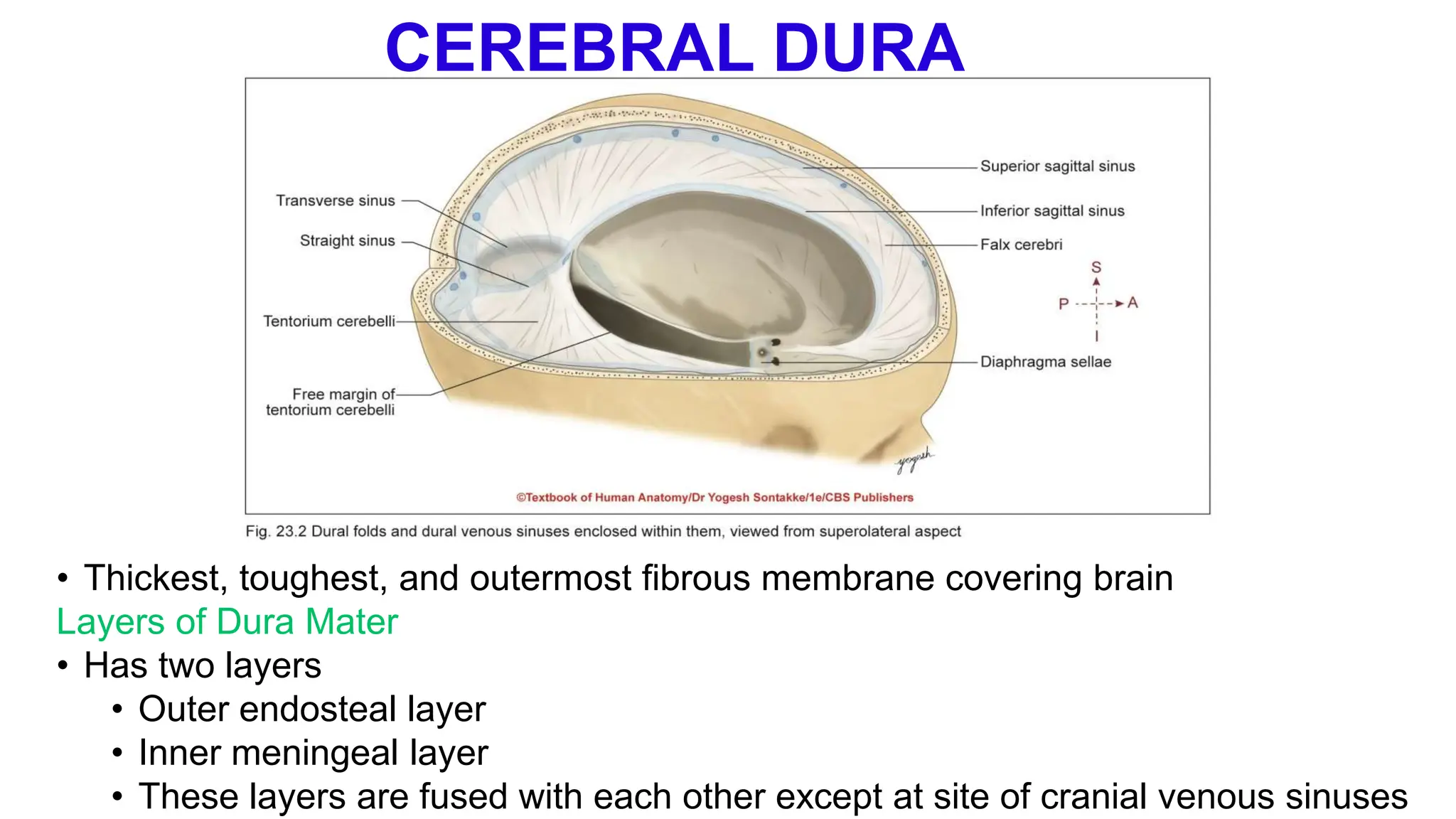
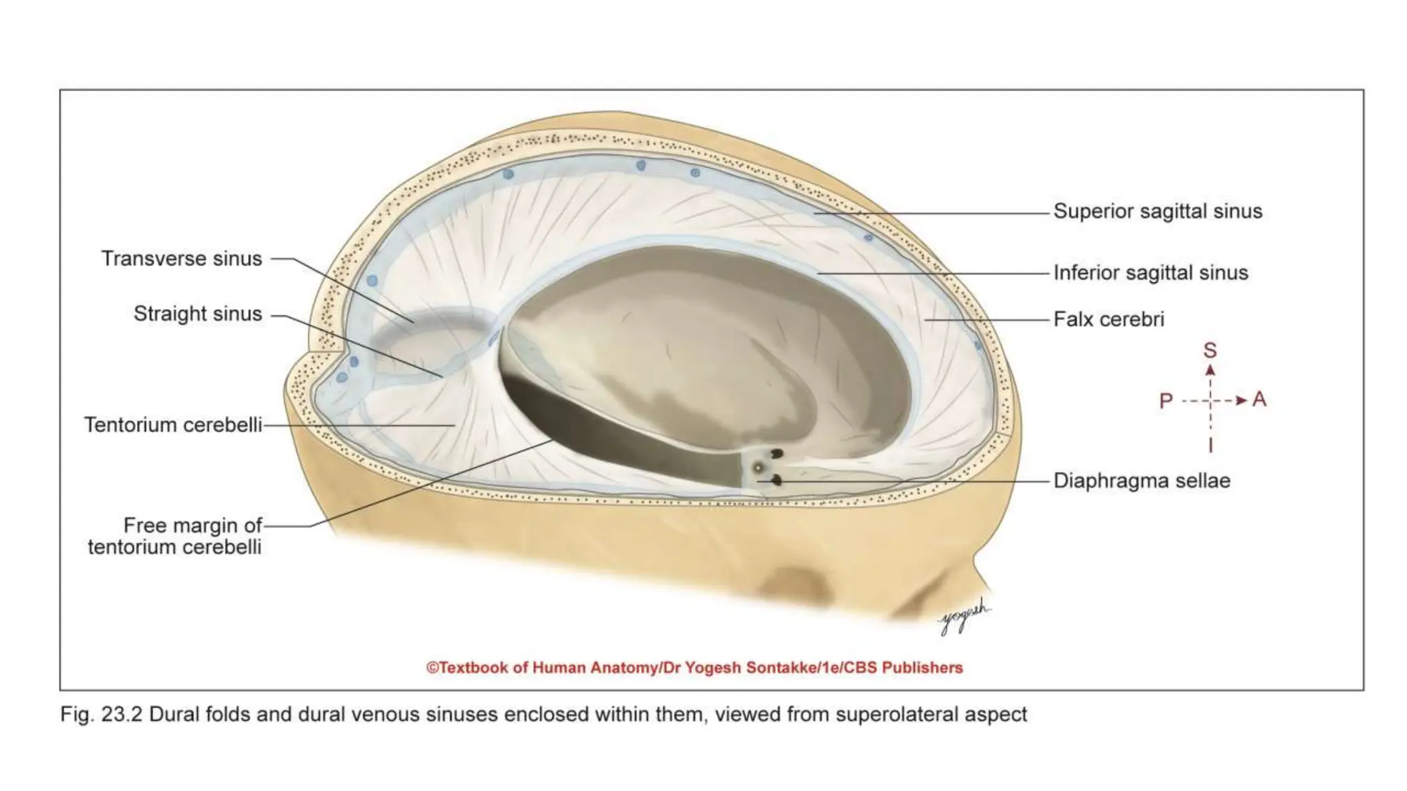
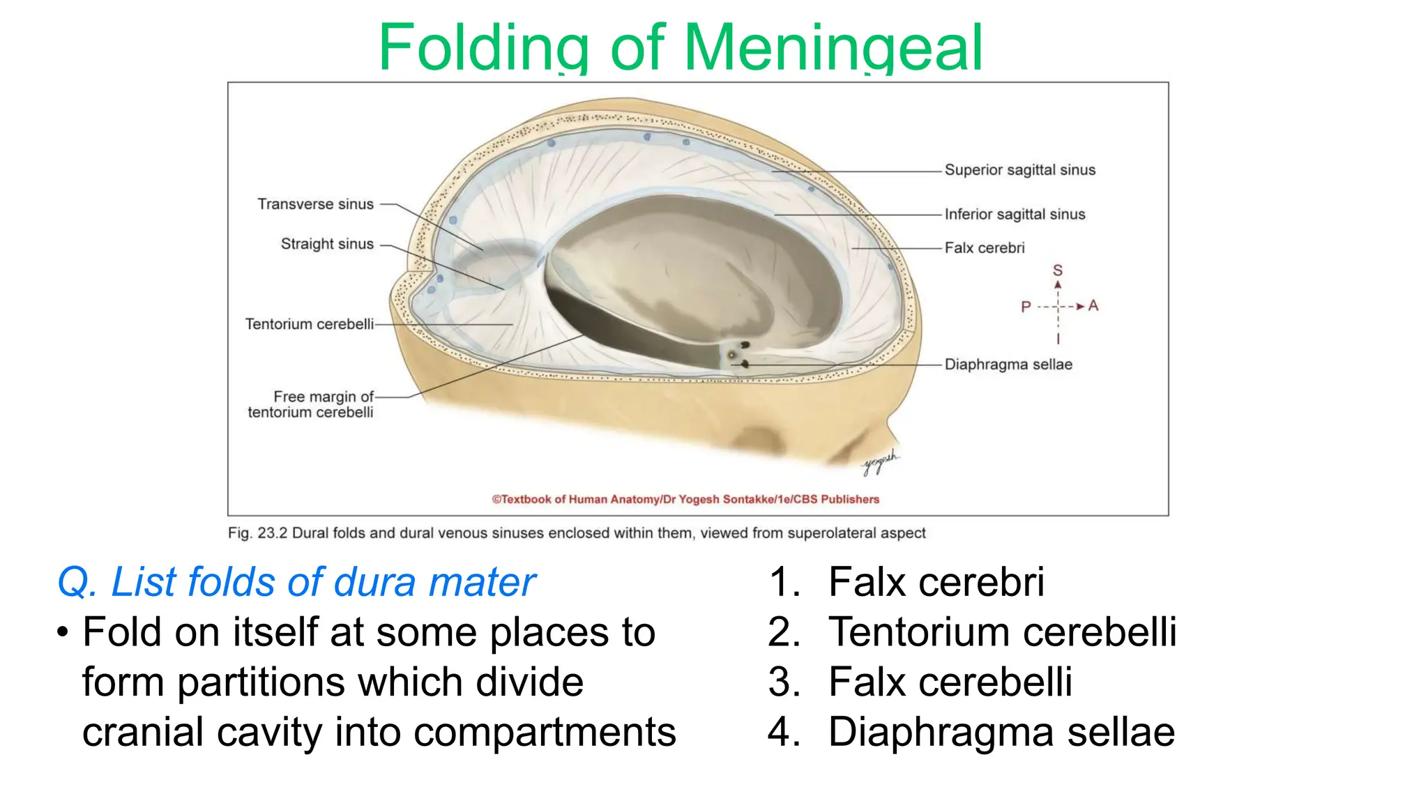
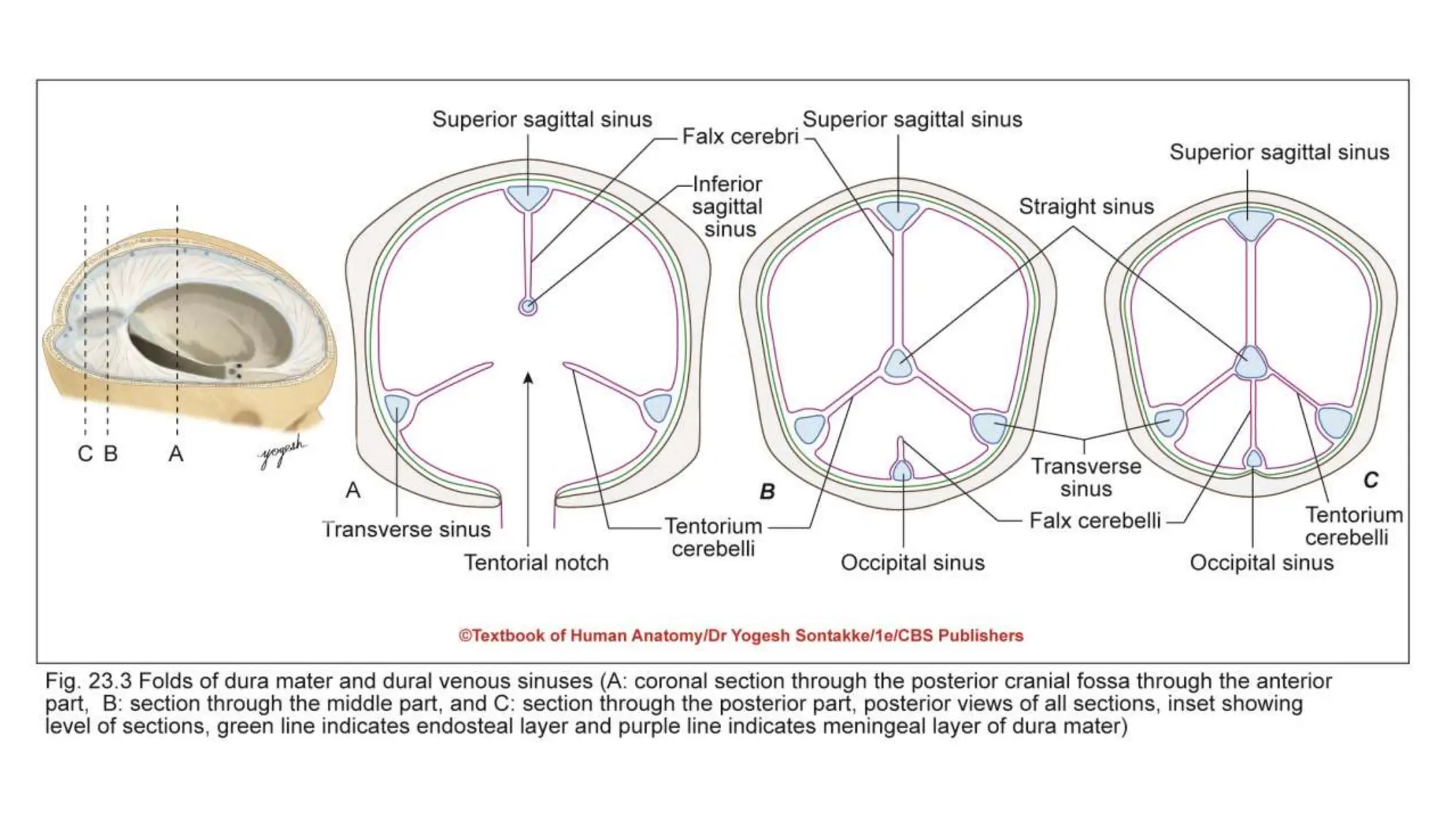
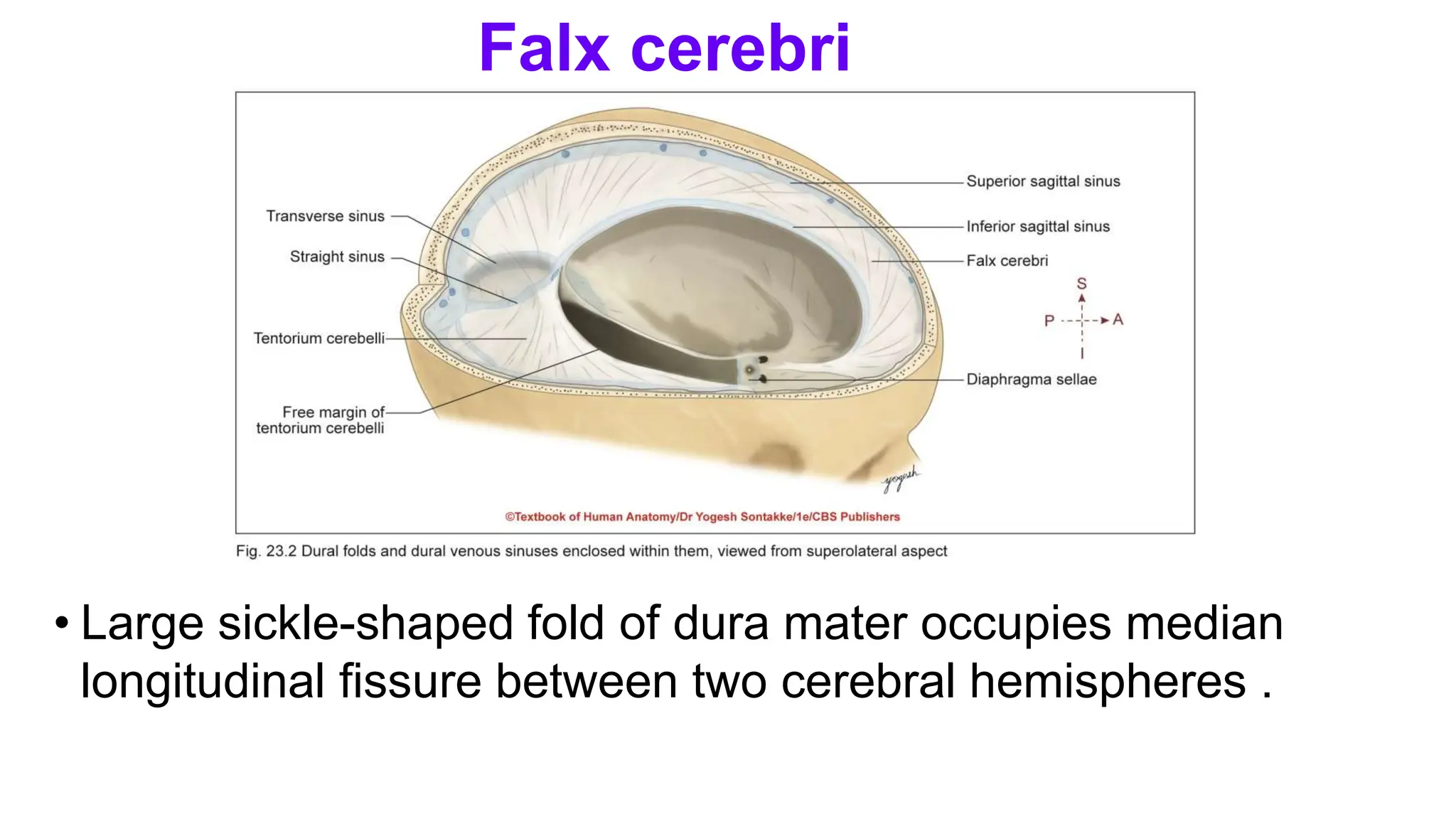
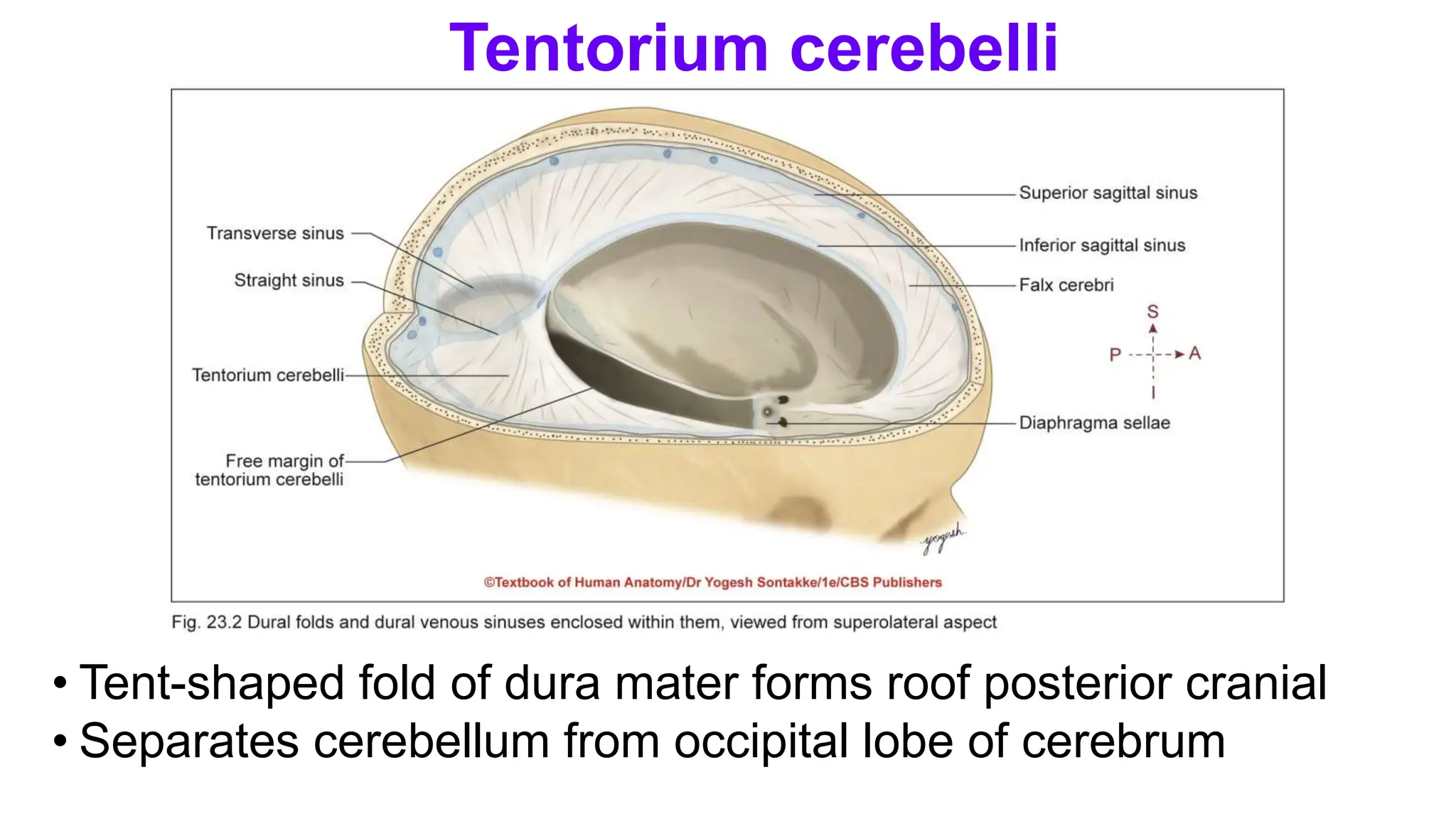
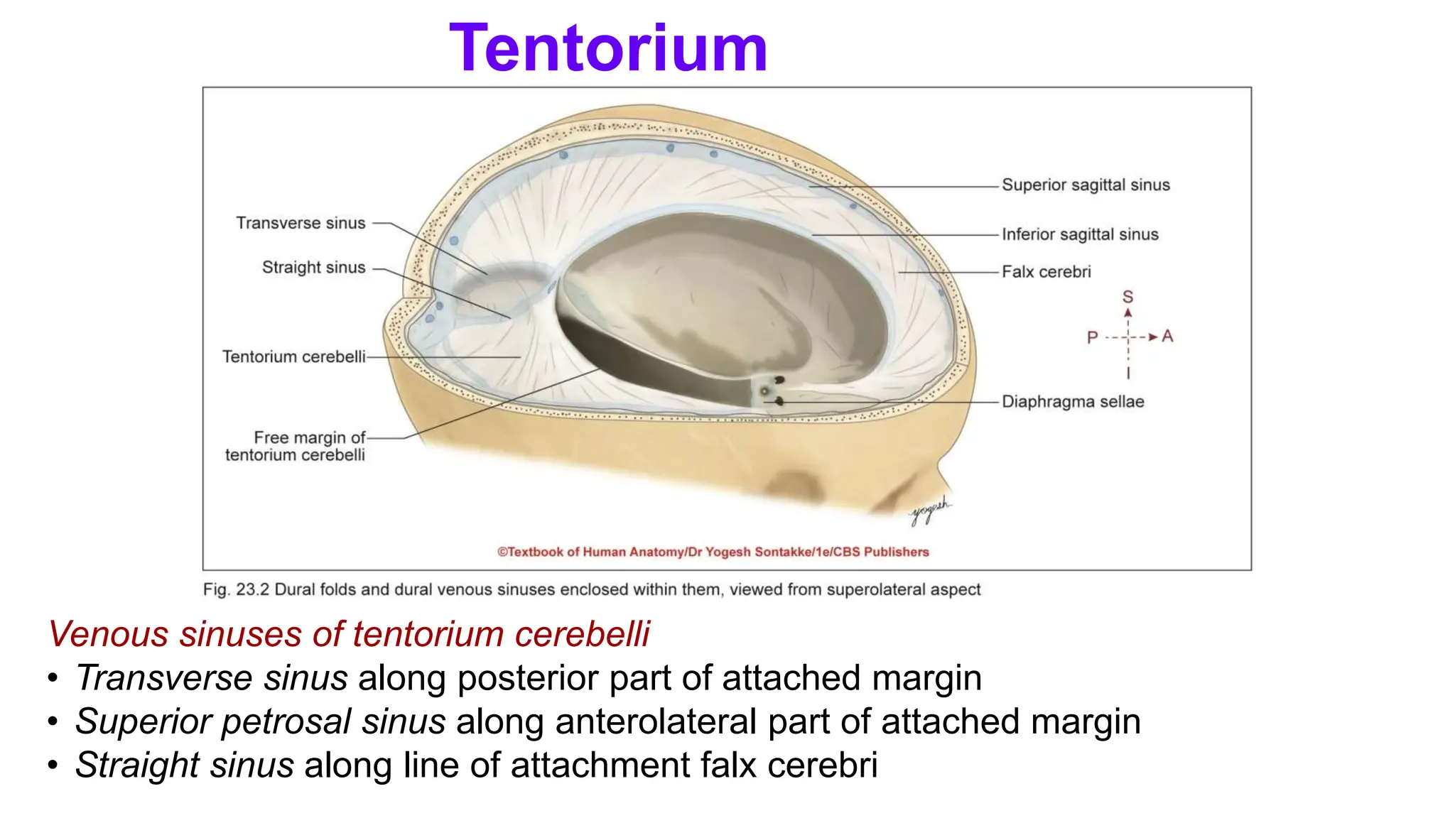
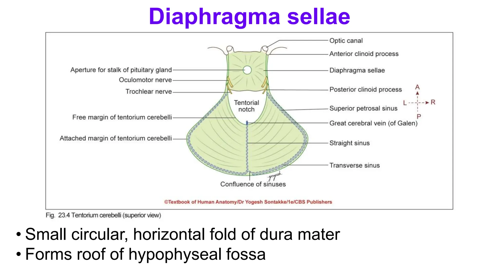
DURAL FOLDS AND DURAL VENOUS SINUSES
DR.VASANTHAKOHILA MBBS
POSTGRADUATE
KILPAUK MEDICAL COLLEGE
DATE: 18.03.2024
AN 30.3 , 30.4.
Human Anatomy/Yogesh Sontakke 1
As per:
Competency based Undergraduate
curriculum
AN 30.3
• Describe and identify dural folds and dural venous sinuses
AN 30.4
• Describe clinical importance of dural venous sinuses
Medical Council of India, Competency based Undergraduate curriculum for the Indian Medical Graduate,
2018. Vol. 1; pg 1-80.
Human Anatomy/Yogesh Sontakke 2
INTRODUCTION
• Cranial cavity – enclosed
within skull and communicates
with exterior through numerous
small and large foramina
• Contents of cranial cavity:
Contains following structures
• Brain
• Meninges –
Outer dura mater
Middle arachnoid mater
Inner pia mater
INTRODUCTION
Contents of cranial cavity:
• Hypophysis cerebri
• Intracranial parts of cranial
nerves
• Dural venous sinuses
including cavernous sinus
• Arteries: Internal carotid
arteries, 4th part of
vertebral arteries, circle of
Willis.
INTRODUCTION
• Interior of cranial cavity – lined by
dura mater and surface of brain –
covered by thin pia mater
• In middle, layer of arachnoid mater
connects the dura with pia mater
• Potential space between dura
mater and arachnoid mater –
subdural space
Subarachnoid space
• Space between arachnoid mater
and pia mater
• Filled with cerebrospinal fluid
(CSF)
• Thickest, toughest, and outermost fibrous membrane covering brain
Layers of Dura Mater
• Has two layers
• Outer endosteal layer
• Inner meningeal layer
• These layers are fused with each other except at site of cranial venous sinuses
CEREBRAL DURA
MATER
Endosteal layer (endocranium)
•Outer layer of dura mater
•Lines inner surfaces of skull bones in form of
periosteum, hence called endocranium
Features
•Continuity
• Continuous with pericranium (on outer surface of skull)
through sutures and foramina of skull.
• Also continuous with periosteal lining of orbit through
superior orbital fissure.
CEREBRAL DURA MATER
Features
•Dural sheaths: Provides dural sheaths for cranial
nerves which fuses with epineurium
•Passage for meningeal vessels: Gives passage to
meningeal vessels
•Adherence: Loosely attached to inner surface of vault
of skull – Easily separable
•Fairly attached to base of skull and margin of foramen
magnum – Difficult to separate
•Dural sheath of optic nerve fuses with sclera of eyeball
CEREBRAL DURA MATER
Meningeal layer
•Inner layer of dura mater
•Encloses brain and continues downward through
foramen magnum with dura mater of spinal cord
(spinal dura)
Some Interesting Facts
Q. List folds of dura mater
• Fold on itself at some places to
form partitions which divide
cranial cavity into compartments
1. Falx cerebri
2. Tentorium cerebelli
3. Falx cerebelli
4. Diaphragma sellae
Folding of Meningeal
Dura Mater
CEREBRAL DURA
MATER
• Large sickle-shaped fold of dura mater occupies median
longitudinal fissure between two cerebral hemispheres .
Falx cerebri
Parts
• Has two ends, two margins, and two surfaces
• Anterior end – narrow and attached to crista galli
• Posterior end – broad and attached to upper surface of tentorium cerebelli in median plane
• Upper margin – convex and attached to lip of sagittal sulcus
• Lower margin – concave and free
• Right and left surfaces of falx cerebri are related to medial surface of corresponding cerebral
hemisphere
Falx cerebri
cerebri
Related dural venous sinuses
• Three important dural venous sinuses are present in relation to falx cerebri:
• Superior sagittal sinus along upper margin
• Inferior sagittal sinus along lower margin
• Straight sinus along line of attachment of falx cerebri to tentorium cerebelli
Falx cerebri
cerebri
• Tent-shaped fold of dura mater forms roof posterior cranial
• Separates cerebellum from occipital lobe of cerebrum
Tentorium cerebelli
Parts
• Has two margins and two surfaces
Anterior or inner margin
• Free margin
• U-shaped and encloses tentorial notch or incisure for passage of midbrain
• Anterior ends of this U-shaped margin – attached to anterior clinoid processes
Tentorium cerebelli
Parts
• Posterior or outer margin
• Attached, convex fixed margin
• Attachments extend from posterior clinoid process, superior border
of temporal bone, posteroinferior angle of parietal bone, and lips of
transverse sulcus on occipital bone
Tentorium
cerebelli
Parts
Surfaces
• Convex surface is slopes to either side of median plane
Gives attachment to falx cerebri in midline.
• Concave surface – concave and gives attachment to falx cerebelli in
posterior part.
Tentorium
cerebelli
Venous sinuses of tentorium cerebelli
• Transverse sinus along posterior part of attached margin
• Superior petrosal sinus along anterolateral part of attached margin
• Straight sinus along line of attachment falx cerebri
Tentorium
cerebelli
Some Interesting Facts
Trigeminal or Meckel’s cave
• Recess of dura mater present in
relation to attached margin of
tentorium
• Formation: Formed by evagination
of meningeal layer of dura mater
over trigeminal impression of
petrous temporal bone
• Trigeminal cave lies below
superior petrosal sinus
• Contents: Contains trigeminal
ganglion and CSF
• Small sickle-shaped folds of dura mater projecting forward into
posterior cerebellar notch
Venous sinuses of falx cerebelli
• Occipital sinus lies along posterior (attached) margin of falx
cerebelli
Falx cerebelli
• Small circular, horizontal fold of dura mater
• Forms roof of hypophyseal fossa
Diaphragma sellae
• Attachments
• Anteriorly – to tuberculum sellae
• Posteriorly – to dorsum sellae
• On each side – continuous with dura mater of middle cranial fossa
• Central aperture: provides passage to stalk of pituitary gland
Diaphragma sellae
Blood supply of endosteal layer of dura mater:
• Richly vascular and provides nourishment to adjacent bone
• Supplied by
• Meningeal branches of ophthalmic, anterior and posterior
ethmoidal arteries in anterior cranial fossa
• Middle and accessory meningeal arteries and meningeal
branches of internal carotid and ascending pharyngeal
arteries in middle cranial fossa
• Meningeal branches of vertebral and occipital arteries in
posterior cranial fossa
Blood Supply of Dura Mater
•Meningeal layer of dura mater – more fibrous and less
vascular
•Meningeal arteries – chief source of blood supply to
the bones of the skull
•Scalping – full thickness soft tissue removal of scalp.
Some Interesting Facts
• Innervation of dura mater – very important for understanding
headache and other clinical conditions
• Following nerves supply dura mater
• Ophthalmic nerve supplies the infratentorial dura mater
• Anterior and posterior ethmoidal nerves and maxillary
nerve supply the dura of anterior cranial fossa.
• Maxillary and mandibular nerves supply dura mater of
middle cranial fossa.
• Recurrent branches of 1st , 2nd , and 3rd cervical spinal
nerves and meningeal branches of 9th and 10th cranial
nerves supply dura mater of posterior cranial fossa.
Nerve Supply of Dura Mater
Clinical Integration
Pain sensitive intracranial structures:
• Dura mater
• Large cranial venous sinuses and their tributaries
• Dural arteries
• Intracranial arteries
Cause of headache
• Infection or inflammation of dura mater
• Inflammation of extracranial structures supplied by
nerves supplying dura mater (referred pain)
Clinical Integration
• Headache is due to
a)Dilatation of intracranial arteries,
b)Raised intracranial pressure by
increased CSF quantity or space
occupying lesion (SOL),
c)Traction of intracranial structures due to
decreased quantity of CSF
Extradural hemorrhage and subdural
hemorrhage are differentiated in the
following table
Differences between extradural and subdural hemorrhages
Feature Extradural (epidural)
hemorrhage
Subdural hemorrhage
Location Between skull and
endosteal layer of dura
mater
Between dura mater
and arachnoid mater
Involved vessels Arteries (middle
meningeal artery)
Veins
Symptoms Rapid onset of
symptoms (sudden
onset of paralysis)
Gradual increasing
symptoms
Blood in CSF Absent Present
CT scan appearance of
hematoma
Biconvex lens Crescent-shaped
•Definition: Dural venous sinuses – venous
spaces or channels present in dura mater
•Are lined by endothelium.
•Are formed by two ways
•By separation of two layers of dura mater
•By folding of meningeal layer on itself.
INTRACRANIAL DURAL VENOUS SINUSES
Features of dural venous
•Muscle coat is absent in their walls.
•Devoid of valves in their lumen.
•Receive blood and CSF both.
•Communicates with veins outside skull through
emissary veins.
•Helps to maintain constant blood pressure in sinuses.
INTRACRANIAL DURAL VENOUS
SINUSES
• Paired sinuses
• Cavernous
• Superior petrosal
• Inferior petrosal
• Transverse
• Sigmoid
• Sphenoparietal
• Petrosquamous
• Unpaired sinuses (lie in midline)
• Superior sagittal
• Inferior sagittal
• Straight
• Occipital
• Anterior intercavernous
• Posterior intercavernous
• Basilar plexus of veins
INTRACRANIAL DURAL VENOUS SINUSES
Classification of dural venous sinuses
Q. List dural venous sinuses
Are about 21 dural venous sinuses
• Large dural venous sinus lies between two layers of dura
mater .
• Size: Length 2 cm, width: 1 cm.
• Location: Each cavernous sinus lies in middle cranial fossa,
on either side of body of sphenoid bone.
CAVERNOUS SINUS
Parts (Structure)
• Lies between two layers of dura mater
• Each cavernous sinus has roof, floor, and two walls (medial and lateral)
• Roof and lateral wall – formed by meningeal layer of dura mater
• Floor and medial wall – formed by endosteal layer of dura mater
CAVERNOUS
SINUS
Interior of cavernous sinus
• Divided into number of small spaces or caverns by
trabeculae, hence called cavernous sinus.
• Trabeculae are less conspicuous in living than in dead.
Some Interesting
Facts
Extent
• Each sinus extends
• Anteriorly: Up to medial end of superior orbital fissure
• Posteriorly: Up to apex petrous part of temporal bone
CAVERNOUS
SINUS
Relations
• Superior
• Optic chiasma
• Optic tract
• Olfactory tract
• Internal carotid artery
• Anterior perforates substance
CAVERNOUS
SINUS
Inferior
• Foramen lacerum
• Junction of the body and greater wing of sphenoid bone
Medial
• Pituitary gland
• Sphenoidal air sinus
CAVERNOUS
SINUS
Laterally
• Temporal lobe with uncus
Anteriorly
• Superior orbital fissure
Posteriorly
• Apex of petrous part of temporal bone
CAVERNOUS
SINUS
Q. List structures in walls of cavernous sinus
• Structures within lateral walls (from above downward)
• Oculomotor nerve
• Trochlear nerve
• Ophthalmic nerve
Structures passing through sinus
• Internal carotid artery and sympathetic plexus around abducent nerve
Structures in Walls of Cavernous
Sinus
• Recent concept
• Maxillary nerve does not run through lateral wall of cavernous sinus
• This nerve traverses beneath dura mater of middle cranial fossa below level of
cavernous sinus
Structures in Walls of Cavernous
Sinus
Some Interesting Facts
•Ophthalmic nerve enters sinus by piercing cavum
trigeminal and before it enters superior orbital fissure,
divides into lacrimal, frontal, and nasociliary nerves
(mnemonics: LFN)
Dorello’s canal:
•Abducent nerve enters cavernous sinus by passing
through dural tunnel called Dorello’s canal
contd
Infratrochlear or
Parkinson’s triangle
• Space between
trochlear nerve and
ophthalmic nerve (V1)
in lateral wall of
cavernous sinus
• Deep to this triangle,
following structures
are located:
• Cavernous segment of
internal carotid artery
• Abducent nerve
• Receives blood from three source as follows
• From orbit
• Superior ophthalmic vein
• Inferior ophthalmic vein
• Central vein of retina (sometimes)
• From brain
• Superficial middle cerebral vein
• Inferior cerebral veins from temporal
lobe (few)
• From meninges
• Sphenoparietal sinus
• Frontal (anterior) trunk of middle
meningeal vein
Tributaries of Cavernous Sinus
• Communicates with (drains into) following
veins/sinuses or plexuses
• Transverse sinus via superior petrosal
sinus
• Internal jugular vein via inferior petrosal
sinus
• Pterygoid venous plexus via emissary
veins passing through foramen ovale,
foramen lacerum, and emissary
sphenoidal foramen
• Facial vein through
• Superior ophthalmic vein and then
angular vein
• Emissary veins to pterygoid venous
plexus and then deep facial vein
Communication of Cavernous Sinus
• Superior sagittal sinus via superficial
middle cerebral and superior
anastomotic veins
• Internal vertebral venous plexus via
basilar venous plexus
• Opposite cavernous sinus via
anterior and posterior
intercavernous sinuses
Note
• All these communications are
valveless
• So, blood can flow in either
direction
Communication of Cavernous Sinus
Factors Regulating Blood Flow from Cavernous Sinus
•Following factors regulate (help in expulsion) of blood
flow through cavernous sinus
•Pulsation of internal carotid artery,
•Gravity,
•Change in position of head.
Communication of Cavernous Sinus
Cavernous sinus thrombosis
• Common due to numerous communications
• May be caused by infection of
• Dangerous area of face
• Nasal cavities
• Paranasal air sinuses
• Sign and symptoms: May cause
a) Involvement of ophthalmic nerve - serve pain in eye and
forehead.
b) Involvement of IIIrd, IVth and VIth cranial nerves -
ophthalmoplegia (paralysis of ocular muscles)
c) Congestion of orbital veins edema of eyelids and
Clinical Integration
Carotid-cavernous sinus fistula
• Direct communication between internal carotid artery and
cavernous sinus.
• May occur due to direct head injury or rupture of aneurysm.
• Results in pulsating exophthalmos (protruding and pulsatile
eyeball), ptosis, periorbital edema, diplopia .
• May cause paralysis of 3rd, 4th, and 6th cranial nerves
• Tumors may arise with in cavernous sinus (such as
meningiomas, hemangiomas) or may extend into cavernous
sinus from adjacent region such as pituitary adenomas.
Clinical Integration
Superior sagittal sinus
• Location: Lies between two layers of falx cerebri along upper
convex and attached margin .
• Beginning: Begins anteriorly at crista galli by union of tiny
meningeal veins.
Superior sagittal sinus
• Termination: At internal occipital protuberance, usually continued as
right transverse sinus which later becomes sigmoid sinus
Clinical Integration
• Thrombosis of superior sagittal sinus
• May occur due to dangerous area of face, scalp, and diploe
Inferior sagittal sinus
• Location: Small channel lies in between low layers of lower
free concave margin of falx cerebri .
• Termination: Ends by joining great cerebral vein of Galen to
form straight sinus.
Straight sinus
• Location: Lies in median plane, along junction of falx cerebri
and tentorium cerebelli .
• Beginning: Formed by fusion of inferior sagittal sinus with
great cerebral vein.
Straight sinus
• Termination: At internal occipital protuberance, straight sinus
continues as transverse sinus, usually left.
Transverse sinuses
• Location: Situated along posterior attached margin of
tentorium cerebelli
• Lodges in groove extending from internal occipital
protuberance to mastoid angle of parietal bone.
Transverse sinuses
• Formation: Usually in right, superior sagittal sinus continues
as transverse sinus, and on left, inferior sagittal sinus
continues as transverse sinus.
Confluence of sinuses
• Confluence of sinuses (torcular Herophili) – connecting point
of 3 sinuses: Superior sagittal sinus, straight sinus, and
occipital sinus
• Lies at internal occipital protuberance
Sigmoid sinuses
• Formation: Each sigmoid sinus – continuation of transverse sinus at mastoid angle of
parietal bone
Shape/course
• Sigmoid sinus – sigmoid or S-shaped, hence name
• Extends from posteroinferior angle of parietal bone to posterior part of jugular foramen
Sigmoid sinuses
• Termination: Passes through jugular foramen and becomes superior bulb of
internal jugular vein
Sigmoid sinuses/ Clinical integration
• Thrombosis of sigmoid sinus.
• Sigmoid sinus – separated from mastoid antrum and air cells by thin plate of bone.
• Hence, infection of mastoiditis or middle ear (otitis media) may spread to sigmoid
sinus and produces thrombosis.
• During surgical procedures at mastoid process, care should be taken to avoid
injuries to sigmoid sinus.
Occipital sinus
• Small sinus situated along attached margin of falx cerebelli .
• Begins posterior margin of foramen magnum and opens in confluence of sinuses.
Some Interesting Facts
Sphenoparietal sinuses
•They lie along posterior
free margin of lesser wing
of sphenoid bone and drain
into anterior part of
cavernous sinus
Superior petrosal sinuses
•Each sinus lodges in groove
along upper border of petrous
part of temporal bone, along
attached margin of tentorium
cerebelli
•Tributaries: Inferior cerebral
veins and cerebellar veins
•Connects cavernous sinus
with transverse sinus
Some Interesting Facts
Inferior Petrosal Sinuses
• Each sinus lies in groove
along petro-occipital fissure
• Connects cavernous sinus
with superior bulb of internal
jugular vein
Petrosquamous sinus
• Each petrosquamous sinus
lies in peterosquamous
suture and drains into
transverse sinus
Some Interesting Facts
Anterior and posterior intercavernous sinuses
• They connect right and left cavernous sinuses.
• They pass through diaphragm sellae, one in front and
other behind infundibulum of pituitary, respectively .
Circular sinus
• Intercavernous sinuses and cavernous sinuses together
form circular sinus.
Some Interesting Facts
Middle meningeal veins (sinuses)
• They form two trunks
• Frontal or anterior trunk: Terminates into the pterygoid
venous plexus through foramen ovale
• Parietal or posterior trunk: Terminates into pterygoid venous
plexus through foramen spinosum
• Anterior and posterior trunks lie along corresponding branch
of middle meningeal artery
Some Interesting Facts
Middle meningeal veins
(sinuses)
• Lies superficially in lateral
sulcus of brain
• Anteriorly, runs forward to drain
into cavernous sinus while
posteriorly, communicates with
superior sagittal sinus via
superior anastomotic vein (of
Trolard) and with transverse
sinus via inferior anastomotic
•Gasserian ganglion – sensory ganglion of trigeminal
nerve
•Homologous with dorsal root ganglion of spinal nerve
Structures
•Contains bodies of pseudounipolar neurons
•Each of these neurons has central and another
peripheral processes
•Shape: Crescentic or semilunar in shape with
convexity directed anterolaterally
TRIGEMINAL GANGLION
•Situation: Situated in trigeminal impression above
apex of petrous part of temporal bone in floor of middle
cranial fossa
Meningeal relations
•Lies within dural pouch called Meckel’s cave or cavum
trigeminale
•Ganglion and motor root of trigeminal nerve both lies in
CSF within cave which lined by pia mater and
arachnoid mater
TRIGEMINAL GANGLION
Relations
•Superiorly: Temporal lobe (parahippocampal gyrus)
•Inferiorly: Motor root of trigeminal nerve
Greater petrosal nerve
Apex of petrous parts of temporal bone
Foramen lacerum
•Medially: Cavernous sinus
Internal carotid artery
•Laterally: Temporal lobe (uncus)
TRIGEMINAL GANGLION
Branches
•Central branches: Central process of ganglion cells
form sensory root of trigeminal nerve which attached to
pons
•Peripheral branches: Form three branches
1.Ophthalmic (V1)
2.Maxillary (V2)
3.Mandibular (V3)
TRIGEMINAL GANGLION
Thank
you…

DURAL FOLDS AND DURAL VENOUS SINUS ,APPLIED ASPECTS

  • 1.
    DURAL FOLDS ANDDURAL VENOUS SINUSES DR.VASANTHAKOHILA MBBS POSTGRADUATE KILPAUK MEDICAL COLLEGE DATE: 18.03.2024 AN 30.3 , 30.4. Human Anatomy/Yogesh Sontakke 1
  • 2.
    As per: Competency basedUndergraduate curriculum AN 30.3 • Describe and identify dural folds and dural venous sinuses AN 30.4 • Describe clinical importance of dural venous sinuses Medical Council of India, Competency based Undergraduate curriculum for the Indian Medical Graduate, 2018. Vol. 1; pg 1-80. Human Anatomy/Yogesh Sontakke 2
  • 3.
    INTRODUCTION • Cranial cavity– enclosed within skull and communicates with exterior through numerous small and large foramina • Contents of cranial cavity: Contains following structures • Brain • Meninges – Outer dura mater Middle arachnoid mater Inner pia mater
  • 4.
    INTRODUCTION Contents of cranialcavity: • Hypophysis cerebri • Intracranial parts of cranial nerves • Dural venous sinuses including cavernous sinus • Arteries: Internal carotid arteries, 4th part of vertebral arteries, circle of Willis.
  • 5.
    INTRODUCTION • Interior ofcranial cavity – lined by dura mater and surface of brain – covered by thin pia mater • In middle, layer of arachnoid mater connects the dura with pia mater • Potential space between dura mater and arachnoid mater – subdural space Subarachnoid space • Space between arachnoid mater and pia mater • Filled with cerebrospinal fluid (CSF)
  • 6.
    • Thickest, toughest,and outermost fibrous membrane covering brain Layers of Dura Mater • Has two layers • Outer endosteal layer • Inner meningeal layer • These layers are fused with each other except at site of cranial venous sinuses CEREBRAL DURA MATER
  • 7.
    Endosteal layer (endocranium) •Outerlayer of dura mater •Lines inner surfaces of skull bones in form of periosteum, hence called endocranium Features •Continuity • Continuous with pericranium (on outer surface of skull) through sutures and foramina of skull. • Also continuous with periosteal lining of orbit through superior orbital fissure. CEREBRAL DURA MATER
  • 8.
    Features •Dural sheaths: Providesdural sheaths for cranial nerves which fuses with epineurium •Passage for meningeal vessels: Gives passage to meningeal vessels •Adherence: Loosely attached to inner surface of vault of skull – Easily separable •Fairly attached to base of skull and margin of foramen magnum – Difficult to separate •Dural sheath of optic nerve fuses with sclera of eyeball CEREBRAL DURA MATER
  • 10.
    Meningeal layer •Inner layerof dura mater •Encloses brain and continues downward through foramen magnum with dura mater of spinal cord (spinal dura) Some Interesting Facts
  • 11.
    Q. List foldsof dura mater • Fold on itself at some places to form partitions which divide cranial cavity into compartments 1. Falx cerebri 2. Tentorium cerebelli 3. Falx cerebelli 4. Diaphragma sellae Folding of Meningeal Dura Mater CEREBRAL DURA MATER
  • 13.
    • Large sickle-shapedfold of dura mater occupies median longitudinal fissure between two cerebral hemispheres . Falx cerebri
  • 14.
    Parts • Has twoends, two margins, and two surfaces • Anterior end – narrow and attached to crista galli • Posterior end – broad and attached to upper surface of tentorium cerebelli in median plane • Upper margin – convex and attached to lip of sagittal sulcus • Lower margin – concave and free • Right and left surfaces of falx cerebri are related to medial surface of corresponding cerebral hemisphere Falx cerebri cerebri
  • 15.
    Related dural venoussinuses • Three important dural venous sinuses are present in relation to falx cerebri: • Superior sagittal sinus along upper margin • Inferior sagittal sinus along lower margin • Straight sinus along line of attachment of falx cerebri to tentorium cerebelli Falx cerebri cerebri
  • 17.
    • Tent-shaped foldof dura mater forms roof posterior cranial • Separates cerebellum from occipital lobe of cerebrum Tentorium cerebelli
  • 18.
    Parts • Has twomargins and two surfaces Anterior or inner margin • Free margin • U-shaped and encloses tentorial notch or incisure for passage of midbrain • Anterior ends of this U-shaped margin – attached to anterior clinoid processes Tentorium cerebelli
  • 19.
    Parts • Posterior orouter margin • Attached, convex fixed margin • Attachments extend from posterior clinoid process, superior border of temporal bone, posteroinferior angle of parietal bone, and lips of transverse sulcus on occipital bone Tentorium cerebelli
  • 20.
    Parts Surfaces • Convex surfaceis slopes to either side of median plane Gives attachment to falx cerebri in midline. • Concave surface – concave and gives attachment to falx cerebelli in posterior part. Tentorium cerebelli
  • 21.
    Venous sinuses oftentorium cerebelli • Transverse sinus along posterior part of attached margin • Superior petrosal sinus along anterolateral part of attached margin • Straight sinus along line of attachment falx cerebri Tentorium cerebelli
  • 24.
    Some Interesting Facts Trigeminalor Meckel’s cave • Recess of dura mater present in relation to attached margin of tentorium • Formation: Formed by evagination of meningeal layer of dura mater over trigeminal impression of petrous temporal bone • Trigeminal cave lies below superior petrosal sinus • Contents: Contains trigeminal ganglion and CSF
  • 25.
    • Small sickle-shapedfolds of dura mater projecting forward into posterior cerebellar notch Venous sinuses of falx cerebelli • Occipital sinus lies along posterior (attached) margin of falx cerebelli Falx cerebelli
  • 26.
    • Small circular,horizontal fold of dura mater • Forms roof of hypophyseal fossa Diaphragma sellae
  • 27.
    • Attachments • Anteriorly– to tuberculum sellae • Posteriorly – to dorsum sellae • On each side – continuous with dura mater of middle cranial fossa • Central aperture: provides passage to stalk of pituitary gland Diaphragma sellae
  • 28.
    Blood supply ofendosteal layer of dura mater: • Richly vascular and provides nourishment to adjacent bone • Supplied by • Meningeal branches of ophthalmic, anterior and posterior ethmoidal arteries in anterior cranial fossa • Middle and accessory meningeal arteries and meningeal branches of internal carotid and ascending pharyngeal arteries in middle cranial fossa • Meningeal branches of vertebral and occipital arteries in posterior cranial fossa Blood Supply of Dura Mater
  • 29.
    •Meningeal layer ofdura mater – more fibrous and less vascular •Meningeal arteries – chief source of blood supply to the bones of the skull •Scalping – full thickness soft tissue removal of scalp. Some Interesting Facts
  • 30.
    • Innervation ofdura mater – very important for understanding headache and other clinical conditions • Following nerves supply dura mater • Ophthalmic nerve supplies the infratentorial dura mater • Anterior and posterior ethmoidal nerves and maxillary nerve supply the dura of anterior cranial fossa. • Maxillary and mandibular nerves supply dura mater of middle cranial fossa. • Recurrent branches of 1st , 2nd , and 3rd cervical spinal nerves and meningeal branches of 9th and 10th cranial nerves supply dura mater of posterior cranial fossa. Nerve Supply of Dura Mater
  • 31.
    Clinical Integration Pain sensitiveintracranial structures: • Dura mater • Large cranial venous sinuses and their tributaries • Dural arteries • Intracranial arteries Cause of headache • Infection or inflammation of dura mater • Inflammation of extracranial structures supplied by nerves supplying dura mater (referred pain)
  • 32.
    Clinical Integration • Headacheis due to a)Dilatation of intracranial arteries, b)Raised intracranial pressure by increased CSF quantity or space occupying lesion (SOL), c)Traction of intracranial structures due to decreased quantity of CSF Extradural hemorrhage and subdural hemorrhage are differentiated in the following table
  • 33.
    Differences between extraduraland subdural hemorrhages Feature Extradural (epidural) hemorrhage Subdural hemorrhage Location Between skull and endosteal layer of dura mater Between dura mater and arachnoid mater Involved vessels Arteries (middle meningeal artery) Veins Symptoms Rapid onset of symptoms (sudden onset of paralysis) Gradual increasing symptoms Blood in CSF Absent Present CT scan appearance of hematoma Biconvex lens Crescent-shaped
  • 34.
    •Definition: Dural venoussinuses – venous spaces or channels present in dura mater •Are lined by endothelium. •Are formed by two ways •By separation of two layers of dura mater •By folding of meningeal layer on itself. INTRACRANIAL DURAL VENOUS SINUSES
  • 35.
    Features of duralvenous •Muscle coat is absent in their walls. •Devoid of valves in their lumen. •Receive blood and CSF both. •Communicates with veins outside skull through emissary veins. •Helps to maintain constant blood pressure in sinuses. INTRACRANIAL DURAL VENOUS SINUSES
  • 36.
    • Paired sinuses •Cavernous • Superior petrosal • Inferior petrosal • Transverse • Sigmoid • Sphenoparietal • Petrosquamous • Unpaired sinuses (lie in midline) • Superior sagittal • Inferior sagittal • Straight • Occipital • Anterior intercavernous • Posterior intercavernous • Basilar plexus of veins INTRACRANIAL DURAL VENOUS SINUSES Classification of dural venous sinuses Q. List dural venous sinuses Are about 21 dural venous sinuses
  • 39.
    • Large duralvenous sinus lies between two layers of dura mater . • Size: Length 2 cm, width: 1 cm. • Location: Each cavernous sinus lies in middle cranial fossa, on either side of body of sphenoid bone. CAVERNOUS SINUS
  • 40.
    Parts (Structure) • Liesbetween two layers of dura mater • Each cavernous sinus has roof, floor, and two walls (medial and lateral) • Roof and lateral wall – formed by meningeal layer of dura mater • Floor and medial wall – formed by endosteal layer of dura mater CAVERNOUS SINUS
  • 41.
    Interior of cavernoussinus • Divided into number of small spaces or caverns by trabeculae, hence called cavernous sinus. • Trabeculae are less conspicuous in living than in dead. Some Interesting Facts
  • 42.
    Extent • Each sinusextends • Anteriorly: Up to medial end of superior orbital fissure • Posteriorly: Up to apex petrous part of temporal bone CAVERNOUS SINUS
  • 43.
    Relations • Superior • Opticchiasma • Optic tract • Olfactory tract • Internal carotid artery • Anterior perforates substance CAVERNOUS SINUS
  • 44.
    Inferior • Foramen lacerum •Junction of the body and greater wing of sphenoid bone Medial • Pituitary gland • Sphenoidal air sinus CAVERNOUS SINUS
  • 45.
    Laterally • Temporal lobewith uncus Anteriorly • Superior orbital fissure Posteriorly • Apex of petrous part of temporal bone CAVERNOUS SINUS
  • 48.
    Q. List structuresin walls of cavernous sinus • Structures within lateral walls (from above downward) • Oculomotor nerve • Trochlear nerve • Ophthalmic nerve Structures passing through sinus • Internal carotid artery and sympathetic plexus around abducent nerve Structures in Walls of Cavernous Sinus
  • 49.
    • Recent concept •Maxillary nerve does not run through lateral wall of cavernous sinus • This nerve traverses beneath dura mater of middle cranial fossa below level of cavernous sinus Structures in Walls of Cavernous Sinus
  • 50.
    Some Interesting Facts •Ophthalmicnerve enters sinus by piercing cavum trigeminal and before it enters superior orbital fissure, divides into lacrimal, frontal, and nasociliary nerves (mnemonics: LFN) Dorello’s canal: •Abducent nerve enters cavernous sinus by passing through dural tunnel called Dorello’s canal
  • 51.
    contd Infratrochlear or Parkinson’s triangle •Space between trochlear nerve and ophthalmic nerve (V1) in lateral wall of cavernous sinus • Deep to this triangle, following structures are located: • Cavernous segment of internal carotid artery • Abducent nerve
  • 52.
    • Receives bloodfrom three source as follows • From orbit • Superior ophthalmic vein • Inferior ophthalmic vein • Central vein of retina (sometimes) • From brain • Superficial middle cerebral vein • Inferior cerebral veins from temporal lobe (few) • From meninges • Sphenoparietal sinus • Frontal (anterior) trunk of middle meningeal vein Tributaries of Cavernous Sinus
  • 55.
    • Communicates with(drains into) following veins/sinuses or plexuses • Transverse sinus via superior petrosal sinus • Internal jugular vein via inferior petrosal sinus • Pterygoid venous plexus via emissary veins passing through foramen ovale, foramen lacerum, and emissary sphenoidal foramen • Facial vein through • Superior ophthalmic vein and then angular vein • Emissary veins to pterygoid venous plexus and then deep facial vein Communication of Cavernous Sinus
  • 56.
    • Superior sagittalsinus via superficial middle cerebral and superior anastomotic veins • Internal vertebral venous plexus via basilar venous plexus • Opposite cavernous sinus via anterior and posterior intercavernous sinuses Note • All these communications are valveless • So, blood can flow in either direction Communication of Cavernous Sinus
  • 57.
    Factors Regulating BloodFlow from Cavernous Sinus •Following factors regulate (help in expulsion) of blood flow through cavernous sinus •Pulsation of internal carotid artery, •Gravity, •Change in position of head. Communication of Cavernous Sinus
  • 59.
    Cavernous sinus thrombosis •Common due to numerous communications • May be caused by infection of • Dangerous area of face • Nasal cavities • Paranasal air sinuses • Sign and symptoms: May cause a) Involvement of ophthalmic nerve - serve pain in eye and forehead. b) Involvement of IIIrd, IVth and VIth cranial nerves - ophthalmoplegia (paralysis of ocular muscles) c) Congestion of orbital veins edema of eyelids and Clinical Integration
  • 60.
    Carotid-cavernous sinus fistula •Direct communication between internal carotid artery and cavernous sinus. • May occur due to direct head injury or rupture of aneurysm. • Results in pulsating exophthalmos (protruding and pulsatile eyeball), ptosis, periorbital edema, diplopia . • May cause paralysis of 3rd, 4th, and 6th cranial nerves • Tumors may arise with in cavernous sinus (such as meningiomas, hemangiomas) or may extend into cavernous sinus from adjacent region such as pituitary adenomas. Clinical Integration
  • 61.
    Superior sagittal sinus •Location: Lies between two layers of falx cerebri along upper convex and attached margin . • Beginning: Begins anteriorly at crista galli by union of tiny meningeal veins.
  • 62.
    Superior sagittal sinus •Termination: At internal occipital protuberance, usually continued as right transverse sinus which later becomes sigmoid sinus Clinical Integration • Thrombosis of superior sagittal sinus • May occur due to dangerous area of face, scalp, and diploe
  • 63.
    Inferior sagittal sinus •Location: Small channel lies in between low layers of lower free concave margin of falx cerebri . • Termination: Ends by joining great cerebral vein of Galen to form straight sinus.
  • 64.
    Straight sinus • Location:Lies in median plane, along junction of falx cerebri and tentorium cerebelli . • Beginning: Formed by fusion of inferior sagittal sinus with great cerebral vein.
  • 65.
    Straight sinus • Termination:At internal occipital protuberance, straight sinus continues as transverse sinus, usually left.
  • 66.
    Transverse sinuses • Location:Situated along posterior attached margin of tentorium cerebelli • Lodges in groove extending from internal occipital protuberance to mastoid angle of parietal bone.
  • 67.
    Transverse sinuses • Formation:Usually in right, superior sagittal sinus continues as transverse sinus, and on left, inferior sagittal sinus continues as transverse sinus.
  • 68.
    Confluence of sinuses •Confluence of sinuses (torcular Herophili) – connecting point of 3 sinuses: Superior sagittal sinus, straight sinus, and occipital sinus • Lies at internal occipital protuberance
  • 69.
    Sigmoid sinuses • Formation:Each sigmoid sinus – continuation of transverse sinus at mastoid angle of parietal bone Shape/course • Sigmoid sinus – sigmoid or S-shaped, hence name • Extends from posteroinferior angle of parietal bone to posterior part of jugular foramen
  • 70.
    Sigmoid sinuses • Termination:Passes through jugular foramen and becomes superior bulb of internal jugular vein
  • 71.
    Sigmoid sinuses/ Clinicalintegration • Thrombosis of sigmoid sinus. • Sigmoid sinus – separated from mastoid antrum and air cells by thin plate of bone. • Hence, infection of mastoiditis or middle ear (otitis media) may spread to sigmoid sinus and produces thrombosis. • During surgical procedures at mastoid process, care should be taken to avoid injuries to sigmoid sinus.
  • 72.
    Occipital sinus • Smallsinus situated along attached margin of falx cerebelli . • Begins posterior margin of foramen magnum and opens in confluence of sinuses.
  • 75.
    Some Interesting Facts Sphenoparietalsinuses •They lie along posterior free margin of lesser wing of sphenoid bone and drain into anterior part of cavernous sinus
  • 76.
    Superior petrosal sinuses •Eachsinus lodges in groove along upper border of petrous part of temporal bone, along attached margin of tentorium cerebelli •Tributaries: Inferior cerebral veins and cerebellar veins •Connects cavernous sinus with transverse sinus Some Interesting Facts
  • 77.
    Inferior Petrosal Sinuses •Each sinus lies in groove along petro-occipital fissure • Connects cavernous sinus with superior bulb of internal jugular vein Petrosquamous sinus • Each petrosquamous sinus lies in peterosquamous suture and drains into transverse sinus Some Interesting Facts
  • 78.
    Anterior and posteriorintercavernous sinuses • They connect right and left cavernous sinuses. • They pass through diaphragm sellae, one in front and other behind infundibulum of pituitary, respectively . Circular sinus • Intercavernous sinuses and cavernous sinuses together form circular sinus. Some Interesting Facts
  • 79.
    Middle meningeal veins(sinuses) • They form two trunks • Frontal or anterior trunk: Terminates into the pterygoid venous plexus through foramen ovale • Parietal or posterior trunk: Terminates into pterygoid venous plexus through foramen spinosum • Anterior and posterior trunks lie along corresponding branch of middle meningeal artery Some Interesting Facts
  • 81.
    Some Interesting Facts Middlemeningeal veins (sinuses) • Lies superficially in lateral sulcus of brain • Anteriorly, runs forward to drain into cavernous sinus while posteriorly, communicates with superior sagittal sinus via superior anastomotic vein (of Trolard) and with transverse sinus via inferior anastomotic
  • 82.
    •Gasserian ganglion –sensory ganglion of trigeminal nerve •Homologous with dorsal root ganglion of spinal nerve Structures •Contains bodies of pseudounipolar neurons •Each of these neurons has central and another peripheral processes •Shape: Crescentic or semilunar in shape with convexity directed anterolaterally TRIGEMINAL GANGLION
  • 83.
    •Situation: Situated intrigeminal impression above apex of petrous part of temporal bone in floor of middle cranial fossa Meningeal relations •Lies within dural pouch called Meckel’s cave or cavum trigeminale •Ganglion and motor root of trigeminal nerve both lies in CSF within cave which lined by pia mater and arachnoid mater TRIGEMINAL GANGLION
  • 84.
    Relations •Superiorly: Temporal lobe(parahippocampal gyrus) •Inferiorly: Motor root of trigeminal nerve Greater petrosal nerve Apex of petrous parts of temporal bone Foramen lacerum •Medially: Cavernous sinus Internal carotid artery •Laterally: Temporal lobe (uncus) TRIGEMINAL GANGLION
  • 85.
    Branches •Central branches: Centralprocess of ganglion cells form sensory root of trigeminal nerve which attached to pons •Peripheral branches: Form three branches 1.Ophthalmic (V1) 2.Maxillary (V2) 3.Mandibular (V3) TRIGEMINAL GANGLION
  • 87.