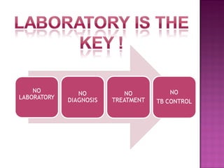
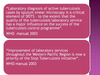
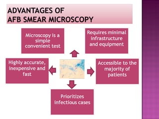
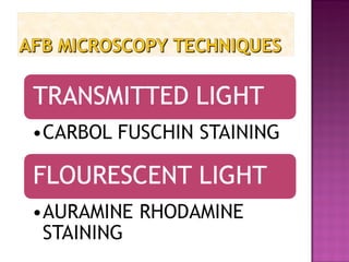
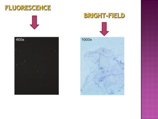
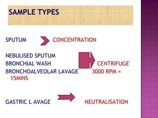
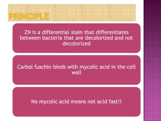
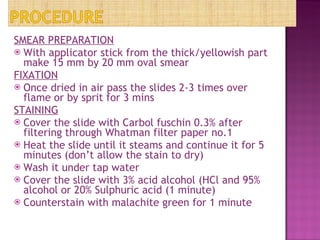
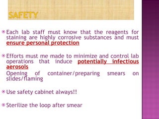
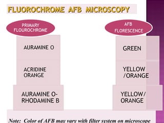
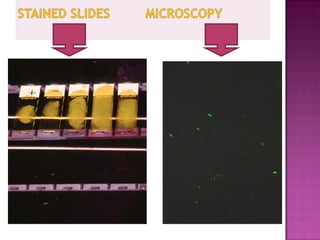
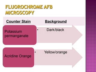
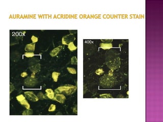
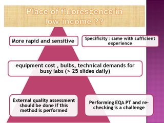
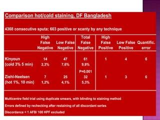
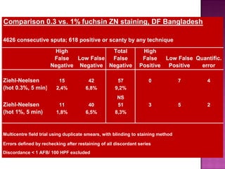
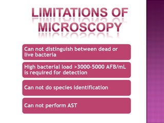
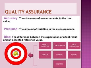
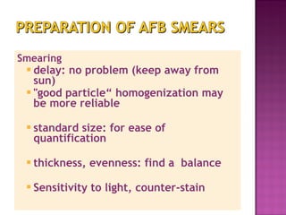
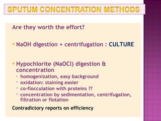
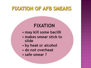
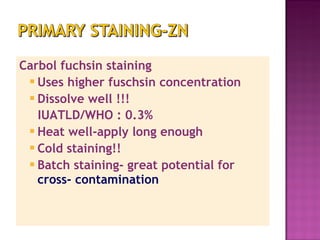
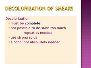
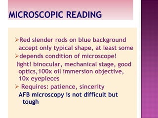
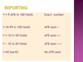
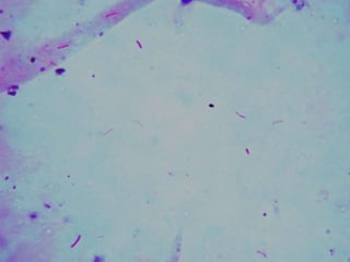
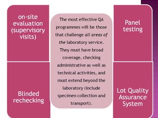
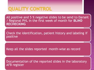
SPUTUM CONCENTRATION NEBULISED SPUTUM BRONCHIAL WASH  CENTRIFUGE BRONCHOALVEOLAR LAVAGE  3000 RPM × 15MINS GASTRIC L AVAGE  NEUTRALISATION
less laborious less robust higher concentration fuchsin longer staining time errors !! NOT RECOMMENDED  for low-income countries
 
SMEAR PREPARATION With applicator stick from the thick/yellowish part make 15 mm by 20 mm oval smear FIXATION Once dried in air pass the slides 2-3 times over flame or by sprit for 3 mins STAINING Cover the slide with Carbol fuschin 0.3% after filtering through Whatman filter paper no.1 Heat the slide until it steams and continue it for 5 minutes (don’t allow the stain to dry) Wash it under tap water Cover the slide with 3% acid alcohol (HCl and 95% alcohol or 20% Sulphuric acid (1 minute) Counterstain with malachite green for 1 minute
Each lab staff must know that the reagents for staining are highly corrosive substances and must  ensure personal protection Efforts must me made to minimize and control lab operations that induce  potentially infectious aerosols Opening of container/preparing smears on slides/flaming Use safety cabinet always!! Sterilize the loop after smear
Note:  Color of AFB may vary with filter system on microscope
 
Counter Stain Background


Accuracy :   The closeness of measurements to the true value. Precision :   The amount of variation in the measurements.  Bias:   The difference between the expectation of a test result  and an accepted reference value.
Location Category False Negative False positive Pre-laboratory Administrative Specimen quality Specimen labeling Patient identification Transport conditions Specimen Labeling Patient identification Specimen container Laboratory Administrative specimen handling specimen registration recording and/or reporting result specimen registration recording and/or reporting result Technical smear preparation stain formulations staining technique microscope performance smear examination technique smear preparation stain formulations staining technique smear examination technique
What is a good sample? What is saliva?  Good sample = yellow? mucous fluid?  Discharge from the bronchial tree May contain solid or purulent substances Minimal amounts of oral/ nasal material May contain macrophages and other cells indicative of infectious disease Leakage– ask for new sample Mucopurulent or purulent required; NOT SALIVA Purulent:thick sticky greenish Mucopurulent: thick brown or green Mucoid:viscous and clear Mucosalivary: mucoid Minimum 2 ml Correct labeling
Smearing delay: no problem (keep away from sun) "good particle“ homogenization may be more reliable standard size: for ease of quantification thickness, evenness: find a  balance Sensitivity to light, counter-stain
Are they worth the effort? NaOH digestion + centrifugation :  CULTURE Hypochlorite (NaOCl) digestion & concentration homogenization, easy background oxidation: staining easier co-flocculation with proteins ?? concentration by sedimentation, centrifugation, filtration or flotation  Contradictory reports on efficiency
 
Carbol fuchsin staining Uses higher fuschsin concentration  Dissolve well !!! IUATLD/WHO : 0.3% Heat well-apply long enough  Cold staining!! Batch staining- great potential for  cross- contamination
Decolorization must be  complete not possible to de-stain too much repeat as needed use strong acids alcohol not absolutely needed
Provides  good contrast  for observation of AFB background for focusing, not too strong methylene blue 0.3% ? diluted or < 1 min use of malachite green?
Red slender rods on blue background accept only typical shape, at least some depends condition of microscope! light! binocular, mechanical stage, good optics,100x oil immersion objective, 10x eyepieces Requires: patience, sincerity AFB microscopy is not difficult but tough
1-9 AFB in 100 fields  Exact  number 10-99 in 100 fields  AFB seen + 1-10 in 50 fields AFB seen ++ > 10 in 20 fields AFB seen +++ NO bacilli No AFB seen


Thank you for your kind attention

Afb microscopy and quality assurance copy

  • 1.
  • 2.
  • 3.
  • 4.
  • 5.
  • 6.
  • 7.
    SPUTUM CONCENTRATION NEBULISEDSPUTUM BRONCHIAL WASH CENTRIFUGE BRONCHOALVEOLAR LAVAGE 3000 RPM × 15MINS GASTRIC L AVAGE NEUTRALISATION
  • 8.
    less laborious lessrobust higher concentration fuchsin longer staining time errors !! NOT RECOMMENDED for low-income countries
  • 9.
  • 10.
    SMEAR PREPARATION Withapplicator stick from the thick/yellowish part make 15 mm by 20 mm oval smear FIXATION Once dried in air pass the slides 2-3 times over flame or by sprit for 3 mins STAINING Cover the slide with Carbol fuschin 0.3% after filtering through Whatman filter paper no.1 Heat the slide until it steams and continue it for 5 minutes (don’t allow the stain to dry) Wash it under tap water Cover the slide with 3% acid alcohol (HCl and 95% alcohol or 20% Sulphuric acid (1 minute) Counterstain with malachite green for 1 minute
  • 11.
    Each lab staffmust know that the reagents for staining are highly corrosive substances and must ensure personal protection Efforts must me made to minimize and control lab operations that induce potentially infectious aerosols Opening of container/preparing smears on slides/flaming Use safety cabinet always!! Sterilize the loop after smear
  • 12.
    Note: Colorof AFB may vary with filter system on microscope
  • 13.
  • 14.
  • 15.
  • 16.
  • 17.
  • 18.
  • 19.
  • 20.
    Accuracy : The closeness of measurements to the true value. Precision : The amount of variation in the measurements. Bias: The difference between the expectation of a test result and an accepted reference value.
  • 21.
    Location Category FalseNegative False positive Pre-laboratory Administrative Specimen quality Specimen labeling Patient identification Transport conditions Specimen Labeling Patient identification Specimen container Laboratory Administrative specimen handling specimen registration recording and/or reporting result specimen registration recording and/or reporting result Technical smear preparation stain formulations staining technique microscope performance smear examination technique smear preparation stain formulations staining technique smear examination technique
  • 22.
    What is agood sample? What is saliva? Good sample = yellow? mucous fluid? Discharge from the bronchial tree May contain solid or purulent substances Minimal amounts of oral/ nasal material May contain macrophages and other cells indicative of infectious disease Leakage– ask for new sample Mucopurulent or purulent required; NOT SALIVA Purulent:thick sticky greenish Mucopurulent: thick brown or green Mucoid:viscous and clear Mucosalivary: mucoid Minimum 2 ml Correct labeling
  • 23.
    Smearing delay: noproblem (keep away from sun) &quot;good particle“ homogenization may be more reliable standard size: for ease of quantification thickness, evenness: find a balance Sensitivity to light, counter-stain
  • 24.
    Are they worththe effort? NaOH digestion + centrifugation : CULTURE Hypochlorite (NaOCl) digestion & concentration homogenization, easy background oxidation: staining easier co-flocculation with proteins ?? concentration by sedimentation, centrifugation, filtration or flotation Contradictory reports on efficiency
  • 25.
  • 26.
    Carbol fuchsin stainingUses higher fuschsin concentration Dissolve well !!! IUATLD/WHO : 0.3% Heat well-apply long enough Cold staining!! Batch staining- great potential for cross- contamination
  • 27.
    Decolorization must be complete not possible to de-stain too much repeat as needed use strong acids alcohol not absolutely needed
  • 28.
    Provides goodcontrast for observation of AFB background for focusing, not too strong methylene blue 0.3% ? diluted or < 1 min use of malachite green?
  • 29.
    Red slender rodson blue background accept only typical shape, at least some depends condition of microscope! light! binocular, mechanical stage, good optics,100x oil immersion objective, 10x eyepieces Requires: patience, sincerity AFB microscopy is not difficult but tough
  • 30.
    1-9 AFB in100 fields Exact number 10-99 in 100 fields AFB seen + 1-10 in 50 fields AFB seen ++ > 10 in 20 fields AFB seen +++ NO bacilli No AFB seen
  • 31.
  • 32.
  • 33.
  • 34.
  • 35.
  • 36.
  • 37.
  • 38.
    Thank you foryour kind attention