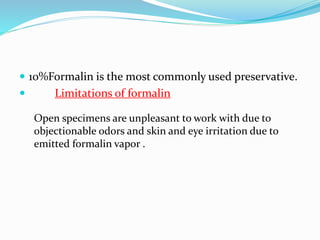
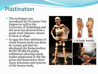
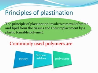
Archa Anil
HS
SSDC
 The study of gross specimens is an integral part in learning
oral pathology. Unfortunately their storage and handling
using traditional formalin is discouragingly difficult. This
session describes an alternative approach called
“plastination” to study and teach gross specimens using
resin polymers.
 Decay is a vital process in nature but an impediment to
morphological studies, teaching, and research.This is
particularly true for biological specimens that shrink
considerably when exposed to normal atmospheric
conditions.
 Therefore their preservation is necessary.
 10%Formalin is the most commonly used preservative.
 Limitations of formalin
Open specimens are unpleasant to work with due to
objectionable odors and skin and eye irritation due to
emitted formalin vapor .
Specimens preserved in glass jars
or Perspex “pots” may overcome
these problems but are bulky and
fragile, need frequent regular
maintenance to avoid the fluid
becoming cloudy which may
obscure the details. In addition,
color preservation is poor and the
parallax error caused by spatial
separation from the pot surface
leads to difficulties in pointing out
features.
Plastination
 The technique was
introduced by Dr.Gunter Von
Hagens in 1978 in the
University of Heidelberg and
the term was derived from a
greek word ‘plassien’ means
to form or shape.
 In 1995 the first exhibition of
whole human body was done
by Gunter and later he
developed the Korperwelton
BODY WORLDS public
exhibitions showing whole
bodies plastinated in life like
poses and dissected to show
many structures and systems
of the human body.
 Later many universities introduced this technique for
medical and dental practices.
Plastin
ated
bovine
tongue
Principles of plastination
The principle of plastination involves removal of water
and lipid from the tissues and their replacement by a
plastic (curable polymer).
Commonly used polymers are
Ideal requirements of the resin
 must have lowest possible viscosity in uncured
state and should be easy to handle.
 Its refractive index must differ from that of
tissue (unless a transparent specimen is desired
outcome).
 Resin activator mixture (base and catalyst)
must have long working time or a relatively
long liquid phase life to allow time for
impregnation of the tissues.
 Curing should not be inhibited by presence of tissue.
 It should have appropriate mechanical properties
when cured, that is, rubber like to simulate a natural
state, or firm to permit its surface to be ground.
 It should be affordable.
 Considering these requirements, the polymer that
has enjoyed widest acceptance in the preparation of
specimens for teaching is silicone rubber
Process
fixation
Dehydration and defatting
Forced impregnation with
polymer
Curing of the polymer
Fixation
 Biological material must be fixed before plastination
to prevent putrefaction and stop the action of other
enzymes.
 Formalin of 5% -20% is used.
 For colour preservation cold(4 degree) Kaiserling
solution containing 5% formalin.Depending on the
size of the oral specimen and fixation method used,
this step may take 24–48 hours.
 Even old specimens stored in
formalin can also be
plastinated.
 Specimens stored in glycerol
should be thoroughly washed
since glycerol interferes with
curing of silicones
 Cysts or cavitatory defects in jaws should be fixed by
inflation with fixative under some pressure.
 solid organs such as tongue should be infiltrated with
fixative in addition to being immersed in the same
solution.
 For display purposes, coloring material may be
injected to illustrate vascular system, although this can
only be done successfully with large, fresh specimens,
before fixation.
Dehydration
Removes water from the tissues.
alcohols and acetone are commonly used.
 Cryosubstitution is the method of choice. An ethanol series
may be used but usually takes longer and is detrimental to
coloration and results in greater shrinkage.
 The cooled fixed specimen is immersed in acetone in
-25°C, whereupon it rapidly freezes retaining its shape. Over
a period of time acetone replaces the water crystals within
the tissue.
 The volume of the acetone must be at least 10 times that of
specimen and the duration depends on the size of the
specimen
 The acetone solution should
be monitored for purity once
in every week
 If acetone purity is 100%and
specimen:acetone =1:10 then
dehydration can completed in
3 weeks
 Larger specimens require
about 5 weeks.
Defatting/degreasing
 It is the removal of fat/lipids in the specimen.
 They do not impregnate resin.
 Once the acetone purity has reached 95% after
dehydration the specimen basket should be taken out
to room temperature to allow defatting.
 Check weekly colour change of acetone .Transparent
acetone turns yellow and specimen fat colour changes
from white to opaque after degreasing .
 Usually takes 2 weeks
Forced impregnation
Performed in a vacuum chamber where the acetone
saturated specimens are submerged into bath of liquid
polymer.
It involves the replacement of a low boiling point, high
vapor pressure acetone by a high boiling point, low vapor
pressure polymer.
Since the reaction mixture has high viscosity vacuum
pumps are used to impregnate the specimen with silicon
 The extraction of acetone creates a vaccum inside the
specimens which forces the penetration of polymer
into them, down to their microscopic level.If the
applied vacuum is too high, the acetone is removed
much before the more viscous polymer can infiltrate
the tissue and the specimen collapses.
 Controlling the vacuum
with a regulator and
regular visual monitoring
are therefore essential.
With the correct vacuum
continuous bubbles should
be seen arising from the
specimen surface. Over a
period of time the pressure
should be gradually
reduced, when bubbling
should have ceased the
specimen can be removed
and allowed to drain.
Curing
 Once impregnation step is completed, the specimens
are removed from polymer bath. The residual polymer
is still liquid and can be reused.
Three different curing techniques depending on the
polymer that is used.
 In case of silicone specimens, the polymer/hardener
mix used in impregnation will eventually harden over a
period of days to weeks.
 An accelerator in the form of vapour or gas may be
used to speed up the process.
 Specimens are placed on a platform above the volatile
accelerator in an airtight container.
 A desiccant is used because any moisture causes
whitish discoloration of the specimen surface.
 Initially polymer expands and oozes out from surface
which must be wiped off until surface gets hardened.
 Curing of epoxy resin-impregnated specimens is
performed by heat treatment at 45°C. Final curing in
an oven takes few days to complete.
 Specimens impregnated with polyester is cured by a
45-minute UV light exposure and completed in an
oven at 45°C for few days
Finishing and storing
 Plastinated specimens can
be made more appealing
by trimming the unwanted
areas or flash polymer with
a scalpel, a dilute detergent
or a lubricant can be
applied to clean, and the
surface can be made
smooth by buffing to get a
display specimen
 Specimens can be
mounted in perspex pots
or stored in plastic bags.
HISTOLOGIC EXAMINATION OF SILICONE-
IMPREGNATED SPECIMENS
 Its interesting to note that the histological qualities of
the plastinated tissues remained intact under
microscopic examination .
 the key to this is “deplastination”which is the ability
of sodium ions to depolymerise the silicon rubber .
Tissue samples to be deplastinated are exposed to a
saturated solution of sodium methylate in anhydrous
methanol until free of polymerized silicone rubber and
then subjected to standard histologic techniques
Plastinated AOT
impacted canine
Plastinated radicular cyst specimen held in
bare hands
Block plastination of a dilacerated molar
tooth
Plastinated specimens as teaching aids
 Preservation of autopsy or surgical tissue samples in a
form useful for teaching.
 Plastinated specimens are generally easier to interpret
than the usual “pots”, so students are more interested in
examining plastinated specimens than those preserved
in formalin jars.
 Can be easily carried to class rooms during lectures
 used as spotters in undergraduate practical
examinations
 Plastinated specimens may be pinned and/or
photographed and labeled to highlight salient
landmarks which should make student to identify easily
 act as patient educative tools
 They are nontoxic, noninfectious, and do not exude
fumes or fluids
 Preparation of tissue sample for use as evidence.
 They can be stored in simple plastic bags.
 Plastinated specimens require little storage and no
maintenance.
Limitations/disadvantages of plastination
 Process is technique sensitive, time consuming, and hence
needs a dedicated pathologist.
 Beginner has to do a lot of trial and error during the
process to achieve the desired outcome which might
lead to consumption/wastage of rare and unusual
specimens.
 Slightly more expensive and needs more equipment than
the conventional laboratory methods
 Process needs lot of postcuring works such as trimming,
polishing, coloring, and mounting to obtain a good
display specimen.
 Learning anatomy on only plastinated specimens is a
compromise because of its limitations in terms of tactile
and emotional experience that is provided by wet
cadavers.
 Deplastination is not possible with all types of resins.
Conclusion
 Plastination , when tried for many oral specimens
successfully, is superior to those preserved in
formalin, both in terms of user acceptance and ease of
maintenance. . In the process, many indigenous
methods can be tried upon to successfully achieve a
beautiful plastinated specimen. Although it appears
time consuming, plastination is recommended to any
oral pathology department for maintenance of student
museum, for preservation of rare specimens, or for
postgraduate use.
Bibliography
 Journal of the International Society of Plastination –
K Dejong andW H Henry
• Plastination a novel innovative teaching adjunct-
Spoorthi Bhavana and Vidhya Manohar Bhatt
• www.wikipedia .com
Plastination

Plastination

  • 1.
  • 2.
     The studyof gross specimens is an integral part in learning oral pathology. Unfortunately their storage and handling using traditional formalin is discouragingly difficult. This session describes an alternative approach called “plastination” to study and teach gross specimens using resin polymers.
  • 3.
     Decay isa vital process in nature but an impediment to morphological studies, teaching, and research.This is particularly true for biological specimens that shrink considerably when exposed to normal atmospheric conditions.  Therefore their preservation is necessary.
  • 4.
     10%Formalin isthe most commonly used preservative.  Limitations of formalin Open specimens are unpleasant to work with due to objectionable odors and skin and eye irritation due to emitted formalin vapor .
  • 5.
    Specimens preserved inglass jars or Perspex “pots” may overcome these problems but are bulky and fragile, need frequent regular maintenance to avoid the fluid becoming cloudy which may obscure the details. In addition, color preservation is poor and the parallax error caused by spatial separation from the pot surface leads to difficulties in pointing out features.
  • 6.
    Plastination  The techniquewas introduced by Dr.Gunter Von Hagens in 1978 in the University of Heidelberg and the term was derived from a greek word ‘plassien’ means to form or shape.  In 1995 the first exhibition of whole human body was done by Gunter and later he developed the Korperwelton BODY WORLDS public exhibitions showing whole bodies plastinated in life like poses and dissected to show many structures and systems of the human body.
  • 8.
     Later manyuniversities introduced this technique for medical and dental practices. Plastin ated bovine tongue
  • 9.
    Principles of plastination Theprinciple of plastination involves removal of water and lipid from the tissues and their replacement by a plastic (curable polymer). Commonly used polymers are
  • 10.
    Ideal requirements ofthe resin  must have lowest possible viscosity in uncured state and should be easy to handle.  Its refractive index must differ from that of tissue (unless a transparent specimen is desired outcome).  Resin activator mixture (base and catalyst) must have long working time or a relatively long liquid phase life to allow time for impregnation of the tissues.
  • 11.
     Curing shouldnot be inhibited by presence of tissue.  It should have appropriate mechanical properties when cured, that is, rubber like to simulate a natural state, or firm to permit its surface to be ground.  It should be affordable.  Considering these requirements, the polymer that has enjoyed widest acceptance in the preparation of specimens for teaching is silicone rubber
  • 12.
    Process fixation Dehydration and defatting Forcedimpregnation with polymer Curing of the polymer
  • 13.
    Fixation  Biological materialmust be fixed before plastination to prevent putrefaction and stop the action of other enzymes.  Formalin of 5% -20% is used.  For colour preservation cold(4 degree) Kaiserling solution containing 5% formalin.Depending on the size of the oral specimen and fixation method used, this step may take 24–48 hours.
  • 14.
     Even oldspecimens stored in formalin can also be plastinated.  Specimens stored in glycerol should be thoroughly washed since glycerol interferes with curing of silicones
  • 15.
     Cysts orcavitatory defects in jaws should be fixed by inflation with fixative under some pressure.  solid organs such as tongue should be infiltrated with fixative in addition to being immersed in the same solution.  For display purposes, coloring material may be injected to illustrate vascular system, although this can only be done successfully with large, fresh specimens, before fixation.
  • 16.
    Dehydration Removes water fromthe tissues. alcohols and acetone are commonly used.  Cryosubstitution is the method of choice. An ethanol series may be used but usually takes longer and is detrimental to coloration and results in greater shrinkage.  The cooled fixed specimen is immersed in acetone in -25°C, whereupon it rapidly freezes retaining its shape. Over a period of time acetone replaces the water crystals within the tissue.  The volume of the acetone must be at least 10 times that of specimen and the duration depends on the size of the specimen
  • 17.
     The acetonesolution should be monitored for purity once in every week  If acetone purity is 100%and specimen:acetone =1:10 then dehydration can completed in 3 weeks  Larger specimens require about 5 weeks.
  • 18.
    Defatting/degreasing  It isthe removal of fat/lipids in the specimen.  They do not impregnate resin.  Once the acetone purity has reached 95% after dehydration the specimen basket should be taken out to room temperature to allow defatting.  Check weekly colour change of acetone .Transparent acetone turns yellow and specimen fat colour changes from white to opaque after degreasing .  Usually takes 2 weeks
  • 21.
    Forced impregnation Performed ina vacuum chamber where the acetone saturated specimens are submerged into bath of liquid polymer. It involves the replacement of a low boiling point, high vapor pressure acetone by a high boiling point, low vapor pressure polymer. Since the reaction mixture has high viscosity vacuum pumps are used to impregnate the specimen with silicon
  • 22.
     The extractionof acetone creates a vaccum inside the specimens which forces the penetration of polymer into them, down to their microscopic level.If the applied vacuum is too high, the acetone is removed much before the more viscous polymer can infiltrate the tissue and the specimen collapses.
  • 23.
     Controlling thevacuum with a regulator and regular visual monitoring are therefore essential. With the correct vacuum continuous bubbles should be seen arising from the specimen surface. Over a period of time the pressure should be gradually reduced, when bubbling should have ceased the specimen can be removed and allowed to drain.
  • 25.
    Curing  Once impregnationstep is completed, the specimens are removed from polymer bath. The residual polymer is still liquid and can be reused. Three different curing techniques depending on the polymer that is used.  In case of silicone specimens, the polymer/hardener mix used in impregnation will eventually harden over a period of days to weeks.  An accelerator in the form of vapour or gas may be used to speed up the process.
  • 26.
     Specimens areplaced on a platform above the volatile accelerator in an airtight container.  A desiccant is used because any moisture causes whitish discoloration of the specimen surface.  Initially polymer expands and oozes out from surface which must be wiped off until surface gets hardened.
  • 27.
     Curing ofepoxy resin-impregnated specimens is performed by heat treatment at 45°C. Final curing in an oven takes few days to complete.  Specimens impregnated with polyester is cured by a 45-minute UV light exposure and completed in an oven at 45°C for few days
  • 28.
    Finishing and storing Plastinated specimens can be made more appealing by trimming the unwanted areas or flash polymer with a scalpel, a dilute detergent or a lubricant can be applied to clean, and the surface can be made smooth by buffing to get a display specimen  Specimens can be mounted in perspex pots or stored in plastic bags.
  • 29.
    HISTOLOGIC EXAMINATION OFSILICONE- IMPREGNATED SPECIMENS  Its interesting to note that the histological qualities of the plastinated tissues remained intact under microscopic examination .  the key to this is “deplastination”which is the ability of sodium ions to depolymerise the silicon rubber . Tissue samples to be deplastinated are exposed to a saturated solution of sodium methylate in anhydrous methanol until free of polymerized silicone rubber and then subjected to standard histologic techniques
  • 30.
    Plastinated AOT impacted canine Plastinatedradicular cyst specimen held in bare hands
  • 31.
    Block plastination ofa dilacerated molar tooth
  • 32.
    Plastinated specimens asteaching aids  Preservation of autopsy or surgical tissue samples in a form useful for teaching.  Plastinated specimens are generally easier to interpret than the usual “pots”, so students are more interested in examining plastinated specimens than those preserved in formalin jars.  Can be easily carried to class rooms during lectures
  • 33.
     used asspotters in undergraduate practical examinations  Plastinated specimens may be pinned and/or photographed and labeled to highlight salient landmarks which should make student to identify easily  act as patient educative tools  They are nontoxic, noninfectious, and do not exude fumes or fluids
  • 34.
     Preparation oftissue sample for use as evidence.  They can be stored in simple plastic bags.  Plastinated specimens require little storage and no maintenance.
  • 35.
    Limitations/disadvantages of plastination Process is technique sensitive, time consuming, and hence needs a dedicated pathologist.  Beginner has to do a lot of trial and error during the process to achieve the desired outcome which might lead to consumption/wastage of rare and unusual specimens.  Slightly more expensive and needs more equipment than the conventional laboratory methods
  • 36.
     Process needslot of postcuring works such as trimming, polishing, coloring, and mounting to obtain a good display specimen.  Learning anatomy on only plastinated specimens is a compromise because of its limitations in terms of tactile and emotional experience that is provided by wet cadavers.  Deplastination is not possible with all types of resins.
  • 37.
    Conclusion  Plastination ,when tried for many oral specimens successfully, is superior to those preserved in formalin, both in terms of user acceptance and ease of maintenance. . In the process, many indigenous methods can be tried upon to successfully achieve a beautiful plastinated specimen. Although it appears time consuming, plastination is recommended to any oral pathology department for maintenance of student museum, for preservation of rare specimens, or for postgraduate use.
  • 38.
    Bibliography  Journal ofthe International Society of Plastination – K Dejong andW H Henry • Plastination a novel innovative teaching adjunct- Spoorthi Bhavana and Vidhya Manohar Bhatt • www.wikipedia .com