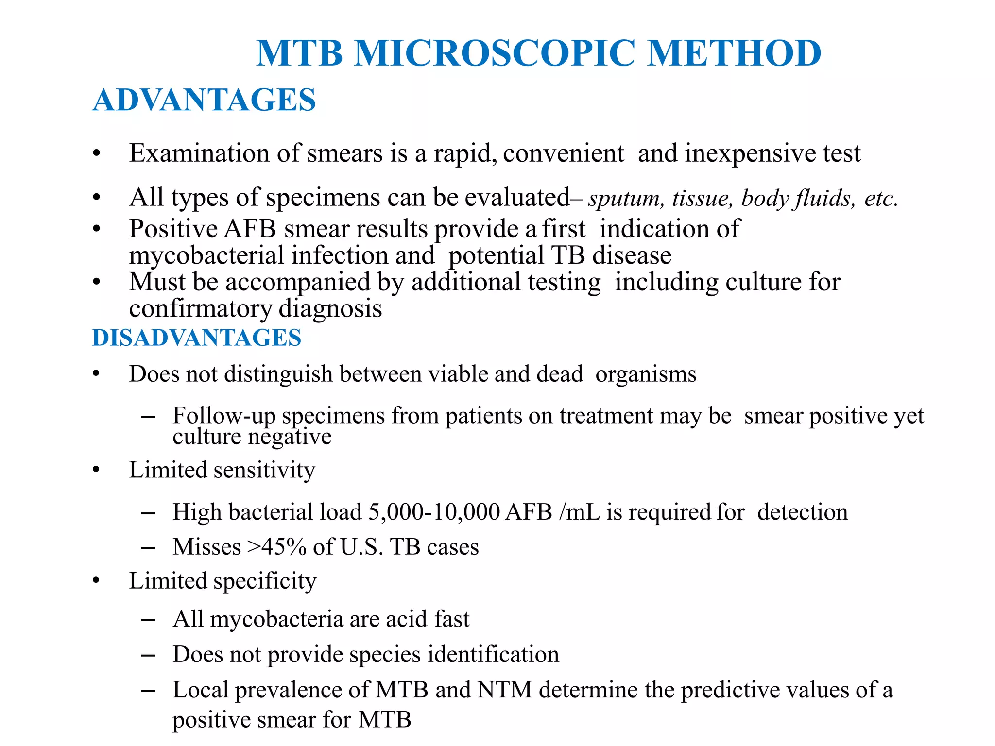
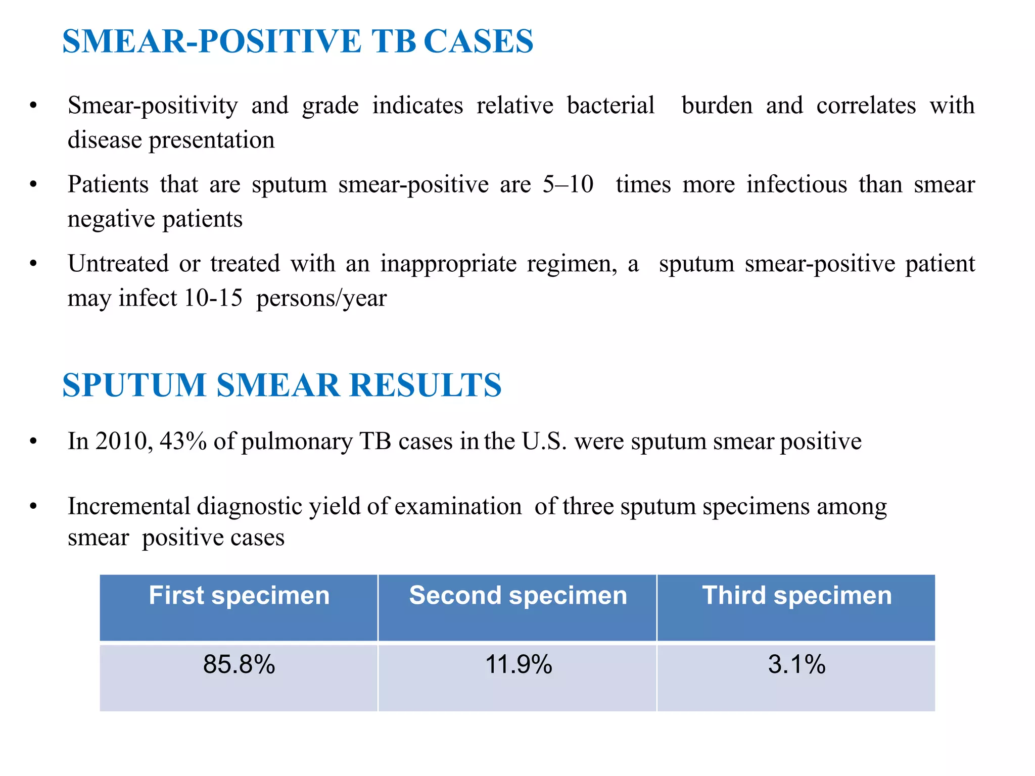
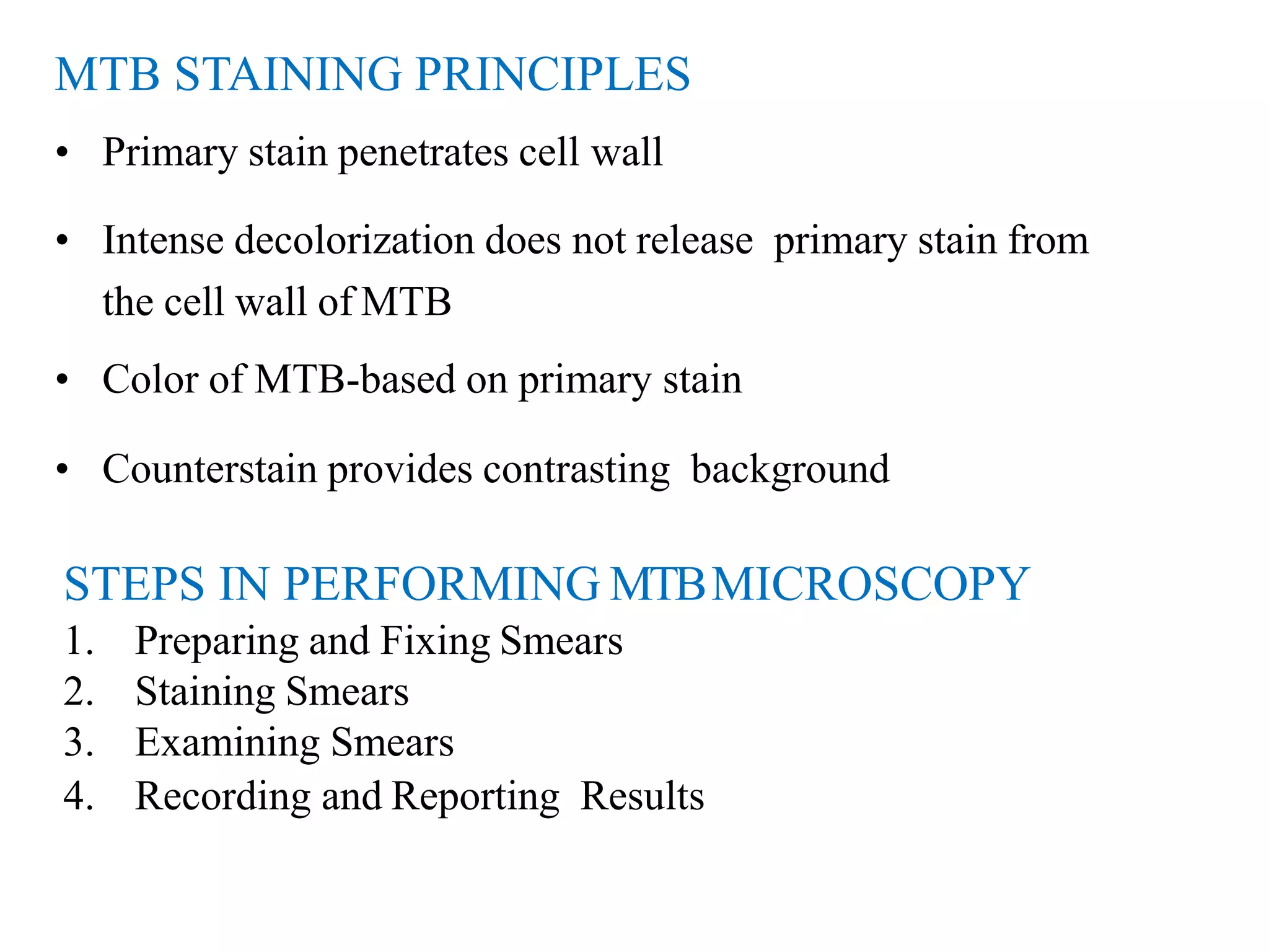
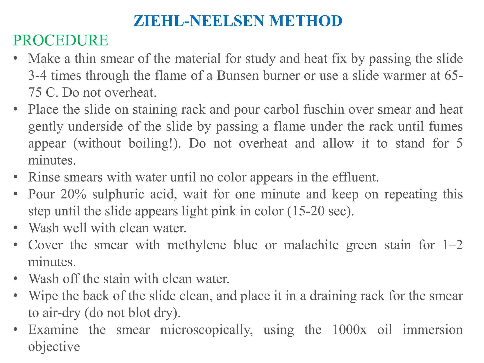
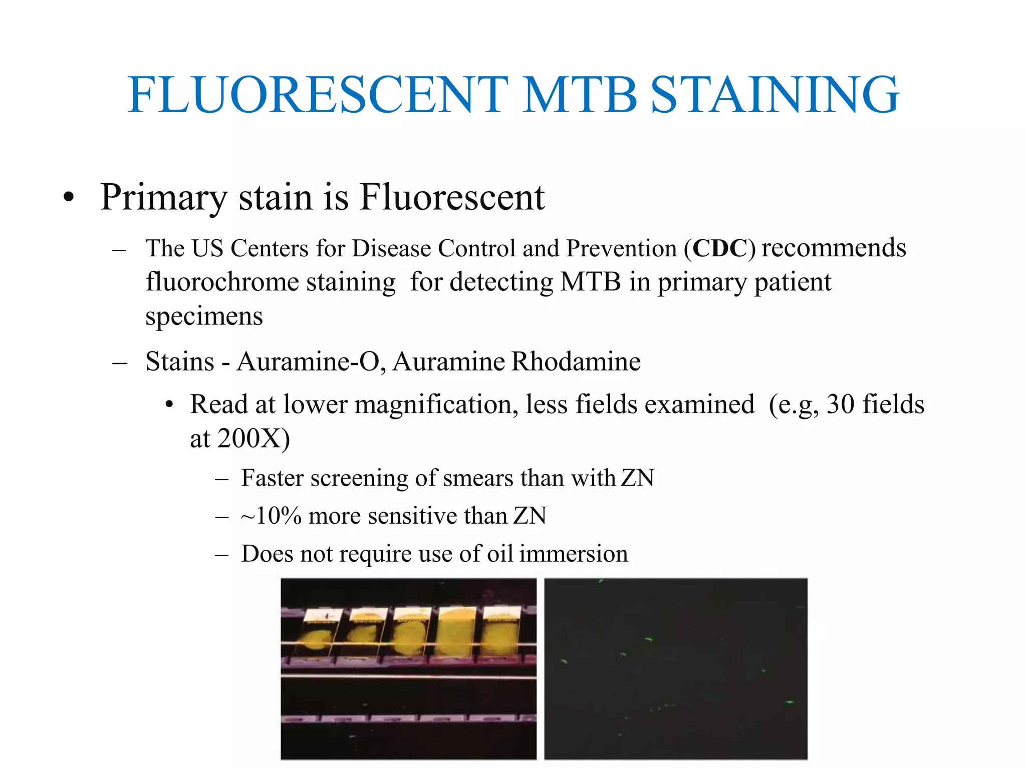
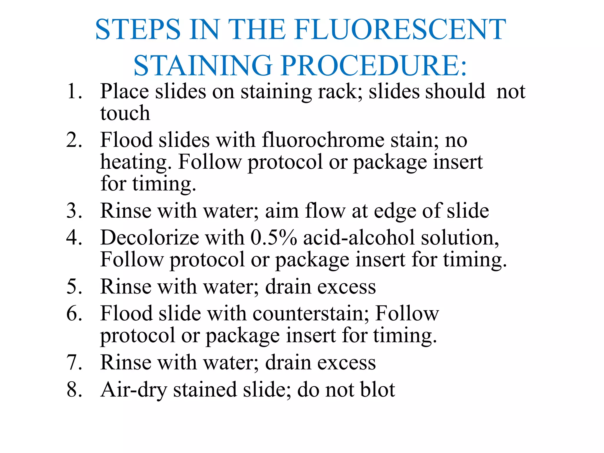
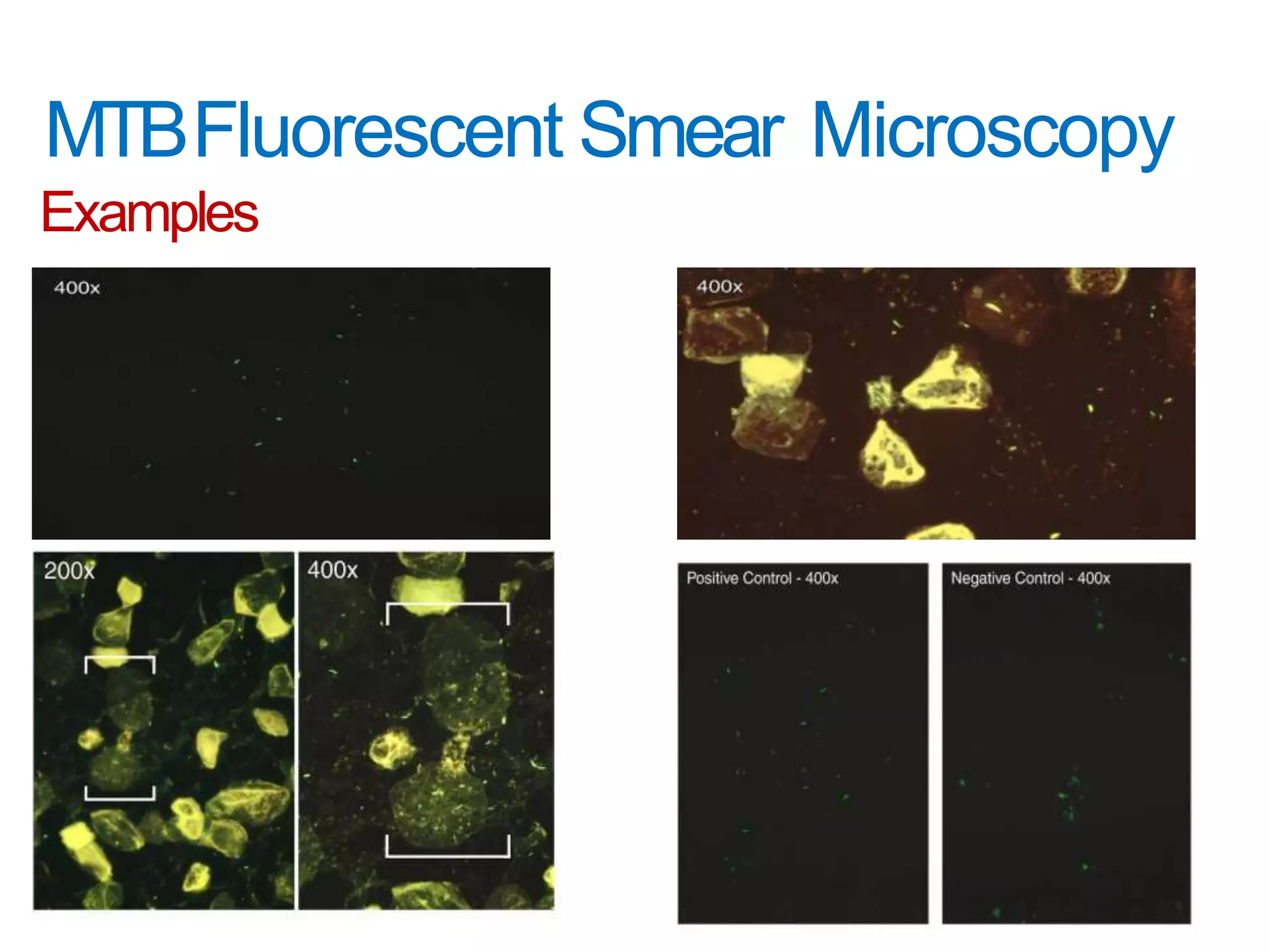
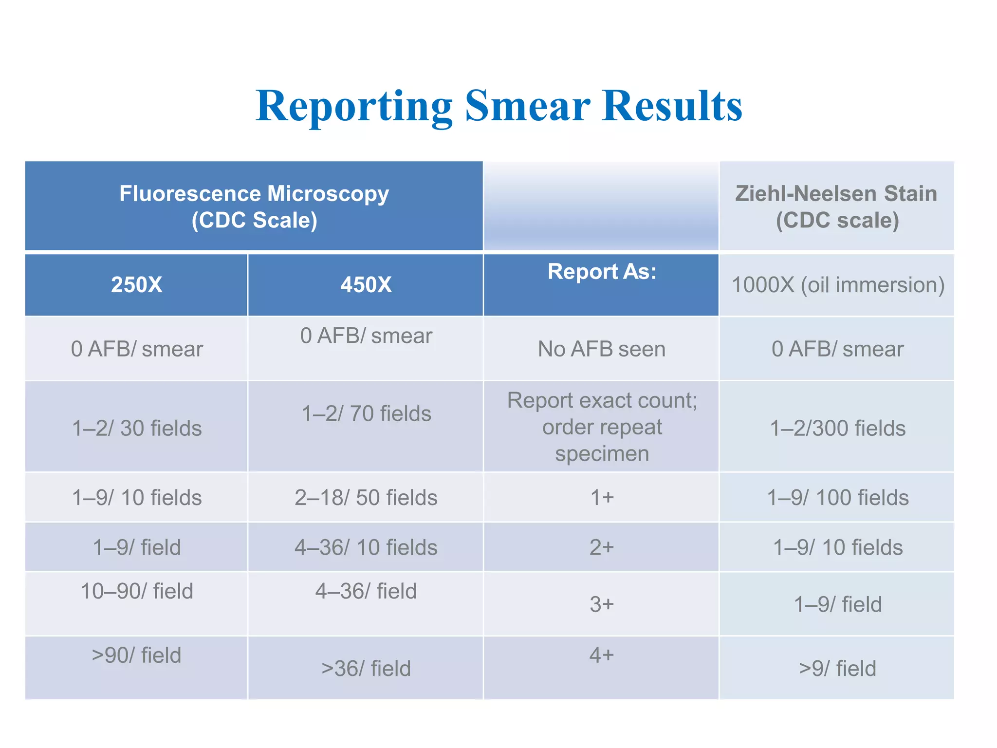
MTB SMEAR
MICROSCOPIC
METHOD
BY,
ROUF ABDUR
GROUP NO 7
8TH SEMESTER
INTRODUCTION
• MTB Smear Microscopy: Microscopic
examination of specially stained smears to detect
acid-fast organisms such as Mycobacterium
tuberculosis(MTB) and non- tuberculous
mycobacteria (NTM)
• Acid Fast Bacilli (AFB): organisms (including
mycobacteria) that resist decolorization with acid
alcohol due to the lipid-rich mycolic acids in the
cell wall thereby retaining the primary stain
ADVANTAGES
• Examination of smears is a rapid, convenient and inexpensive test
• All types of specimens can be evaluated– sputum, tissue, body fluids, etc.
• Positive AFB smear results provide afirst indication of
mycobacterial infection and potential TB disease
• Must be accompanied by additional testing including culture for
confirmatory diagnosis
MTB MICROSCOPIC METHOD
DISADVANTAGES
• Does not distinguish between viable and dead organisms
– Follow-up specimens from patients on treatment may be smear positive yet
culture negative
• Limited sensitivity
– High bacterial load 5,000-10,000 AFB /mL is required for detection
– Misses >45% of U.S. TB cases
• Limited specificity
– All mycobacteria are acid fast
– Does not provide species identification
– Local prevalence of MTB and NTM determine the predictive values of a
positive smear for MTB
MTB MICROSCOPY RESULTS
• Clinical management
– Patient therapy may be initiated for TB based on smear result and
clinical presentation
– Changes in smear status important for monitoring response
to therapy
• Laboratory testing
– Algorithms for use of nucleic acid amplification tests are often
based on smear positivity
• Public health interventions
– Smear status and grade useful for identifying the most
infectious cases
– Contact investigations prioritized based on smear result
– Decisions regarding respiratory isolation based on smear
result
SMEAR-POSITIVE TB CASES
• Smear-positivity and grade indicates relative bacterial burden and correlates with
disease presentation
• Patients that are sputum smear-positive are 5–10 times more infectious than smear
negative patients
• Untreated or treated with an inappropriate regimen, a sputum smear-positive patient
may infect 10-15 persons/year
SPUTUM SMEAR RESULTS
• In 2010, 43% of pulmonary TB cases in the U.S. were sputum smear positive
• Incremental diagnostic yield of examination of three sputum specimens among
smear positive cases
First specimen Second specimen Third specimen
85.8% 11.9% 3.1%
STEPS IN PERFORMING MTBMICROSCOPY
1. Preparing and Fixing Smears
2. Staining Smears
3. Examining Smears
4. Recording and Reporting Results
MTB STAINING PRINCIPLES
• Primary stain penetrates cell wall
• Intense decolorization does not release primary stain from
the cell wall of MTB
• Color of MTB-based on primary stain
• Counterstain provides contrasting background
MTB MICROSCOPY STAINING TECHNIQUES
Two basic techniques: same
principle:
• Fluorescence: Auramine staining
– Also known as Fluorochrome staining
– Contrast light & dark
• Brightfield: carbol fuchsin staining
– Contrast red AFB on blue orgreen
background
CARBOL-FUCHSIN MTB STAINING
• Primary stain is Carbol Fuchsin
– Ziehl-Neelson (ZN) Method
• Requires heat during staining
• Requires higher magnification
• More fields examined (e.g. 300 fields at 1000X)
• Requires more time to read
• Requires use of oil immersion
• Stains all NTM well
– Kinyoun Method
• Cold carbol fuchsin method
• Less toxic fumes
• Neither method is reccomended for staining primary
specimens
ZIEHL-NEELSEN METHOD
PROCEDURE
• Make a thin smear of the material for study and heat fix by passing the slide
3-4 times through the flame of a Bunsen burner or use a slide warmer at 65-
75 C. Do not overheat.
• Place the slide on staining rack and pour carbol fuschin over smear and heat
gently underside of the slide by passing a flame under the rack until fumes
appear (without boiling!). Do not overheat and allow it to stand for 5
minutes.
• Rinse smears with water until no color appears in the effluent.
• Pour 20% sulphuric acid, wait for one minute and keep on repeating this
step until the slide appears light pink in color (15-20 sec).
• Wash well with clean water.
• Cover the smear with methylene blue or malachite green stain for 1–2
minutes.
• Wash off the stain with clean water.
• Wipe the back of the slide clean, and place it in a draining rack for the smear
to air-dry (do not blot dry).
• Examine the smear microscopically, using the 1000x oil immersion
objective
FLUORESCENT MTB STAINING
• Primary stain is Fluorescent
– The US Centers for Disease Control and Prevention (CDC) recommends
fluorochrome staining for detecting MTB in primary patient
specimens
– Stains - Auramine-O, Auramine Rhodamine
• Read at lower magnification, less fields examined (e.g, 30 fields
at 200X)
– Faster screening of smears than with ZN
– ~10% more sensitive than ZN
– Does not require use of oil immersion
STEPS IN THE FLUORESCENT
STAINING PROCEDURE:
1. Place slides on staining rack; slides should not
touch
2. Flood slides with fluorochrome stain; no
heating. Follow protocol or package insert
for timing.
3. Rinse with water; aim flow at edge of slide
4. Decolorize with 0.5% acid-alcohol solution,
Follow protocol or package insert for timing.
5. Rinse with water; drain excess
6. Flood slide with counterstain; Follow
protocol or package insert for timing.
7. Rinse with water; drain excess
8. Air-dry stained slide; do not blot
FLUORESCENCE MICROSCOPY
• A fluorescence microscope is required for examining
fluorochrome-stained smears:
– Mercury vapor or halogen bulb light source (about 150 hours
of use)
– Newer mercury bulbs (about 2,000 hours of use)
– LED Bulbs (about 15,000 hours of use)
– Excitation and emission (barrier) filters are necessary for
visualization of the fluorescently-stained smear (specific to
the staining method used. Check package insert)
• LED-based Fluorescent Microscopy
– LED modules used to adapt light microscopes for reading
fluorescently-stained smears
• May be useful in low income settings
• More research is needed to evaluate performance
MTBFluorescent Smear Microscopy
Examples
SYSTEMATIC EXAMINATION OF SMEARS
Whichever method
youuse,BECONSISTEN!
Number of Fields to Examine
Magnification a Number of Fields b
200x
250x
400x
450x
a) This final magnification represents the objective lens magnification multiplied
by the eyepiece magnification
b) The minimum number of fields to examine before reporting a smear as
negative for acid-fast organisms.
30
30
55
70
EXAMINING SMEARS FOR MTB
• MTB will be rod-shaped, 1–10 µm in length and 0.2–
0.6 µm wide
• Appearance is generally long and slender but may also
appear bent
• Bacilli may contain heavily stained areas called
beads
• Count a clump of bacilli that are touching as one
• Debris, some species of the genera Nocardia and
Corynebacterium, and some fungal spores may appear
acid fast
Reporting Smear Results
Fluorescence Microscopy
(CDC Scale)
Ziehl-Neelsen Stain
(CDC scale)
250X 450X
Report As:
1000X (oil immersion)
0 AFB/ smear
0 AFB/ smear
No AFB seen 0 AFB/ smear
1–2/ 30 fields
1–2/ 70 fields
Report exact count;
order repeat
specimen
1–2/300 fields
1–9/ 10 fields 2–18/ 50 fields 1+ 1–9/ 100 fields
1–9/ field 4–36/ 10 fields 2+ 1–9/ 10 fields
10–90/ field 4–36/ field
3+ 1–9/ field
>90/ field
>36/ field
4+
>9/ field
Maintaining Proficiency in Microscopic Smear
Examination:
• Smears should be examined by an experienced
microscopist
– Microscopists should meet a level of competency before being allowed
to report smear results.
• Mycobacteriology laboratories should participate in an approved
proficiency testing program that includes smear microscopy
• Tomaintain proficiency, laboratories should process at least 15 AFB
smears per week
• Other procifiency testing activities
– Participate in multiple proficiency testing programs
– Develop an internal proficiency testing program
– Establish a QAprogram
Achieving Reliable Results
• Obtain good quality specimens is essential
• Prevent contamination of testing reagents
and adjacent slides when staining
• Follow established procedures &
recommendations
• Ensure accurate reporting and record keeping
THANK YOU

Mycobacterium tuberculosis smear microscopy

  • 1.
  • 2.
    INTRODUCTION • MTB SmearMicroscopy: Microscopic examination of specially stained smears to detect acid-fast organisms such as Mycobacterium tuberculosis(MTB) and non- tuberculous mycobacteria (NTM) • Acid Fast Bacilli (AFB): organisms (including mycobacteria) that resist decolorization with acid alcohol due to the lipid-rich mycolic acids in the cell wall thereby retaining the primary stain
  • 3.
    ADVANTAGES • Examination ofsmears is a rapid, convenient and inexpensive test • All types of specimens can be evaluated– sputum, tissue, body fluids, etc. • Positive AFB smear results provide afirst indication of mycobacterial infection and potential TB disease • Must be accompanied by additional testing including culture for confirmatory diagnosis MTB MICROSCOPIC METHOD DISADVANTAGES • Does not distinguish between viable and dead organisms – Follow-up specimens from patients on treatment may be smear positive yet culture negative • Limited sensitivity – High bacterial load 5,000-10,000 AFB /mL is required for detection – Misses >45% of U.S. TB cases • Limited specificity – All mycobacteria are acid fast – Does not provide species identification – Local prevalence of MTB and NTM determine the predictive values of a positive smear for MTB
  • 4.
    MTB MICROSCOPY RESULTS •Clinical management – Patient therapy may be initiated for TB based on smear result and clinical presentation – Changes in smear status important for monitoring response to therapy • Laboratory testing – Algorithms for use of nucleic acid amplification tests are often based on smear positivity • Public health interventions – Smear status and grade useful for identifying the most infectious cases – Contact investigations prioritized based on smear result – Decisions regarding respiratory isolation based on smear result
  • 5.
    SMEAR-POSITIVE TB CASES •Smear-positivity and grade indicates relative bacterial burden and correlates with disease presentation • Patients that are sputum smear-positive are 5–10 times more infectious than smear negative patients • Untreated or treated with an inappropriate regimen, a sputum smear-positive patient may infect 10-15 persons/year SPUTUM SMEAR RESULTS • In 2010, 43% of pulmonary TB cases in the U.S. were sputum smear positive • Incremental diagnostic yield of examination of three sputum specimens among smear positive cases First specimen Second specimen Third specimen 85.8% 11.9% 3.1%
  • 6.
    STEPS IN PERFORMINGMTBMICROSCOPY 1. Preparing and Fixing Smears 2. Staining Smears 3. Examining Smears 4. Recording and Reporting Results MTB STAINING PRINCIPLES • Primary stain penetrates cell wall • Intense decolorization does not release primary stain from the cell wall of MTB • Color of MTB-based on primary stain • Counterstain provides contrasting background
  • 7.
    MTB MICROSCOPY STAININGTECHNIQUES Two basic techniques: same principle: • Fluorescence: Auramine staining – Also known as Fluorochrome staining – Contrast light & dark • Brightfield: carbol fuchsin staining – Contrast red AFB on blue orgreen background
  • 8.
    CARBOL-FUCHSIN MTB STAINING •Primary stain is Carbol Fuchsin – Ziehl-Neelson (ZN) Method • Requires heat during staining • Requires higher magnification • More fields examined (e.g. 300 fields at 1000X) • Requires more time to read • Requires use of oil immersion • Stains all NTM well – Kinyoun Method • Cold carbol fuchsin method • Less toxic fumes • Neither method is reccomended for staining primary specimens
  • 9.
    ZIEHL-NEELSEN METHOD PROCEDURE • Makea thin smear of the material for study and heat fix by passing the slide 3-4 times through the flame of a Bunsen burner or use a slide warmer at 65- 75 C. Do not overheat. • Place the slide on staining rack and pour carbol fuschin over smear and heat gently underside of the slide by passing a flame under the rack until fumes appear (without boiling!). Do not overheat and allow it to stand for 5 minutes. • Rinse smears with water until no color appears in the effluent. • Pour 20% sulphuric acid, wait for one minute and keep on repeating this step until the slide appears light pink in color (15-20 sec). • Wash well with clean water. • Cover the smear with methylene blue or malachite green stain for 1–2 minutes. • Wash off the stain with clean water. • Wipe the back of the slide clean, and place it in a draining rack for the smear to air-dry (do not blot dry). • Examine the smear microscopically, using the 1000x oil immersion objective
  • 10.
    FLUORESCENT MTB STAINING •Primary stain is Fluorescent – The US Centers for Disease Control and Prevention (CDC) recommends fluorochrome staining for detecting MTB in primary patient specimens – Stains - Auramine-O, Auramine Rhodamine • Read at lower magnification, less fields examined (e.g, 30 fields at 200X) – Faster screening of smears than with ZN – ~10% more sensitive than ZN – Does not require use of oil immersion
  • 11.
    STEPS IN THEFLUORESCENT STAINING PROCEDURE: 1. Place slides on staining rack; slides should not touch 2. Flood slides with fluorochrome stain; no heating. Follow protocol or package insert for timing. 3. Rinse with water; aim flow at edge of slide 4. Decolorize with 0.5% acid-alcohol solution, Follow protocol or package insert for timing. 5. Rinse with water; drain excess 6. Flood slide with counterstain; Follow protocol or package insert for timing. 7. Rinse with water; drain excess 8. Air-dry stained slide; do not blot
  • 12.
    FLUORESCENCE MICROSCOPY • Afluorescence microscope is required for examining fluorochrome-stained smears: – Mercury vapor or halogen bulb light source (about 150 hours of use) – Newer mercury bulbs (about 2,000 hours of use) – LED Bulbs (about 15,000 hours of use) – Excitation and emission (barrier) filters are necessary for visualization of the fluorescently-stained smear (specific to the staining method used. Check package insert) • LED-based Fluorescent Microscopy – LED modules used to adapt light microscopes for reading fluorescently-stained smears • May be useful in low income settings • More research is needed to evaluate performance
  • 13.
  • 14.
    SYSTEMATIC EXAMINATION OFSMEARS Whichever method youuse,BECONSISTEN! Number of Fields to Examine Magnification a Number of Fields b 200x 250x 400x 450x a) This final magnification represents the objective lens magnification multiplied by the eyepiece magnification b) The minimum number of fields to examine before reporting a smear as negative for acid-fast organisms. 30 30 55 70
  • 15.
    EXAMINING SMEARS FORMTB • MTB will be rod-shaped, 1–10 µm in length and 0.2– 0.6 µm wide • Appearance is generally long and slender but may also appear bent • Bacilli may contain heavily stained areas called beads • Count a clump of bacilli that are touching as one • Debris, some species of the genera Nocardia and Corynebacterium, and some fungal spores may appear acid fast
  • 16.
    Reporting Smear Results FluorescenceMicroscopy (CDC Scale) Ziehl-Neelsen Stain (CDC scale) 250X 450X Report As: 1000X (oil immersion) 0 AFB/ smear 0 AFB/ smear No AFB seen 0 AFB/ smear 1–2/ 30 fields 1–2/ 70 fields Report exact count; order repeat specimen 1–2/300 fields 1–9/ 10 fields 2–18/ 50 fields 1+ 1–9/ 100 fields 1–9/ field 4–36/ 10 fields 2+ 1–9/ 10 fields 10–90/ field 4–36/ field 3+ 1–9/ field >90/ field >36/ field 4+ >9/ field
  • 17.
    Maintaining Proficiency inMicroscopic Smear Examination: • Smears should be examined by an experienced microscopist – Microscopists should meet a level of competency before being allowed to report smear results. • Mycobacteriology laboratories should participate in an approved proficiency testing program that includes smear microscopy • Tomaintain proficiency, laboratories should process at least 15 AFB smears per week • Other procifiency testing activities – Participate in multiple proficiency testing programs – Develop an internal proficiency testing program – Establish a QAprogram
  • 18.
    Achieving Reliable Results •Obtain good quality specimens is essential • Prevent contamination of testing reagents and adjacent slides when staining • Follow established procedures & recommendations • Ensure accurate reporting and record keeping
  • 19.