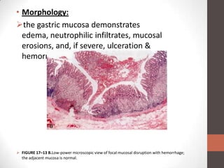
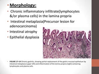
Acute & chronic
gastritis
Etiology, pathogenesis & morphology
Acute Gastritis:
• Gastritis: inflammation of the gastric mucosa.
• Diagnosis mostly histological.

• Acute Gastritis:acute mucosal inflammatory
  process usually of a transient nature.
• neutrophilic infiltration

• Etiology:
Heavy use of NSAID’s, particularly aspirin
Excessive alcohol consumption&Heavy smoking
Treatment with cancer chemotherapeutic drugs Gastric
 irradiation or freezing
 Systemic bacterial or viral infections
 Severe stress& Mechanical trauma
 Ischemia, shock & uremia
 Distal gastrectomy.
• Pathogenesis:
increased acid secretion with back-diffusion,
 decreased production of bicarbonate buffer,
reduced blood flow,
disruption of the adherent mucus layer,
 direct damage to the epithelium.
• Morphology:
the gastric mucosa demonstrates
  edema, neutrophilic infiltrates, mucosal
  erosions, and, if severe, ulceration &
  hemorrhage.


 FIGURE 17–13 B.Low-power microscopic view of focal mucosal disruption with hemorrhage;
  the adjacent mucosa is normal.
Chronic Gastritis:
• Chronic gastritis is defined as the presence of
  chronic mucosal inflammatory changes leading
  eventually to mucosal atrophy and intestinal
  metaplasia, usually in the absence of erosions


• Etiology:
Chronic infection by H. pylori
 Immunologic,(in association with pernicious
 anemia)
Toxic,(alcohol &cigarette smoking)
 Postsurgical,(esp following antrectomy with
 gastroenterostomy)
 Motor and mechanical,(obstruction, bezoars&
 gastric atony)
Radiation
 Granulomatous conditions (e.g., Crohn disease)
 Misc.—amyloidosis, graft-versus-host disease
 ,uremia.
• Pathogenesis:
oChronic atrophic gastritis caused by H.pylori:
• (1) Gram-negative, curved rod
• (2) Produces urease, proteases, cytotoxins
  (a) Urease converts amino groups in proteins to
ammonia
  (b) Secretion products produce chronic gastritis
and PUD,
• (3) Colonizes mucus layer lining
oAutoimmune atrophic gastritis:
• Autoantibodies to parietal cells (H/K ATPase
  proton pump) and intrinsic factor
• Morphology:
• Chronic inflammatory infiltrate(lymphocytes
  &/or plasma cells) in the lamina propria
• Intestinal metaplasia(Precursor lesion for
  adenocarcinoma)
• Intestinal atrophy
• Epithelial dysplasia


• FIGURE 17–14 Chronic gastritis, showing partial replacement of the gastric mucosal epithelium by
  intestinal metaplasia (upper left) and inflammation of the lamina propria (right) containing
  lymphocytes and plasma cells.
Thank You!!

Acute & chronic gastritis

  • 1.
    Acute & chronic gastritis Etiology,pathogenesis & morphology
  • 2.
    Acute Gastritis: • Gastritis:inflammation of the gastric mucosa. • Diagnosis mostly histological. • Acute Gastritis:acute mucosal inflammatory process usually of a transient nature. • neutrophilic infiltration • Etiology: Heavy use of NSAID’s, particularly aspirin Excessive alcohol consumption&Heavy smoking
  • 3.
    Treatment with cancerchemotherapeutic drugs Gastric irradiation or freezing  Systemic bacterial or viral infections  Severe stress& Mechanical trauma  Ischemia, shock & uremia  Distal gastrectomy. • Pathogenesis: increased acid secretion with back-diffusion,  decreased production of bicarbonate buffer, reduced blood flow, disruption of the adherent mucus layer,  direct damage to the epithelium.
  • 4.
    • Morphology: the gastricmucosa demonstrates edema, neutrophilic infiltrates, mucosal erosions, and, if severe, ulceration & hemorrhage.  FIGURE 17–13 B.Low-power microscopic view of focal mucosal disruption with hemorrhage; the adjacent mucosa is normal.
  • 5.
    Chronic Gastritis: • Chronicgastritis is defined as the presence of chronic mucosal inflammatory changes leading eventually to mucosal atrophy and intestinal metaplasia, usually in the absence of erosions • Etiology: Chronic infection by H. pylori  Immunologic,(in association with pernicious anemia)
  • 6.
    Toxic,(alcohol &cigarette smoking) Postsurgical,(esp following antrectomy with gastroenterostomy)  Motor and mechanical,(obstruction, bezoars& gastric atony) Radiation  Granulomatous conditions (e.g., Crohn disease)  Misc.—amyloidosis, graft-versus-host disease ,uremia. • Pathogenesis:
  • 7.
    oChronic atrophic gastritiscaused by H.pylori: • (1) Gram-negative, curved rod • (2) Produces urease, proteases, cytotoxins (a) Urease converts amino groups in proteins to ammonia (b) Secretion products produce chronic gastritis and PUD, • (3) Colonizes mucus layer lining oAutoimmune atrophic gastritis: • Autoantibodies to parietal cells (H/K ATPase proton pump) and intrinsic factor
  • 8.
    • Morphology: • Chronicinflammatory infiltrate(lymphocytes &/or plasma cells) in the lamina propria • Intestinal metaplasia(Precursor lesion for adenocarcinoma) • Intestinal atrophy • Epithelial dysplasia • FIGURE 17–14 Chronic gastritis, showing partial replacement of the gastric mucosal epithelium by intestinal metaplasia (upper left) and inflammation of the lamina propria (right) containing lymphocytes and plasma cells.
  • 9.