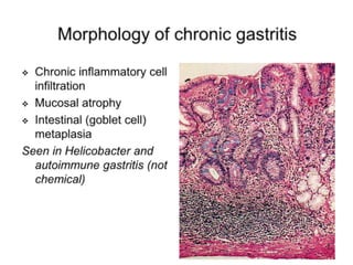
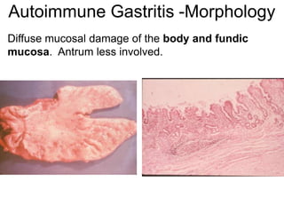
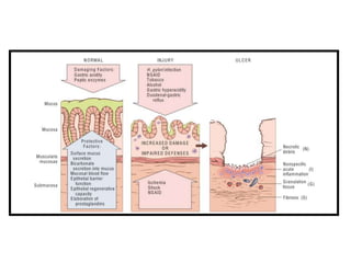
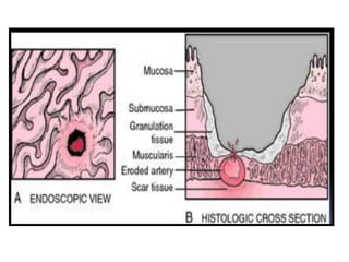
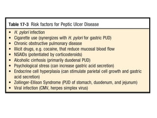
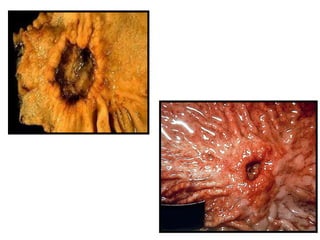
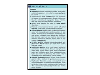
Chronic gastritis and Peptic ulcer
15-04-2020
Outlines
• Gastritis
• Chronic gastritis
• Types, etiopathogenesis and microscopic
findings.
• Peptic ulcer
• Gastritis
Mucosal inflammatory process
Acute and chronic
Acute & Chronic
 1. Acute refers to short term
inflammation.
 Acute refering to neurophilic infiltrate
 2. Chronic referring to long standing
forms
 Chronic referring to mononuclear cell
infiltrate especially lymphocyte and
macrophages
• Chronic Gastritis: ( 2 important causes )
• Helicobacter pylori infection.
• Auto-immune gastritis
Chronic
Gastritis
Type B
Antral
Gastritis
Type A
Autoimmune
gastritis
H.pylori infection
Helicobacter pylori aka (Campylobacter
pyloridis)
Gram-negative bacterium found in the
stomach.
70% to 90% of patients with duodenal
ulcers and in about 70% of those with
gastric ulcers.
Antral predominant inflammation.
Contaminated food and feco-oral
transmission
Intense inflammatory and immune response. There is increased
production of proinflammatory cytokines such as interleukin (IL)-1, IL-
6, tumor necrosis factor (TNF), and, most notably, IL-8
H. pylori secretes a urease that breaks down urea to form toxic
compounds such as ammonium chloride and monochloramine.
Adhesins and Flagellin
Phospholipases that damage surface epithelial cells.
Toxin encoding genes : vacuolating toxin (VacA), cytotoxin-
associated gene A (CagA)
• Complications:
• Long standing inflammation – MALToma.
• Long standing injury – Intestinal metaplasia
and risk of Adenocarcinoma
• Direct method : Endoscopic biopsy
• Indirect method;
• Stool fecal antigen test (sensitive)
• Anti H.pylori antibodies in the saliva
 Autoimmune gastritis
1) Antibodies to parietal cells, gastrin
receptor, intrinsic factor, and H+,K+
ATPase.
2) <10% of cases of chronic gastritis
3) Cardia and Body region affected
Clinical Features
 Usually only a few symptoms: nausea,
vomiting, upper abdominal discomfort
 Autoimmune
Hypo to achlorhydria (severe loss of
parietal glands)
Hypergastrinemia
10% have pernicious anemia
 Autoimmune:
Often seen in association with other
autoimmune disorders (Hashimoto thyroiditis,
Addison disease, and type I diabetes)
Significant risk for the development of gastric
carcinoma (2-4%) and endocrine tumors
(carcinoid tumor)
 Regeneration - constant feature
 Metaplasia - mucosa of antral and body-
fundic regions converts to columnar
absorptive cells and goblet cells (intestinal
metaplasia)
 Atrophy - marked loss of glands
 Dysplasia – precursor lesion to gastric
cancer in atrophic gastritis
Autoimmune Gastritis -Morphology
Diffuse mucosal damage of the body and fundic
mucosa. Antrum less involved.
Peptic Ulcer
• Chronic mucosal ulceration.
• (Break in the continuity of the epithelium)
• Involves distal part of the stomach and /or
duodenum.
DEFENSIVE FORCES
Secretion of mucus and Bicarbonate by surface epithelial cells.
Rapid gastric epithelial regeneration.
Mucosal blood flow, to sweep away hydrogen ions that have back-
diffused into the mucosa from the lumen and to sustain the high cellular
metabolic and regenerative activity.
Prostaglandins, which help maintain mucosal blood flow.
IMPAIRED
DEFENCE
ISCHAEMIA,
SHOCK,
DELAYED GASRIC
EMPTYING,
GASRO-
OESOPHAGEAL
REFLUX
•INCREASED
AGGRESSION
•H.Pylori INFECTION,
NSAIDs (ASPIRIN),
SMOKING, ALCOHOL,
•IMPAIRED REGULATION
OF ACID-PEPSIN
SECRETION,
•BILE ACIDS
NSAIDS
 NSAIDs and aspirin also interfere with the
protective mucus layer by inhibiting
mucosal cyclooxygenase activity, reducing
levels of mucosal prostaglandins
Smoking
 Stimulate gastric acid secretion
 Causes alteration in mucosal blood flow
 Decrease mucus secretion
 Reduces prostaglandin synthesis
 Decrease pancreatic bicarbonate secretion
• Chronic and recurring epigastric pain,
• Burning occuring 1-3 hours after having food,
• Relieves with antacids.
Complications
• Q1) Which type of gastritis will lead to
MALToma?
• Q2) Which type of gastritis will lead to well
differentiated neuroendocrine tumour?
•THANK YOU

Gastritis

  • 1.
    Chronic gastritis andPeptic ulcer 15-04-2020
  • 2.
    Outlines • Gastritis • Chronicgastritis • Types, etiopathogenesis and microscopic findings. • Peptic ulcer
  • 5.
    • Gastritis Mucosal inflammatoryprocess Acute and chronic
  • 6.
    Acute & Chronic 1. Acute refers to short term inflammation.  Acute refering to neurophilic infiltrate  2. Chronic referring to long standing forms  Chronic referring to mononuclear cell infiltrate especially lymphocyte and macrophages
  • 7.
    • Chronic Gastritis:( 2 important causes ) • Helicobacter pylori infection. • Auto-immune gastritis
  • 8.
  • 9.
    H.pylori infection Helicobacter pyloriaka (Campylobacter pyloridis) Gram-negative bacterium found in the stomach. 70% to 90% of patients with duodenal ulcers and in about 70% of those with gastric ulcers. Antral predominant inflammation. Contaminated food and feco-oral transmission
  • 10.
    Intense inflammatory andimmune response. There is increased production of proinflammatory cytokines such as interleukin (IL)-1, IL- 6, tumor necrosis factor (TNF), and, most notably, IL-8 H. pylori secretes a urease that breaks down urea to form toxic compounds such as ammonium chloride and monochloramine. Adhesins and Flagellin Phospholipases that damage surface epithelial cells. Toxin encoding genes : vacuolating toxin (VacA), cytotoxin- associated gene A (CagA)
  • 12.
    • Complications: • Longstanding inflammation – MALToma. • Long standing injury – Intestinal metaplasia and risk of Adenocarcinoma
  • 13.
    • Direct method: Endoscopic biopsy • Indirect method; • Stool fecal antigen test (sensitive) • Anti H.pylori antibodies in the saliva
  • 14.
     Autoimmune gastritis 1)Antibodies to parietal cells, gastrin receptor, intrinsic factor, and H+,K+ ATPase. 2) <10% of cases of chronic gastritis 3) Cardia and Body region affected
  • 15.
    Clinical Features  Usuallyonly a few symptoms: nausea, vomiting, upper abdominal discomfort  Autoimmune Hypo to achlorhydria (severe loss of parietal glands) Hypergastrinemia 10% have pernicious anemia
  • 16.
     Autoimmune: Often seenin association with other autoimmune disorders (Hashimoto thyroiditis, Addison disease, and type I diabetes) Significant risk for the development of gastric carcinoma (2-4%) and endocrine tumors (carcinoid tumor)
  • 17.
     Regeneration -constant feature  Metaplasia - mucosa of antral and body- fundic regions converts to columnar absorptive cells and goblet cells (intestinal metaplasia)  Atrophy - marked loss of glands  Dysplasia – precursor lesion to gastric cancer in atrophic gastritis
  • 19.
    Autoimmune Gastritis -Morphology Diffusemucosal damage of the body and fundic mucosa. Antrum less involved.
  • 20.
    Peptic Ulcer • Chronicmucosal ulceration. • (Break in the continuity of the epithelium) • Involves distal part of the stomach and /or duodenum.
  • 22.
    DEFENSIVE FORCES Secretion ofmucus and Bicarbonate by surface epithelial cells. Rapid gastric epithelial regeneration. Mucosal blood flow, to sweep away hydrogen ions that have back- diffused into the mucosa from the lumen and to sustain the high cellular metabolic and regenerative activity. Prostaglandins, which help maintain mucosal blood flow.
  • 23.
  • 24.
    NSAIDS  NSAIDs andaspirin also interfere with the protective mucus layer by inhibiting mucosal cyclooxygenase activity, reducing levels of mucosal prostaglandins
  • 25.
    Smoking  Stimulate gastricacid secretion  Causes alteration in mucosal blood flow  Decrease mucus secretion  Reduces prostaglandin synthesis  Decrease pancreatic bicarbonate secretion
  • 28.
    • Chronic andrecurring epigastric pain, • Burning occuring 1-3 hours after having food, • Relieves with antacids.
  • 30.
  • 32.
    • Q1) Whichtype of gastritis will lead to MALToma? • Q2) Which type of gastritis will lead to well differentiated neuroendocrine tumour?
  • 33.