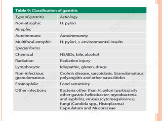
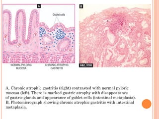
SlideShare a Scribd company logo
GASTRITIS
Subject: Practice Of Medicine
Prepared by Dr. Suman Chaudhary MD (Hom.)
 The term ‘gastritis’ is commonly employed for
any clinical condition with upper abdominal
discomfort like indigestion or dyspepsia in which
the specific clinical signs and radiological
abnormalities are absent.
 The condition is of great importance due to its
relationship with peptic ulcer and gastric cancer.
Broadly speaking, gastritis may be of 2 types—
acute and chronic. Chronic gastritis can further
be of various types.
ACUTE GASTRITIS
 Acute gastritis is a transient acute inflammatory
involvement of the stomach, mainly mucosa
ETIOPATHOGENESIS
 A variety of etiologic agents have been implicated
in the causation of acute gastritis. These are as
follows:
 1. Diet and personal habits:
 Highly spiced food
 Excessive alcohol consumption
 Malnutrition
 Heavy smoking.
 2. Infections:
 Bacterial infections e.g. Helicobacter pylori,
diphtheria, salmonellosis, pneumonia,
staphylococcal food poisoning.
 Viral infections e.g. viral hepatitis, influenza,
infectious mononucleosis.
 3. Drugs: Intake of drugs like non-steroidal anti-
inflammatory drugs (NSAIDs), aspirin, cortisone,
phenylbutazone, indomethacin, preparations of iron,
chemotherapeutic agents.
 4. Chemical and physical agents: Intake of
corrosive chemicals such as caustic soda, phenol, lysol
Gastric irradiation Freezing.
 5. Severe stress:
 Emotional factors like shock, anger, resentment etc.
 Extensive burns
 Trauma
 Surgery.
 The mucosal injury and subsequent acute
inflammation in acute gastritis occurs by one of
the following mechanisms:
 1. Reduced blood flow, resulting in mucosal
hypoperfusion due to ischaemia.
 2. Increased acid secretion and its accumulation
due to H. pylori infection resulting in damage to
epithelial barrier.
 3. Decreased production of bicarbonate buffer.
MANIFESTATION
 Epigastric discomfort
 Abdominal tenderness
 Cramping
 Belching
 Reflux
 Severe nausea and vomiting
 Hematemesis
 Sometimes GI bleeding is the only manifestation
 When contaminated food is the cause of gastritis,
diarrhea usually develops within 5 hours of
ingestion
DIAGNOSIS
 Diagnosis is based on a detailed history of food
intake, medications taken, and any disorder
related to gastritis.
 The physician may also perform a gastroscopic
examination with endoscopy.
 Histological examination by biopsy of a sample.
MANAGEMENT
 Anti – emetic drugs like Inj. Perinorm or Tab.
Domperidone are frequently effective in vomiting.
 Antacids like cimetidine, Ranitidine, or
Famotidine are effective to reduce the pain.
 If ingestion of NSAIDs is a problem, a
prostaglandin E1 (PGE1) analog may be
prescribed to protect the stomach mucosa and
inhibit gastric acid secretion.
 Initially foods and fluids are withheld until
nausea and vomiting subside.
 Once the client tolerates food, the diet includes
decaffeinated tea, gelatin, toast, and simple
bland foods.
 The client should avoid spicy foods, caffeine and
large, heavy meals.
 In the continued absence of nausea, vomiting
and bloating, the client can slowly return to a
normal diet.
CHRONIC GASTRITIS
 The condition occurs more frequently with
advancing age; average age for symptomatic
chronic gastritis being 45 years which
corresponds well with the age incidence of gastric
ulcer.
ETIOPATHOGENESIS
 All the causative factors of acute gastritis
described above may result in chronic gastritis
too. Recurrent attacks of acute gastritis may
result in chronic gastritis. Some additional
causes are as under:
 1. Reflux of duodenal contents into the stomach,
especially in cases which have undergone surgical
intervention in the region of pylorus.
 2. Infection with H. pylori is strongly implicated
in the etiology of chronic gastritis and is more
common.
 3. Associated disease of the stomach and
duodenum, such as gastric or duodenal ulcer,
gastric carcinoma.
 4. Chronic hypochromic anaemia, especially
associated with atrophic gastritis.
 5. Immunological factors such as autoantibodies
to gastric parietal cells in atrophic gastritis and
autoantibodies against intrinsic factor.
CLASSIFICATION
 1. Type A gastritis (Autoimmune gastritis)
 Type A gastritis involves mainly the body-fundic mucosa. It
is also called autoimmune gastritis due to the presence of
circulating antibodies and is sometimes associated with
other autoimmune diseases such as Hashimoto’s thyroiditis
and Addison’s disease.
 As a result of the antibodies against parietal cells and
intrinsic factor, there is depletion of parietal cells and
impaired secretion of intrinsic factor. These changes may
lead to significant gastric atrophy where intestinal
metaplasia may occur, and a small proportion of these
patients may develop pernicious anaemia.
 Due to depletion of gastric acid-producing mucosal area,
there is hypo- or achlorhydria, and hyperplasia of gastrin-
producing G cells in the antrum resulting in
hypergastrinaemia.
 2. Type B gastritis (H. pylori-related). Type
B gastritis mainly involves the region of antral
mucosa and is more common.
 It is also called hypersecretory gastritis due to
excessive secretion of acid, commonly due to
infection with H. pylori. These patients may have
associated peptic ulcer.
 3. Type AB gastritis (Mixed gastritis,
Environmental gastritis, Chronic atrophic
gastritis). Type AB gastritis affects the
mucosal region of A as well as B types (body-
fundic and antral mucosa). This is the most
common type of gastritis in all age groups.
 following simple morphologic classification has
been proposed:
 1. Chronic superficial gastritis
 2. Chronic atrophic gastritis
 3. Gastric atrophy
 4. Chronic hypertrophic gastritis (Ménétrier’s
disease)
 5. Uncommon forms of chronic gastritis.
1. CHRONIC SUPERFICIAL
GASTRITIS
 As the name suggests there is inflammatory
infiltrate consisting of plasma cells and
lymphocytes in the superficial layer of the gastric
mucosa, but there are no histological changes in
the deep layer of mucosa containing gastric
glands. Chronic superficial gastritis may resolve
completely or may progress to chronic gastric
atrophy.
 Although most patients of chronic superficial
gastritis due to H. pylori remain asymptomatic,
they may develop chronic atrophic gastritis,
gastric atrophy, peptic ulcer disease.
 H. pylori infection is now considered an
independent risk factor for gastric cancer: 3-6
fold increased risk for gastric adenocarcinoma
and 6-50 times risk of MALT lymphoma
2. CHRONIC ATROPHIC
GASTRITIS
 In this stage, there is inflammatory cell infiltrate
in the deeper layer of the mucosa and atrophy of
the epithelial elements including destruction of
the glands. Two types of metaplasia are
commonly associated with atrophic gastritis:
 i) Intestinal metaplasia. Intestinal
metaplasia is more common and involves
antral mucosa more frequently. Characteristic
histologic feature is the presence of intestinal
type mucus-goblet cells; Paneth cells and
endocrine cells may also be present.
A, Chronic atrophic gastritis (right) contrasted with normal pyloric
mucosa (left). There is marked gastric atrophy with disappearance
of gastric glands and appearance of goblet cells (intestinal metaplasia).
B, Photomicrograph showing chronic atrophic gastritis with intestinal
metaplasia.
 ii) Pseudopyloric metaplasia. It involves the
body glands which are replaced by proliferated
mucus neck cells, conforming in appearance to
normal pyloric glands. Its significance is not
known.
 3. GASTRIC ATROPHY. In this, there is
thinning of the gastric mucosa with loss of
glands but no inflammation though lymphoid
aggregates may be present.
4. CHRONIC HYPERTROPHIC
GASTRITIS (MÉNÉ-
TRIER’S DISEASE).
 This is an uncommon condition characterised
pathologically by enormous thickening of gastric
rugal folds resembling cerebral convolutions, affecting
mainly the region of fundic-body mucosa and
characteristically sparing antral mucosa.
 The patients present with dyspepsia, haematemesis,
melaena or protein-losing enteropathy.
 Histologically, the gastric pits are elongated
and are tortuous. The mucosa is markedly thickened
and parts of muscularis mucosae may extend into the
thickened folds. Epithelium-lined cysts are commonly
seen in the glandular layer. Inflammatory infiltrate is
usually mild but lymphoid follicles may be present.
5. MISCELLANEOUS FORMS OF
CHRONIC GASTRITIS
 i) Eosinophilic gastritis. This condition is
characterised by diffuse thickening of the
pyloric antrum due to oedema and extensive
infiltration by eosinophils in all the layers of the
wall of antrum. Eosinophilic gastritis probabl has
an allergic basis.
 iii) Chronic follicular gastritis. This is a
variant of chronic atrophic gastritis in which
numerous lymphoid follicles are present in the
mucosa and submucosa of the stomach.
 iv) Haemorrhagic (Erosive) gastritis. In this
condition, there are superficial erosions and
mucosal haemorrhages, usually following severe
haematemesis.
 The causes for such erosions and haemorrhages
are duodenal-gastric reflux, administration of
non-steroidal anti-inflammatory drugs (NSAIDs),
portal hypertension.
 v) Granulomatous gastritis. Rarely,
granulomas may be present in the gastric
mucosa such as in tuberculosis, sarcoidosis,
Crohn’s disease, syphilis, various mycoses, and as
a reaction to endogenous substance or foreign
material.
MANIFESTATIONS
 Manifestations are vague and may be absent
because the problem does not cause an increase
in hydrochloric acid.
 Assessment may reveal
 Anorexia
 Feeling of fullness
 Dyspepsia
 Belching
 Vague epigastric pain
 Nausea
 Vomiting
 Intolerance of spicy and fatty foods
COMPLICATIONS
 Bleeding
 Pernicious anemia
 Gastric cancer
TREATMENT
 Discomfort may lessen with a bland diet, small
frequent meals, antacids, H2 receptor
antagonists, proton pump inhibitors, and
avoidance of food that cause manifestations.
 If H.pylori bacteria are present, anti-biotics and
other medications are administered to eliminate
the bacteria.
 If 1 week of this regimen does not succeed in
eliminating the bacteria, the regimen may be
repeated for an additional week.
 If pernicious anemia develops, intramuscular
injections of vitamin B12 may be administered
monthly for the remainder of the client’s life.
 Thank you

More Related Content

What's hot

Malabsorption syndrome ppt
Malabsorption syndrome pptMalabsorption syndrome ppt
Malabsorption syndrome ppt
missmarimo
 

What's hot (20)

Aetiopathophysiology of peptic ulcer diesese
Aetiopathophysiology of peptic ulcer dieseseAetiopathophysiology of peptic ulcer diesese
Aetiopathophysiology of peptic ulcer diesese
 
Gastritis
Gastritis Gastritis
Gastritis
 
L3 esophagitis
L3 esophagitisL3 esophagitis
L3 esophagitis
 
peptic ulcer
peptic ulcerpeptic ulcer
peptic ulcer
 
Peptic ulcer disease causes and treatment
Peptic ulcer disease causes and treatmentPeptic ulcer disease causes and treatment
Peptic ulcer disease causes and treatment
 
1. Gastritis.pptx
1. Gastritis.pptx1. Gastritis.pptx
1. Gastritis.pptx
 
Gsatritis
Gsatritis  Gsatritis
Gsatritis
 
Peptic ulcer disease
Peptic ulcer diseasePeptic ulcer disease
Peptic ulcer disease
 
Gastrointestinal disease lecture(ppt)
Gastrointestinal disease lecture(ppt)Gastrointestinal disease lecture(ppt)
Gastrointestinal disease lecture(ppt)
 
Esophageal disorders
Esophageal disordersEsophageal disorders
Esophageal disorders
 
Peptic ulcer Disease
Peptic ulcer DiseasePeptic ulcer Disease
Peptic ulcer Disease
 
GASTRITIS.pptx
GASTRITIS.pptxGASTRITIS.pptx
GASTRITIS.pptx
 
Ulcerative colitis
Ulcerative colitisUlcerative colitis
Ulcerative colitis
 
Ulcerative Colitis (IBD)
Ulcerative Colitis (IBD)Ulcerative Colitis (IBD)
Ulcerative Colitis (IBD)
 
Gastritis- Mr. panneh
Gastritis- Mr. pannehGastritis- Mr. panneh
Gastritis- Mr. panneh
 
Ulcerative Colitis (UC)
Ulcerative Colitis (UC) Ulcerative Colitis (UC)
Ulcerative Colitis (UC)
 
Gastritis
GastritisGastritis
Gastritis
 
Esophagitis: Symptoms, Causes and Treatment
Esophagitis: Symptoms, Causes and Treatment Esophagitis: Symptoms, Causes and Treatment
Esophagitis: Symptoms, Causes and Treatment
 
Pep ulcer
Pep ulcerPep ulcer
Pep ulcer
 
Malabsorption syndrome ppt
Malabsorption syndrome pptMalabsorption syndrome ppt
Malabsorption syndrome ppt
 

Similar to Gastritis

Similar to Gastritis (20)

Peptic ulcer modified 2
Peptic ulcer modified 2Peptic ulcer modified 2
Peptic ulcer modified 2
 
GASTRITIS.pptx
GASTRITIS.pptxGASTRITIS.pptx
GASTRITIS.pptx
 
Gastritis.pptx
Gastritis.pptxGastritis.pptx
Gastritis.pptx
 
Lect 4- gastric disorder
Lect 4- gastric disorderLect 4- gastric disorder
Lect 4- gastric disorder
 
Topic: Gastritis Nursing Lecture ppt.ppt
Topic: Gastritis Nursing Lecture ppt.pptTopic: Gastritis Nursing Lecture ppt.ppt
Topic: Gastritis Nursing Lecture ppt.ppt
 
Gastritis
GastritisGastritis
Gastritis
 
3 gastritis.pptx
3 gastritis.pptx3 gastritis.pptx
3 gastritis.pptx
 
Gastritis
GastritisGastritis
Gastritis
 
Gastritis.pptx Gastro Intestinal Disorder
Gastritis.pptx Gastro Intestinal DisorderGastritis.pptx Gastro Intestinal Disorder
Gastritis.pptx Gastro Intestinal Disorder
 
Gastritis.pdf
Gastritis.pdfGastritis.pdf
Gastritis.pdf
 
Lect 4- gastric disorder
Lect 4- gastric disorderLect 4- gastric disorder
Lect 4- gastric disorder
 
Lect 4- gastric disord
Lect 4- gastric disordLect 4- gastric disord
Lect 4- gastric disord
 
Gastritis definition etc by Dr MJBK.pptx
Gastritis definition etc by Dr MJBK.pptxGastritis definition etc by Dr MJBK.pptx
Gastritis definition etc by Dr MJBK.pptx
 
Bohomolets Surgery 4th year Lecture #7
Bohomolets Surgery 4th year Lecture #7Bohomolets Surgery 4th year Lecture #7
Bohomolets Surgery 4th year Lecture #7
 
Git 4th 5th Gastritis.
Git 4th 5th Gastritis.Git 4th 5th Gastritis.
Git 4th 5th Gastritis.
 
Peptic ulcer disease
Peptic ulcer diseasePeptic ulcer disease
Peptic ulcer disease
 
Peptic Ulcer disease
Peptic Ulcer disease Peptic Ulcer disease
Peptic Ulcer disease
 
Gastritis NCM 103
Gastritis NCM 103Gastritis NCM 103
Gastritis NCM 103
 
ACUTE GASTRITIS.pptx
ACUTE GASTRITIS.pptxACUTE GASTRITIS.pptx
ACUTE GASTRITIS.pptx
 
Gastritis
GastritisGastritis
Gastritis
 

Recently uploaded

THORACOTOMY . SURGICAL PERSPECTIVES VOL 1
THORACOTOMY . SURGICAL PERSPECTIVES VOL 1THORACOTOMY . SURGICAL PERSPECTIVES VOL 1
THORACOTOMY . SURGICAL PERSPECTIVES VOL 1
DR SETH JOTHAM
 
Alcohol_Dr. Jeenal Mistry MD Pharmacology.pdf
Alcohol_Dr. Jeenal Mistry MD Pharmacology.pdfAlcohol_Dr. Jeenal Mistry MD Pharmacology.pdf
Alcohol_Dr. Jeenal Mistry MD Pharmacology.pdf
Dr Jeenal Mistry
 

Recently uploaded (20)

5cl adbb 5cladba cheap and fine Telegram: +85297504341
5cl adbb 5cladba cheap and fine Telegram: +852975043415cl adbb 5cladba cheap and fine Telegram: +85297504341
5cl adbb 5cladba cheap and fine Telegram: +85297504341
 
Couples presenting to the infertility clinic- Do they really have infertility...
Couples presenting to the infertility clinic- Do they really have infertility...Couples presenting to the infertility clinic- Do they really have infertility...
Couples presenting to the infertility clinic- Do they really have infertility...
 
DIGITAL RADIOGRAPHY-SABBU KHATOON .pptx
DIGITAL RADIOGRAPHY-SABBU KHATOON  .pptxDIGITAL RADIOGRAPHY-SABBU KHATOON  .pptx
DIGITAL RADIOGRAPHY-SABBU KHATOON .pptx
 
A thorough review of supernormal conduction.pptx
A thorough review of supernormal conduction.pptxA thorough review of supernormal conduction.pptx
A thorough review of supernormal conduction.pptx
 
THORACOTOMY . SURGICAL PERSPECTIVES VOL 1
THORACOTOMY . SURGICAL PERSPECTIVES VOL 1THORACOTOMY . SURGICAL PERSPECTIVES VOL 1
THORACOTOMY . SURGICAL PERSPECTIVES VOL 1
 
Compare home pulse pressure components collected directly from home
Compare home pulse pressure components collected directly from homeCompare home pulse pressure components collected directly from home
Compare home pulse pressure components collected directly from home
 
Alcohol_Dr. Jeenal Mistry MD Pharmacology.pdf
Alcohol_Dr. Jeenal Mistry MD Pharmacology.pdfAlcohol_Dr. Jeenal Mistry MD Pharmacology.pdf
Alcohol_Dr. Jeenal Mistry MD Pharmacology.pdf
 
Non-Invasive assessment of arterial stiffness in advanced heart failure patie...
Non-Invasive assessment of arterial stiffness in advanced heart failure patie...Non-Invasive assessment of arterial stiffness in advanced heart failure patie...
Non-Invasive assessment of arterial stiffness in advanced heart failure patie...
 
Is preeclampsia and spontaneous preterm delivery associate with vascular and ...
Is preeclampsia and spontaneous preterm delivery associate with vascular and ...Is preeclampsia and spontaneous preterm delivery associate with vascular and ...
Is preeclampsia and spontaneous preterm delivery associate with vascular and ...
 
Aptopadesha Pramana / Pariksha: The Verbal Testimony
Aptopadesha Pramana / Pariksha: The Verbal TestimonyAptopadesha Pramana / Pariksha: The Verbal Testimony
Aptopadesha Pramana / Pariksha: The Verbal Testimony
 
Factors Affecting child behavior in Pediatric Dentistry
Factors Affecting child behavior in Pediatric DentistryFactors Affecting child behavior in Pediatric Dentistry
Factors Affecting child behavior in Pediatric Dentistry
 
Temporal, Infratemporal & Pterygopalatine BY Dr.RIG.pptx
Temporal, Infratemporal & Pterygopalatine BY Dr.RIG.pptxTemporal, Infratemporal & Pterygopalatine BY Dr.RIG.pptx
Temporal, Infratemporal & Pterygopalatine BY Dr.RIG.pptx
 
"Central Hypertension"‚ in China: Towards the nation-wide use of SphygmoCor t...
"Central Hypertension"‚ in China: Towards the nation-wide use of SphygmoCor t..."Central Hypertension"‚ in China: Towards the nation-wide use of SphygmoCor t...
"Central Hypertension"‚ in China: Towards the nation-wide use of SphygmoCor t...
 
1130525--家醫計畫2.0糖尿病照護研討會-社團法人高雄市醫師公會.pdf
1130525--家醫計畫2.0糖尿病照護研討會-社團法人高雄市醫師公會.pdf1130525--家醫計畫2.0糖尿病照護研討會-社團法人高雄市醫師公會.pdf
1130525--家醫計畫2.0糖尿病照護研討會-社團法人高雄市醫師公會.pdf
 
TEST BANK For Advanced Practice Nursing in the Care of Older Adults, 2nd Edit...
TEST BANK For Advanced Practice Nursing in the Care of Older Adults, 2nd Edit...TEST BANK For Advanced Practice Nursing in the Care of Older Adults, 2nd Edit...
TEST BANK For Advanced Practice Nursing in the Care of Older Adults, 2nd Edit...
 
CURRENT HEALTH PROBLEMS AND ITS SOLUTION BY AYURVEDA.pptx
CURRENT HEALTH PROBLEMS AND ITS SOLUTION BY AYURVEDA.pptxCURRENT HEALTH PROBLEMS AND ITS SOLUTION BY AYURVEDA.pptx
CURRENT HEALTH PROBLEMS AND ITS SOLUTION BY AYURVEDA.pptx
 
Multiple sclerosis diet.230524.ppt3.pptx
Multiple sclerosis diet.230524.ppt3.pptxMultiple sclerosis diet.230524.ppt3.pptx
Multiple sclerosis diet.230524.ppt3.pptx
 
Introducing VarSeq Dx as a Medical Device in the European Union
Introducing VarSeq Dx as a Medical Device in the European UnionIntroducing VarSeq Dx as a Medical Device in the European Union
Introducing VarSeq Dx as a Medical Device in the European Union
 
Anuman- An inference for helpful in diagnosis and treatment
Anuman- An inference for helpful in diagnosis and treatmentAnuman- An inference for helpful in diagnosis and treatment
Anuman- An inference for helpful in diagnosis and treatment
 
The POPPY STUDY (Preconception to post-partum cardiovascular function in prim...
The POPPY STUDY (Preconception to post-partum cardiovascular function in prim...The POPPY STUDY (Preconception to post-partum cardiovascular function in prim...
The POPPY STUDY (Preconception to post-partum cardiovascular function in prim...
 

Gastritis

  • 1. GASTRITIS Subject: Practice Of Medicine Prepared by Dr. Suman Chaudhary MD (Hom.)
  • 2.  The term ‘gastritis’ is commonly employed for any clinical condition with upper abdominal discomfort like indigestion or dyspepsia in which the specific clinical signs and radiological abnormalities are absent.  The condition is of great importance due to its relationship with peptic ulcer and gastric cancer. Broadly speaking, gastritis may be of 2 types— acute and chronic. Chronic gastritis can further be of various types.
  • 3.
  • 4. ACUTE GASTRITIS  Acute gastritis is a transient acute inflammatory involvement of the stomach, mainly mucosa
  • 5. ETIOPATHOGENESIS  A variety of etiologic agents have been implicated in the causation of acute gastritis. These are as follows:  1. Diet and personal habits:  Highly spiced food  Excessive alcohol consumption  Malnutrition  Heavy smoking.  2. Infections:  Bacterial infections e.g. Helicobacter pylori, diphtheria, salmonellosis, pneumonia, staphylococcal food poisoning.
  • 6.  Viral infections e.g. viral hepatitis, influenza, infectious mononucleosis.  3. Drugs: Intake of drugs like non-steroidal anti- inflammatory drugs (NSAIDs), aspirin, cortisone, phenylbutazone, indomethacin, preparations of iron, chemotherapeutic agents.  4. Chemical and physical agents: Intake of corrosive chemicals such as caustic soda, phenol, lysol Gastric irradiation Freezing.  5. Severe stress:  Emotional factors like shock, anger, resentment etc.  Extensive burns  Trauma  Surgery.
  • 7.  The mucosal injury and subsequent acute inflammation in acute gastritis occurs by one of the following mechanisms:  1. Reduced blood flow, resulting in mucosal hypoperfusion due to ischaemia.  2. Increased acid secretion and its accumulation due to H. pylori infection resulting in damage to epithelial barrier.  3. Decreased production of bicarbonate buffer.
  • 8. MANIFESTATION  Epigastric discomfort  Abdominal tenderness  Cramping  Belching  Reflux  Severe nausea and vomiting  Hematemesis  Sometimes GI bleeding is the only manifestation  When contaminated food is the cause of gastritis, diarrhea usually develops within 5 hours of ingestion
  • 9. DIAGNOSIS  Diagnosis is based on a detailed history of food intake, medications taken, and any disorder related to gastritis.  The physician may also perform a gastroscopic examination with endoscopy.  Histological examination by biopsy of a sample.
  • 10. MANAGEMENT  Anti – emetic drugs like Inj. Perinorm or Tab. Domperidone are frequently effective in vomiting.  Antacids like cimetidine, Ranitidine, or Famotidine are effective to reduce the pain.  If ingestion of NSAIDs is a problem, a prostaglandin E1 (PGE1) analog may be prescribed to protect the stomach mucosa and inhibit gastric acid secretion.
  • 11.  Initially foods and fluids are withheld until nausea and vomiting subside.  Once the client tolerates food, the diet includes decaffeinated tea, gelatin, toast, and simple bland foods.  The client should avoid spicy foods, caffeine and large, heavy meals.  In the continued absence of nausea, vomiting and bloating, the client can slowly return to a normal diet.
  • 12. CHRONIC GASTRITIS  The condition occurs more frequently with advancing age; average age for symptomatic chronic gastritis being 45 years which corresponds well with the age incidence of gastric ulcer.
  • 13. ETIOPATHOGENESIS  All the causative factors of acute gastritis described above may result in chronic gastritis too. Recurrent attacks of acute gastritis may result in chronic gastritis. Some additional causes are as under:  1. Reflux of duodenal contents into the stomach, especially in cases which have undergone surgical intervention in the region of pylorus.  2. Infection with H. pylori is strongly implicated in the etiology of chronic gastritis and is more common.
  • 14.  3. Associated disease of the stomach and duodenum, such as gastric or duodenal ulcer, gastric carcinoma.  4. Chronic hypochromic anaemia, especially associated with atrophic gastritis.  5. Immunological factors such as autoantibodies to gastric parietal cells in atrophic gastritis and autoantibodies against intrinsic factor.
  • 15. CLASSIFICATION  1. Type A gastritis (Autoimmune gastritis)  Type A gastritis involves mainly the body-fundic mucosa. It is also called autoimmune gastritis due to the presence of circulating antibodies and is sometimes associated with other autoimmune diseases such as Hashimoto’s thyroiditis and Addison’s disease.  As a result of the antibodies against parietal cells and intrinsic factor, there is depletion of parietal cells and impaired secretion of intrinsic factor. These changes may lead to significant gastric atrophy where intestinal metaplasia may occur, and a small proportion of these patients may develop pernicious anaemia.  Due to depletion of gastric acid-producing mucosal area, there is hypo- or achlorhydria, and hyperplasia of gastrin- producing G cells in the antrum resulting in hypergastrinaemia.
  • 16.  2. Type B gastritis (H. pylori-related). Type B gastritis mainly involves the region of antral mucosa and is more common.  It is also called hypersecretory gastritis due to excessive secretion of acid, commonly due to infection with H. pylori. These patients may have associated peptic ulcer.
  • 17.  3. Type AB gastritis (Mixed gastritis, Environmental gastritis, Chronic atrophic gastritis). Type AB gastritis affects the mucosal region of A as well as B types (body- fundic and antral mucosa). This is the most common type of gastritis in all age groups.
  • 18.  following simple morphologic classification has been proposed:  1. Chronic superficial gastritis  2. Chronic atrophic gastritis  3. Gastric atrophy  4. Chronic hypertrophic gastritis (Ménétrier’s disease)  5. Uncommon forms of chronic gastritis.
  • 19. 1. CHRONIC SUPERFICIAL GASTRITIS  As the name suggests there is inflammatory infiltrate consisting of plasma cells and lymphocytes in the superficial layer of the gastric mucosa, but there are no histological changes in the deep layer of mucosa containing gastric glands. Chronic superficial gastritis may resolve completely or may progress to chronic gastric atrophy.
  • 20.  Although most patients of chronic superficial gastritis due to H. pylori remain asymptomatic, they may develop chronic atrophic gastritis, gastric atrophy, peptic ulcer disease.  H. pylori infection is now considered an independent risk factor for gastric cancer: 3-6 fold increased risk for gastric adenocarcinoma and 6-50 times risk of MALT lymphoma
  • 21. 2. CHRONIC ATROPHIC GASTRITIS  In this stage, there is inflammatory cell infiltrate in the deeper layer of the mucosa and atrophy of the epithelial elements including destruction of the glands. Two types of metaplasia are commonly associated with atrophic gastritis:  i) Intestinal metaplasia. Intestinal metaplasia is more common and involves antral mucosa more frequently. Characteristic histologic feature is the presence of intestinal type mucus-goblet cells; Paneth cells and endocrine cells may also be present.
  • 22. A, Chronic atrophic gastritis (right) contrasted with normal pyloric mucosa (left). There is marked gastric atrophy with disappearance of gastric glands and appearance of goblet cells (intestinal metaplasia). B, Photomicrograph showing chronic atrophic gastritis with intestinal metaplasia.
  • 23.  ii) Pseudopyloric metaplasia. It involves the body glands which are replaced by proliferated mucus neck cells, conforming in appearance to normal pyloric glands. Its significance is not known.
  • 24.  3. GASTRIC ATROPHY. In this, there is thinning of the gastric mucosa with loss of glands but no inflammation though lymphoid aggregates may be present.
  • 25. 4. CHRONIC HYPERTROPHIC GASTRITIS (MÉNÉ- TRIER’S DISEASE).  This is an uncommon condition characterised pathologically by enormous thickening of gastric rugal folds resembling cerebral convolutions, affecting mainly the region of fundic-body mucosa and characteristically sparing antral mucosa.  The patients present with dyspepsia, haematemesis, melaena or protein-losing enteropathy.  Histologically, the gastric pits are elongated and are tortuous. The mucosa is markedly thickened and parts of muscularis mucosae may extend into the thickened folds. Epithelium-lined cysts are commonly seen in the glandular layer. Inflammatory infiltrate is usually mild but lymphoid follicles may be present.
  • 26. 5. MISCELLANEOUS FORMS OF CHRONIC GASTRITIS  i) Eosinophilic gastritis. This condition is characterised by diffuse thickening of the pyloric antrum due to oedema and extensive infiltration by eosinophils in all the layers of the wall of antrum. Eosinophilic gastritis probabl has an allergic basis.  iii) Chronic follicular gastritis. This is a variant of chronic atrophic gastritis in which numerous lymphoid follicles are present in the mucosa and submucosa of the stomach.
  • 27.  iv) Haemorrhagic (Erosive) gastritis. In this condition, there are superficial erosions and mucosal haemorrhages, usually following severe haematemesis.  The causes for such erosions and haemorrhages are duodenal-gastric reflux, administration of non-steroidal anti-inflammatory drugs (NSAIDs), portal hypertension.  v) Granulomatous gastritis. Rarely, granulomas may be present in the gastric mucosa such as in tuberculosis, sarcoidosis, Crohn’s disease, syphilis, various mycoses, and as a reaction to endogenous substance or foreign material.
  • 28. MANIFESTATIONS  Manifestations are vague and may be absent because the problem does not cause an increase in hydrochloric acid.  Assessment may reveal  Anorexia  Feeling of fullness  Dyspepsia  Belching  Vague epigastric pain  Nausea  Vomiting  Intolerance of spicy and fatty foods
  • 29. COMPLICATIONS  Bleeding  Pernicious anemia  Gastric cancer
  • 30. TREATMENT  Discomfort may lessen with a bland diet, small frequent meals, antacids, H2 receptor antagonists, proton pump inhibitors, and avoidance of food that cause manifestations.  If H.pylori bacteria are present, anti-biotics and other medications are administered to eliminate the bacteria.  If 1 week of this regimen does not succeed in eliminating the bacteria, the regimen may be repeated for an additional week.  If pernicious anemia develops, intramuscular injections of vitamin B12 may be administered monthly for the remainder of the client’s life.