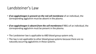
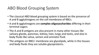
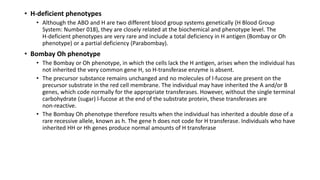
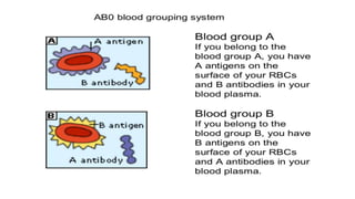
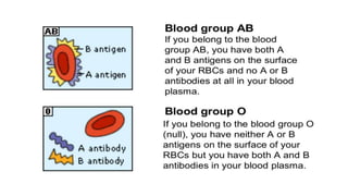
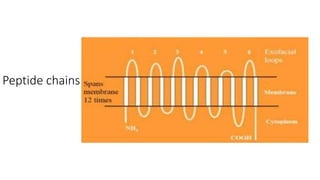
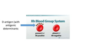
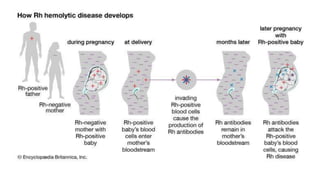
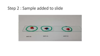
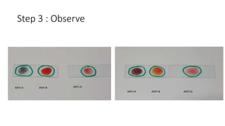
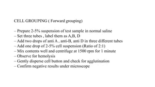
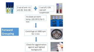
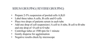
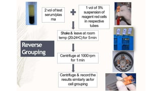
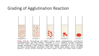
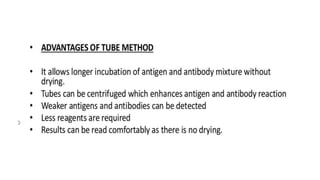
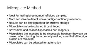
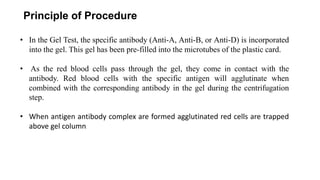
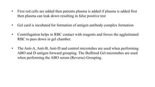
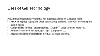
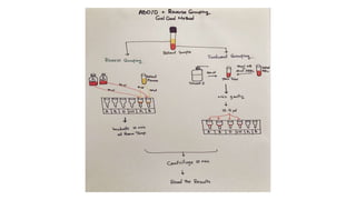
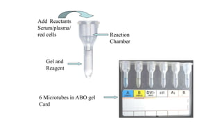
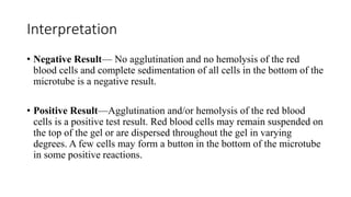
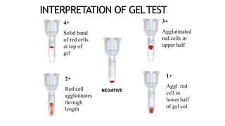
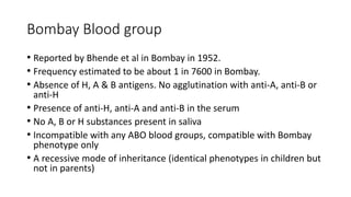
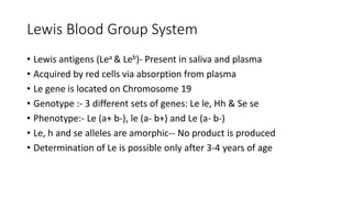
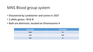
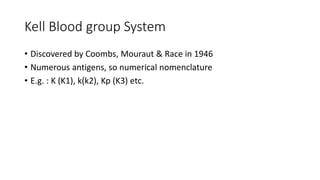
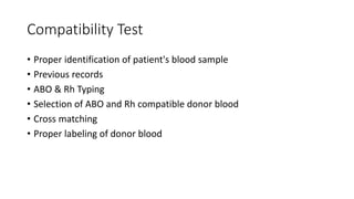
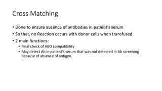
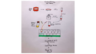
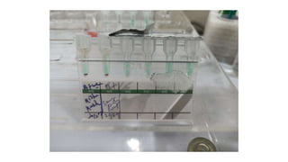
Blood Grouping and Cross
Matching
Dr. Nikhil Sharma
Department of Pathology
Kalinga Institute of Medical Sciences
Bhubaneswar, Odisha
Agglutinogens & Agglutinins
• Agglutinogens refer to the antigens present on the cell membranes of red
blood cells (RBCs). A variety of antigens are present on the cell membrane,
but only a few of them are of practical significance.
• Agglutinins refer to the antibodies against the agglutinogens. These are
present in the plasma.
• Agglutination of RBCs can be caused by the antigens present on their cell
membranes in the presence of suitable agglutinins (antibodies). That is why
these antigens are called agglutinogens.
Blood Grouping
Systems
• 30 blood group systems are
recognized by ISBT
Why do we need Blood Grouping?
• Blood transfusion
• Haemolytic Disease of new born
• Paternity Disputes
• Medico-legal issues
• Disease susceptibility
ABO System
History
• The ABO blood group system discovered by the Austrian scientist
Karl Landsteiner, who found three different blood types in 1900;
he was awarded the Nobel Prize in Physiology or Medicine in 1930
for his work.
• Jan Jansky also pioneered similar classification of blood group but
it remained in obscurity. It is still followed in Russia and former
USSR.
• In America, Moss published his own (very similar) work in 1910.
• Landsteiner described A, B, and O.
• Alfred von Decastello and Adriano Sturli discovered the fourth type,
AB, in 1902.
Landsteiner's Law
• If an agglutinogen is present on the red cell membrane of an individual, the
corresponding agglutinin must be absent in the plasma.
• If an agglutinogen is absent from the cell membraneof RBCs of an individual, the
corresponding agglutinin must be present in the plasma.
• The Landsteiner law is applicable to ABO blood group system only.
• The law is not applicable to other blood group systems because there are no
naturally occurring agglutinins in these systems.
ABO Blood Grouping System
• The classical ABO blood grouping system is based on the presence of
A and B agglutinogens on the cell membrane of RBCs.
• A and B agglutinogens are complex oligosaccharides differing in their
terminal sugars.
• The A and B antigens are also present in many other tissues like
salivary glands, pancreas, kidney, liver, lungs and testis, and also in
body fluids like saliva, semen and amniotic fluid.
• The antigens on RBCs’ membrane are glycolipids, while in the tissues
and body fluids they are soluble glycoproteins.
Production of ABO antigens
• The ABO red cell antigens expressed on the red cells are dependent on the
presence of both the H gene, and the ABO genes.
• The H, A and B genes do not code directly for red cell antigens, but for enzymes
known as transferases.
• The H‐transferase adds the sugar l‐fucose to a precursor substrate, which is a
carbohydrate chain already present on the red cell membrane. Once this has
been performed, the A‐ and B‐transferases can act.
• The A‐transferase adds another sugar called N‐acetyl‐D‐galactosamine, which
results in the expression of A antigen on the red cells.
• Similarly, the B‐transferase adds the sugar D‐galactose and the cells then also
express B antigen. These red cells type as group AB.
• Group A antigen is expressed when H and A transferases are the two
enzymes present; group B antigen is expressed when the H and B
transferases are the enzymes present, and in group O only H
transferase is present.
• The A, B and H antigens are detectable long before birth. The ABH
antigen strength usually peaks at between 2 and 4 years of age and
then remains relatively constant in most individuals.
Gal
Transferase
• H‐deficient phenotypes
• Although the ABO and H are two different blood group systems genetically (H Blood Group
System: Number 018), they are closely related at the biochemical and phenotype level. The
H‐deficient phenotypes are very rare and include a total deficiency in H antigen (Bombay or Oh
phenotype) or a partial deficiency (Parabombay).
• Bombay Oh phenotype
• The Bombay or Oh phenotype, in which the cells lack the H antigen, arises when the individual has
not inherited the very common gene H, so H‐transferase enzyme is absent.
• The precursor substance remains unchanged and no molecules of l‐fucose are present on the
precursor substrate in the red cell membrane. The individual may have inherited the A and/or B
genes, which code normally for the appropriate transferases. However, without the single terminal
carbohydrate (sugar) l‐fucose at the end of the substrate protein, these transferases are
non‐reactive.
• The Bombay Oh phenotype therefore results when the individual has inherited a double dose of a
rare recessive allele, known as h. The gene h does not code for H transferase. Individuals who have
inherited HH or Hh genes produce normal amounts of H transferase
Subgroups
• ABO subgroups differ in amount of antigen present on cell membrane
• A and AB have the following subgroups:
• A1, A2, A1B and A2B
• Depends on reaction with Anti A1 Lectin (Dolichos biflorus)
• 80% of group A and AB are A1 and A1B i.e. they react with Anti A1
• Other rare subgroups: A3, A intermediate, Ax etc.
• Subgroups of B are rare and may be either Bm or Bx
Why are subgroups important?
• Sometimes, people with A2 or A2B have anti A1 Ab in their serum
• This antibody is usually weak and causes no problem
• But it does have an importance in blood transfusion
Secretors and Non secretors
• A, B, H substances are found in all
body secretions (except CSF) in 80%
of individuals
• Ability to secrete these substances
is determined by the presence of
secretor gene (Se) in either
homozygous (SeSe) or heterozygous
(Sese) state.
• Persons secreting A/B and H
substances in saliva are Secretors
Blood Group Substances Secreted
O H
A A & H
B B & H
AB A, B, & H
Oh Nil
Rh Blood group System
• Most important after ABO
• 1939- Levine and Stetson detected irregular Ab in serum of a mother
whose fetus had hemolytic Disease of new born
• 1940- Landsteiner and Weiner raised Ab in rabbits to Rhesus monkey
RBCs which agglutinated red cells of 85% of caucacians
• Antigen was named as “Rh factor”
• Antibody was named as “anti Rh”
Genetics
• Co dominant inheritence
• Chromosome 1
• Three pairs of genes, Cc / Dd / Ee
• Five Antigens: C, c, D, E and e (d produces no antigen)
• D antigen is most prevalant and most immunogenic
• Rh (+) generally means D + and Rh (-) means D (-)
Peptide chains
D-antigen (with
antigenic
determinants
• 'D' positive : D antigen present
• 'D' negative: D antigen absent
• Weak 'D': Density of 'D' antigen is less
• Partial 'D': Part of normal D antigen is missing
• Rh null : Absence of all Rh antigens (More prone to develop Rh
antibodies)
• The importance of weak D lies in the fact that it is much less antigenic
as compared to Rh D and weak D red cells may be destroyed if
transfused to a person having anti D.
• If partial D patient is transfused with D positive red cells, they may
develop anti D to the parts of Ag that is missing
Clinical Significance
• The Rh antigens are highly immunogenic, and most of the Rh
antibodies should be considered as potential causes of hemolytic
transfusion reactions and HDN.
• D antigen, after A and B, is the most important RBC antigen in
transfusion practice.
Transfusion reactions :
• Routine blood typing for Rh D status in both blood donors and
transfusion recipients has reduced the incidence of transfusion
reactions caused by anti-D.
• But sensitization to other Rh antigens can be a problem in transfusion
medicine.
Methods of Blood grouping
• Slide / Tile method
• Tube Method
• Immediate Spin Technique
• Microplate Method
• Gel Card / Solid Phase technology
Slide Testing
 Red cells from the specimen are reacted with reagent antisera(anti
A and anti B).
 Agglutination of red cells indicates presence of corresponding
antigen (agglutinogen) on red cells.
Step 1: Antisera on slide
Step 2 : Sample added to slide
Step 3 : Observe
Tube Testing
• Forward Grouping
• Reverse Grouping
CELL GROUPING ( Forward grouping)
– Prepare 2-5% suspension of test sample in normal saline
– Set three tubes , label them as A,B, D
– Add two drops of anti A , anti-B, anti D in three different tubes
– Add one drop of 2-5% cell suspension (Ratio of 2:1)
– Mix contents well and centrifuge at 1500 rpm for 1 minute
– Observe for hemolysis
– Gently disperse cell button and check for agglutination
– Confirm negative results under microscope
1 vol of 2-5%
red cell
suspension
2 vol of anti- A /
anti-B/ Anti-AB
Forward
Grouping
Incubate at room
temp (20-24oC) for 5
min
Centrifuge at 1000 rpm
for 1 min
Check for agglutination
against well lighted
background
• Prepare 2-5% suspension of pooled cells A,B,O
• Label three tubes A cells, B cells and O cells
• Place two drops of patients serum in each tube
• Add one drop of cell suspension ( A cell to A tube, B cell to B tube
and one drop of O cell to O tube
• Centrifuge tubes at 1500 rpm for 1 minute
• Gently disperse for agglutination
• Negative results check by microscope
SERUMGROUPING(REVERSEGROUPING)
Grading of Agglutination Reaction
Microplate Method
• Ideal for testing large number of blood samples.
• More sensitive to detect weaker antigen-antibody reactions
• Results can be photographed for archival storage
• Microplate can be incubated & centrifuged
• Saves time and cost of disposables and reagents.
• Microplates are intended to be disposable however they can be
reused after cleaning them properly making sure that all foreign
protein are removed.
• Microplates can be adapted for automation
ABO Gel card test
Principle of Procedure
• In the Gel Test, the specific antibody (Anti-A, Anti-B, or Anti-D) is incorporated
into the gel. This gel has been pre-filled into the microtubes of the plastic card.
• As the red blood cells pass through the gel, they come in contact with the
antibody. Red blood cells with the specific antigen will agglutinate when
combined with the corresponding antibody in the gel during the centrifugation
step.
• When antigen antibody complex are formed agglutinated red cells are trapped
above gel column
• First red cells are added then patients plasma is added if plasma is added first
then plasma can leak down resulting in false positive test
• Gel card is incubated for formation of antigen antibody complex formation
• Centrifugation helps in RBC contact with reagents and forces the agglutinated
RBC to pass down in gel chamber.
• The Anti-A, Anti-B, Anti-D and control microtubes are used when performing
ABO and D antigen forward grouping. The Buffered Gel microtubes are used
when performing the ABO serum (Reverse) Grouping.
Uses of Gel Technology
Any immunohaematology test that has haemagglutination as its end point:
• ABO-Rh typing, typing for other blood group systems. Antibody screening and
identification.
• Compatibility testing – crossmatching. DAT/IAT, other Coombs phase test.
• Antibody classification- IgG, IgM, IgA, complement…
• Specialized hematological tests: PNH, Sickle cell anaemia.
Add Reactants
Serum/plasma/
red cells Reaction
Chamber
Gel and
Reagent
6 Microtubes in ABO gel
Card
Procedure
• Bring samples and reagents to room temperature (18–25 °C).
• Label the gel card
• Remove the foil seal.
• Add 50 µL of each of the 0.8% suspensions of reagent A1 and B red blood cells to the
labeled Buffered Gel microtubes
• Dilute the donor or patient red blood delivering 0.5 mL of Diluent into a test tube and
pipet 50 µL whole blood or 25 µL packed red blood cells into the diluent). Mix gently to
resuspend.
• Add 10–12.5 µL of this diluted cells to Anti-A, Anti-B, Anti-D and Control microtubes.
• Centrifuge the gel card
Interpretation
• Negative Result— No agglutination and no hemolysis of the red
blood cells and complete sedimentation of all cells in the bottom of the
microtube is a negative result.
• Positive Result—Agglutination and/or hemolysis of the red blood
cells is a positive test result. Red blood cells may remain suspended on
the top of the gel or are dispersed throughout the gel in varying
degrees. A few cells may form a button in the bottom of the microtube
in some positive reactions.
INTERPRETATION OF GELTEST
4+
Solid band
of red cells
at top of
gel
3+
Agglutinated
red cells in
upper half
2+
Red cell
agglutinates
through
length
1+
Aggl. red
cell in
lower half
of gel col.
NEGATIVE
Advantage of Gel card
• Improved sensitivity and specificity
• Easy to use, simple to read
• Minimal training required
• Reliable, reproducible results
• Easy storage and long shelf life of reagents
• Easy disposal of biodegradable cards
Disadvantage of Gel Card
• Special centrifuge is required to accommodate the microtube cards
• Expensive
• Skilled technical workers to dispense appropriate amount of serum
or red cell suspension
Fully Automated Gel card
GRIFOLS Automated
System for Blood Typing
A fully automated, high-
capacity analyzer designed
to perform pretransfusion
compatibility tests using the
DG Gel card technology.
Efficient
• Manage high-volume workloads efficiently
with automated processes and compatible
products.
• Easy to Use
• Spend less time performing pretransfusion
compatibility tests with intuitive software and
simple design.
• Real-time information about the reagent and
sample status, including system warnings
• High definition color results for results
revision
• Quality control checking at different steps of
the test procedure
• Unique simultaneous perforation and
dispensing technology uses of 100% of the
card wells and avoids cross contamination
• Minimal training required
• Minimal maintenance
Bombay Blood group
• Reported by Bhende et al in Bombay in 1952.
• Frequency estimated to be about 1 in 7600 in Bombay.
• Absence of H, A & B antigens. No agglutination with anti-A, anti-B or
anti-H
• Presence of anti-H, anti-A and anti-B in the serum
• No A, B or H substances present in saliva
• Incompatible with any ABO blood groups, compatible with Bombay
phenotype only
• A recessive mode of inheritance (identical phenotypes in children but
not in parents)
Minor Blood group systems
Lewis Blood Group System
• Lewis antigens (Lea & Leb)- Present in saliva and plasma
• Acquired by red cells via absorption from plasma
• Le gene is located on Chromosome 19
• Genotype :- 3 different sets of genes: Le le, Hh & Se se
• Phenotype:- Le (a+ b-), le (a- b+) and Le (a- b-)
• Le, h and se alleles are amorphic-- No product is produced
• Determination of Le is possible only after 3-4 years of age
MNS Blood group system
• Discovered by Landsteiner and Levine in 1927
• 2 allelic genes:- M & N
• Both are dominant, located on Chromosome 4
Genotype Phenotype
MM M
MN MN
NN N
Kell Blood group System
• Discovered by Coombs, Mouraut & Race in 1946
• Numerous antigens, so numerical nomenclature
• E.g. : K (K1), k(k2), Kp (K3) etc.
Duffy blood group system
• Antigens of this system (Fya & Fyb) are determined by a pair of co
dominanat allele
• 70-75% of Indian population has Fya
• Phenotypes : Fy (a+ b-), Fy (a+ b+), Fy (a- b+), Fy (a- b-)
• Fy (a-b-), common in blacks are resistant to malarial infection. why?
• Because Fya & Fyb act as receptors on RBC membrane for invasion by
vivax merozoites
P blood group system
• Discovered by Landsteiner and Levine in 1927
• we are all either P1 or P2
• Rare phenotypes : P1k, P2k etc
• Anti P1 is found in normal indivduals with P2 phenotypes as IgM type
Compatibility Test
• Proper identification of patient's blood sample
• Previous records
• ABO & Rh Typing
• Selection of ABO and Rh compatible donor blood
• Cross matching
• Proper labeling of donor blood
Cross Matching
• Done to ensure absence of antibodies in patient's serum
• So that, no Reaction occurs with donor cells when transfused
• 2 main functions:
• Final check of ABO compatibility
• May detect Ab in patient's serum that was not detected in Ab screening
because of absence of antigen.
Major and Minor Cross match
• Donor Red Cells + Patient's Serum ==> Major
• Patient's cells + Donor's Serum ==> Minor
• Immediate Spin Crossmatch
• Saline room temperature technique
• Indirect Antiglobulin Crossmatch
• Gel card technique
Immediate Spin / Saline room temperature
Technique
• Rules out ABO grouping error
• Inadequate to detect significant
IgG Antibodies
• Not a good technique for cross
matching
Indirect Antiglobulin Technique
• Widely used
• Procedure
• 2 drops of patient's serum in labelled glass tube
• 1 drop of 2-4% suspension of donor RBCs
• Mix contents and incubate at 37℃for 1 hour
• Centrifuge at 1000 rpm
• Examine for haemolysis or agglutination
• Haemolysis if present, crossmatch is incompatible
• If negative, we continue further
• If negative, wash the cells 4 times with saline and decant completely
• Add one drop of anti human globulin reagent to detect any coating of Ab on
donor red cells
• Centrifuge at 1000 rpm for 1 min
• Look for agglutination and record the results
• If negative, we continue further...
• Add one drop of control IgG coated red cells
• Centrifuge at 1000 rpm for 1 min
• Check for haemolysis/agglutination. If no agglutination, test is invalid. Repeat
the procedure
• Agglutination or haemolysis at any step of the procedure indicates
incompatibility.
• Except after addition of control IgG coated Red cells.
Gel Card technique
Causes of positive result / Incompatibility
• Wrong patient
• Wrong blood group
• An antibody in patient's serum reacting with corresponding antigen
on donor's red cells
• Incompatible with all donors: Antibody against Antigen of high incidence
• Incompatible with only one donor: Ab against Ag of low incidence
• Auto antibody in patient's serum reacting with corresponding
antigens
• Dirty glassware
Blood grouping and Cross matching

Blood grouping and Cross matching

  • 1.
    Blood Grouping andCross Matching Dr. Nikhil Sharma Department of Pathology Kalinga Institute of Medical Sciences Bhubaneswar, Odisha
  • 2.
    Agglutinogens & Agglutinins •Agglutinogens refer to the antigens present on the cell membranes of red blood cells (RBCs). A variety of antigens are present on the cell membrane, but only a few of them are of practical significance. • Agglutinins refer to the antibodies against the agglutinogens. These are present in the plasma. • Agglutination of RBCs can be caused by the antigens present on their cell membranes in the presence of suitable agglutinins (antibodies). That is why these antigens are called agglutinogens.
  • 3.
    Blood Grouping Systems • 30blood group systems are recognized by ISBT
  • 4.
    Why do weneed Blood Grouping? • Blood transfusion • Haemolytic Disease of new born • Paternity Disputes • Medico-legal issues • Disease susceptibility
  • 5.
  • 6.
    History • The ABOblood group system discovered by the Austrian scientist Karl Landsteiner, who found three different blood types in 1900; he was awarded the Nobel Prize in Physiology or Medicine in 1930 for his work. • Jan Jansky also pioneered similar classification of blood group but it remained in obscurity. It is still followed in Russia and former USSR. • In America, Moss published his own (very similar) work in 1910.
  • 7.
    • Landsteiner describedA, B, and O. • Alfred von Decastello and Adriano Sturli discovered the fourth type, AB, in 1902.
  • 8.
    Landsteiner's Law • Ifan agglutinogen is present on the red cell membrane of an individual, the corresponding agglutinin must be absent in the plasma. • If an agglutinogen is absent from the cell membraneof RBCs of an individual, the corresponding agglutinin must be present in the plasma. • The Landsteiner law is applicable to ABO blood group system only. • The law is not applicable to other blood group systems because there are no naturally occurring agglutinins in these systems.
  • 9.
    ABO Blood GroupingSystem • The classical ABO blood grouping system is based on the presence of A and B agglutinogens on the cell membrane of RBCs. • A and B agglutinogens are complex oligosaccharides differing in their terminal sugars. • The A and B antigens are also present in many other tissues like salivary glands, pancreas, kidney, liver, lungs and testis, and also in body fluids like saliva, semen and amniotic fluid. • The antigens on RBCs’ membrane are glycolipids, while in the tissues and body fluids they are soluble glycoproteins.
  • 10.
    Production of ABOantigens • The ABO red cell antigens expressed on the red cells are dependent on the presence of both the H gene, and the ABO genes. • The H, A and B genes do not code directly for red cell antigens, but for enzymes known as transferases. • The H‐transferase adds the sugar l‐fucose to a precursor substrate, which is a carbohydrate chain already present on the red cell membrane. Once this has been performed, the A‐ and B‐transferases can act. • The A‐transferase adds another sugar called N‐acetyl‐D‐galactosamine, which results in the expression of A antigen on the red cells. • Similarly, the B‐transferase adds the sugar D‐galactose and the cells then also express B antigen. These red cells type as group AB.
  • 11.
    • Group Aantigen is expressed when H and A transferases are the two enzymes present; group B antigen is expressed when the H and B transferases are the enzymes present, and in group O only H transferase is present. • The A, B and H antigens are detectable long before birth. The ABH antigen strength usually peaks at between 2 and 4 years of age and then remains relatively constant in most individuals.
  • 12.
  • 14.
    • H‐deficient phenotypes •Although the ABO and H are two different blood group systems genetically (H Blood Group System: Number 018), they are closely related at the biochemical and phenotype level. The H‐deficient phenotypes are very rare and include a total deficiency in H antigen (Bombay or Oh phenotype) or a partial deficiency (Parabombay). • Bombay Oh phenotype • The Bombay or Oh phenotype, in which the cells lack the H antigen, arises when the individual has not inherited the very common gene H, so H‐transferase enzyme is absent. • The precursor substance remains unchanged and no molecules of l‐fucose are present on the precursor substrate in the red cell membrane. The individual may have inherited the A and/or B genes, which code normally for the appropriate transferases. However, without the single terminal carbohydrate (sugar) l‐fucose at the end of the substrate protein, these transferases are non‐reactive. • The Bombay Oh phenotype therefore results when the individual has inherited a double dose of a rare recessive allele, known as h. The gene h does not code for H transferase. Individuals who have inherited HH or Hh genes produce normal amounts of H transferase
  • 17.
    Subgroups • ABO subgroupsdiffer in amount of antigen present on cell membrane • A and AB have the following subgroups: • A1, A2, A1B and A2B • Depends on reaction with Anti A1 Lectin (Dolichos biflorus) • 80% of group A and AB are A1 and A1B i.e. they react with Anti A1 • Other rare subgroups: A3, A intermediate, Ax etc. • Subgroups of B are rare and may be either Bm or Bx
  • 18.
    Why are subgroupsimportant? • Sometimes, people with A2 or A2B have anti A1 Ab in their serum • This antibody is usually weak and causes no problem • But it does have an importance in blood transfusion
  • 19.
    Secretors and Nonsecretors • A, B, H substances are found in all body secretions (except CSF) in 80% of individuals • Ability to secrete these substances is determined by the presence of secretor gene (Se) in either homozygous (SeSe) or heterozygous (Sese) state. • Persons secreting A/B and H substances in saliva are Secretors Blood Group Substances Secreted O H A A & H B B & H AB A, B, & H Oh Nil
  • 20.
    Rh Blood groupSystem • Most important after ABO • 1939- Levine and Stetson detected irregular Ab in serum of a mother whose fetus had hemolytic Disease of new born • 1940- Landsteiner and Weiner raised Ab in rabbits to Rhesus monkey RBCs which agglutinated red cells of 85% of caucacians • Antigen was named as “Rh factor” • Antibody was named as “anti Rh”
  • 21.
    Genetics • Co dominantinheritence • Chromosome 1 • Three pairs of genes, Cc / Dd / Ee • Five Antigens: C, c, D, E and e (d produces no antigen) • D antigen is most prevalant and most immunogenic • Rh (+) generally means D + and Rh (-) means D (-)
  • 22.
  • 23.
  • 24.
    • 'D' positive: D antigen present • 'D' negative: D antigen absent • Weak 'D': Density of 'D' antigen is less • Partial 'D': Part of normal D antigen is missing • Rh null : Absence of all Rh antigens (More prone to develop Rh antibodies)
  • 25.
    • The importanceof weak D lies in the fact that it is much less antigenic as compared to Rh D and weak D red cells may be destroyed if transfused to a person having anti D. • If partial D patient is transfused with D positive red cells, they may develop anti D to the parts of Ag that is missing
  • 26.
    Clinical Significance • TheRh antigens are highly immunogenic, and most of the Rh antibodies should be considered as potential causes of hemolytic transfusion reactions and HDN. • D antigen, after A and B, is the most important RBC antigen in transfusion practice.
  • 27.
    Transfusion reactions : •Routine blood typing for Rh D status in both blood donors and transfusion recipients has reduced the incidence of transfusion reactions caused by anti-D. • But sensitization to other Rh antigens can be a problem in transfusion medicine.
  • 29.
    Methods of Bloodgrouping • Slide / Tile method • Tube Method • Immediate Spin Technique • Microplate Method • Gel Card / Solid Phase technology
  • 30.
    Slide Testing  Redcells from the specimen are reacted with reagent antisera(anti A and anti B).  Agglutination of red cells indicates presence of corresponding antigen (agglutinogen) on red cells.
  • 32.
  • 33.
    Step 2 :Sample added to slide
  • 34.
    Step 3 :Observe
  • 36.
    Tube Testing • ForwardGrouping • Reverse Grouping
  • 37.
    CELL GROUPING (Forward grouping) – Prepare 2-5% suspension of test sample in normal saline – Set three tubes , label them as A,B, D – Add two drops of anti A , anti-B, anti D in three different tubes – Add one drop of 2-5% cell suspension (Ratio of 2:1) – Mix contents well and centrifuge at 1500 rpm for 1 minute – Observe for hemolysis – Gently disperse cell button and check for agglutination – Confirm negative results under microscope
  • 38.
    1 vol of2-5% red cell suspension 2 vol of anti- A / anti-B/ Anti-AB Forward Grouping Incubate at room temp (20-24oC) for 5 min Centrifuge at 1000 rpm for 1 min Check for agglutination against well lighted background
  • 39.
    • Prepare 2-5%suspension of pooled cells A,B,O • Label three tubes A cells, B cells and O cells • Place two drops of patients serum in each tube • Add one drop of cell suspension ( A cell to A tube, B cell to B tube and one drop of O cell to O tube • Centrifuge tubes at 1500 rpm for 1 minute • Gently disperse for agglutination • Negative results check by microscope SERUMGROUPING(REVERSEGROUPING)
  • 41.
  • 43.
    Microplate Method • Idealfor testing large number of blood samples. • More sensitive to detect weaker antigen-antibody reactions • Results can be photographed for archival storage • Microplate can be incubated & centrifuged • Saves time and cost of disposables and reagents. • Microplates are intended to be disposable however they can be reused after cleaning them properly making sure that all foreign protein are removed. • Microplates can be adapted for automation
  • 45.
  • 46.
    Principle of Procedure •In the Gel Test, the specific antibody (Anti-A, Anti-B, or Anti-D) is incorporated into the gel. This gel has been pre-filled into the microtubes of the plastic card. • As the red blood cells pass through the gel, they come in contact with the antibody. Red blood cells with the specific antigen will agglutinate when combined with the corresponding antibody in the gel during the centrifugation step. • When antigen antibody complex are formed agglutinated red cells are trapped above gel column
  • 47.
    • First redcells are added then patients plasma is added if plasma is added first then plasma can leak down resulting in false positive test • Gel card is incubated for formation of antigen antibody complex formation • Centrifugation helps in RBC contact with reagents and forces the agglutinated RBC to pass down in gel chamber. • The Anti-A, Anti-B, Anti-D and control microtubes are used when performing ABO and D antigen forward grouping. The Buffered Gel microtubes are used when performing the ABO serum (Reverse) Grouping.
  • 48.
    Uses of GelTechnology Any immunohaematology test that has haemagglutination as its end point: • ABO-Rh typing, typing for other blood group systems. Antibody screening and identification. • Compatibility testing – crossmatching. DAT/IAT, other Coombs phase test. • Antibody classification- IgG, IgM, IgA, complement… • Specialized hematological tests: PNH, Sickle cell anaemia.
  • 50.
    Add Reactants Serum/plasma/ red cellsReaction Chamber Gel and Reagent 6 Microtubes in ABO gel Card
  • 51.
    Procedure • Bring samplesand reagents to room temperature (18–25 °C). • Label the gel card • Remove the foil seal. • Add 50 µL of each of the 0.8% suspensions of reagent A1 and B red blood cells to the labeled Buffered Gel microtubes • Dilute the donor or patient red blood delivering 0.5 mL of Diluent into a test tube and pipet 50 µL whole blood or 25 µL packed red blood cells into the diluent). Mix gently to resuspend. • Add 10–12.5 µL of this diluted cells to Anti-A, Anti-B, Anti-D and Control microtubes. • Centrifuge the gel card
  • 52.
    Interpretation • Negative Result—No agglutination and no hemolysis of the red blood cells and complete sedimentation of all cells in the bottom of the microtube is a negative result. • Positive Result—Agglutination and/or hemolysis of the red blood cells is a positive test result. Red blood cells may remain suspended on the top of the gel or are dispersed throughout the gel in varying degrees. A few cells may form a button in the bottom of the microtube in some positive reactions.
  • 53.
    INTERPRETATION OF GELTEST 4+ Solidband of red cells at top of gel 3+ Agglutinated red cells in upper half 2+ Red cell agglutinates through length 1+ Aggl. red cell in lower half of gel col. NEGATIVE
  • 54.
    Advantage of Gelcard • Improved sensitivity and specificity • Easy to use, simple to read • Minimal training required • Reliable, reproducible results • Easy storage and long shelf life of reagents • Easy disposal of biodegradable cards
  • 55.
    Disadvantage of GelCard • Special centrifuge is required to accommodate the microtube cards • Expensive • Skilled technical workers to dispense appropriate amount of serum or red cell suspension
  • 56.
  • 57.
    GRIFOLS Automated System forBlood Typing A fully automated, high- capacity analyzer designed to perform pretransfusion compatibility tests using the DG Gel card technology.
  • 58.
    Efficient • Manage high-volumeworkloads efficiently with automated processes and compatible products. • Easy to Use • Spend less time performing pretransfusion compatibility tests with intuitive software and simple design. • Real-time information about the reagent and sample status, including system warnings • High definition color results for results revision • Quality control checking at different steps of the test procedure • Unique simultaneous perforation and dispensing technology uses of 100% of the card wells and avoids cross contamination • Minimal training required • Minimal maintenance
  • 59.
    Bombay Blood group •Reported by Bhende et al in Bombay in 1952. • Frequency estimated to be about 1 in 7600 in Bombay. • Absence of H, A & B antigens. No agglutination with anti-A, anti-B or anti-H • Presence of anti-H, anti-A and anti-B in the serum • No A, B or H substances present in saliva • Incompatible with any ABO blood groups, compatible with Bombay phenotype only • A recessive mode of inheritance (identical phenotypes in children but not in parents)
  • 60.
  • 61.
    Lewis Blood GroupSystem • Lewis antigens (Lea & Leb)- Present in saliva and plasma • Acquired by red cells via absorption from plasma • Le gene is located on Chromosome 19 • Genotype :- 3 different sets of genes: Le le, Hh & Se se • Phenotype:- Le (a+ b-), le (a- b+) and Le (a- b-) • Le, h and se alleles are amorphic-- No product is produced • Determination of Le is possible only after 3-4 years of age
  • 62.
    MNS Blood groupsystem • Discovered by Landsteiner and Levine in 1927 • 2 allelic genes:- M & N • Both are dominant, located on Chromosome 4 Genotype Phenotype MM M MN MN NN N
  • 63.
    Kell Blood groupSystem • Discovered by Coombs, Mouraut & Race in 1946 • Numerous antigens, so numerical nomenclature • E.g. : K (K1), k(k2), Kp (K3) etc.
  • 64.
    Duffy blood groupsystem • Antigens of this system (Fya & Fyb) are determined by a pair of co dominanat allele • 70-75% of Indian population has Fya • Phenotypes : Fy (a+ b-), Fy (a+ b+), Fy (a- b+), Fy (a- b-) • Fy (a-b-), common in blacks are resistant to malarial infection. why? • Because Fya & Fyb act as receptors on RBC membrane for invasion by vivax merozoites
  • 65.
    P blood groupsystem • Discovered by Landsteiner and Levine in 1927 • we are all either P1 or P2 • Rare phenotypes : P1k, P2k etc • Anti P1 is found in normal indivduals with P2 phenotypes as IgM type
  • 66.
    Compatibility Test • Properidentification of patient's blood sample • Previous records • ABO & Rh Typing • Selection of ABO and Rh compatible donor blood • Cross matching • Proper labeling of donor blood
  • 67.
    Cross Matching • Doneto ensure absence of antibodies in patient's serum • So that, no Reaction occurs with donor cells when transfused • 2 main functions: • Final check of ABO compatibility • May detect Ab in patient's serum that was not detected in Ab screening because of absence of antigen.
  • 68.
    Major and MinorCross match • Donor Red Cells + Patient's Serum ==> Major • Patient's cells + Donor's Serum ==> Minor
  • 69.
    • Immediate SpinCrossmatch • Saline room temperature technique • Indirect Antiglobulin Crossmatch • Gel card technique
  • 70.
    Immediate Spin /Saline room temperature Technique • Rules out ABO grouping error • Inadequate to detect significant IgG Antibodies • Not a good technique for cross matching
  • 71.
    Indirect Antiglobulin Technique •Widely used • Procedure • 2 drops of patient's serum in labelled glass tube • 1 drop of 2-4% suspension of donor RBCs • Mix contents and incubate at 37℃for 1 hour • Centrifuge at 1000 rpm • Examine for haemolysis or agglutination • Haemolysis if present, crossmatch is incompatible • If negative, we continue further
  • 72.
    • If negative,wash the cells 4 times with saline and decant completely • Add one drop of anti human globulin reagent to detect any coating of Ab on donor red cells • Centrifuge at 1000 rpm for 1 min • Look for agglutination and record the results • If negative, we continue further... • Add one drop of control IgG coated red cells • Centrifuge at 1000 rpm for 1 min • Check for haemolysis/agglutination. If no agglutination, test is invalid. Repeat the procedure
  • 73.
    • Agglutination orhaemolysis at any step of the procedure indicates incompatibility. • Except after addition of control IgG coated Red cells.
  • 74.
  • 76.
    Causes of positiveresult / Incompatibility • Wrong patient • Wrong blood group • An antibody in patient's serum reacting with corresponding antigen on donor's red cells • Incompatible with all donors: Antibody against Antigen of high incidence • Incompatible with only one donor: Ab against Ag of low incidence • Auto antibody in patient's serum reacting with corresponding antigens • Dirty glassware