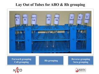
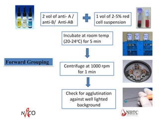
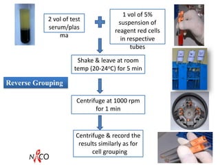
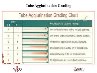
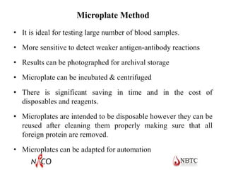
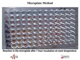
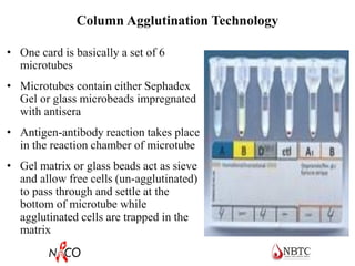
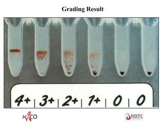
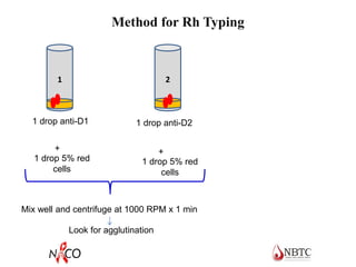
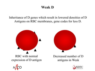
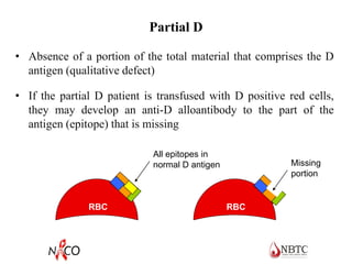
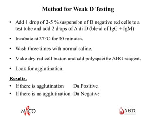
ABO and Rh Blood Grouping & Typing
Teaching Aim
• Understanding inheritance, synthesis, various antigens and
antibodies and their clinical significance in ABO & Rh blood
group systems
• Understanding practical aspects of ABO & Rh blood grouping
Human Blood Groups
• Red cell membranes have antigens (protein / glycoprotein)
on their external surfaces
• These antigens are
o unique to the individual
o recognized as foreign if transfused into another
individual
o promote agglutination of red cells if combine with
antibody
o more than 30 such antigen systems discovered
• Presence or absence of these antigens is used to classify
blood groups
• Major blood groups – ABO & Rh
• Minor blood groups – Kell, Kidd, Duffy etc
ABO Blood Groups
• Most well known & clinically important blood group system.
• Discovered by Karl Landsteiner in 1900
• It was the first to be identified and is the most significant for
transfusion practice
• It is the ONLY system that the reciprocal antibodies are
consistently and predictably present in the sera of people who
have had no exposure to human red cells
• ABO blood group consist of
o two antigens (A & B) on the surface of the RBCs
o two antibodies in the plasma (anti-A & anti-B)
Reciprocal relationship between ABO antigens and
antibodies
Antigens on
RBCs
Antibody in plasma /
serum
Blood group
A Anti-B A
B Anti-A B
AB None AB
None Anti-A, Anti-B O
Development at birth
 All the ABH antigens develop as early as day 37 of fetal life
but do not increase very much in strength during gestational
period
 Red cell of newborn carry 25-50 % of number of antigenic sites
found on adult RBC
 Although cord red cells can be ABO grouped, the reactions may
be a bit weaker than expected
 A or B antigen expression fully developed at 2-4 yrs of age and
remain constant throughout life
Expression of ABO Antigens
 Although the ABO blood group antigens are regarded as
RBC antigens, they are actually expressed on a wide
variety of human tissues and are present on most epithelial
and endothelial cells
 ABH antigens are not only found in humans, but also in
various organisms such as bacteria, plants, and animals
 Present both on red blood cells and in secretions only in
humans and some of the apes (chimpanzee, gorilla)
 In all other mammalian species these substances are found
only in secretions
Anti-A and anti-B antibodies
 Not present in the newborn, appear in the first years of life (4-
6 months usually), reach adult level at 5-10 years of age,
decreases in elderly
 Naturally occurring as they do not need any antigenic stimulus
 However, some food & environmental antigens (bacterial,
viral or plant antigens) are similar enough to A and B
glycoprotein antigens and may stimulate antibody
development
Anti-A and anti-B antibodies (contd…)
 Immunocompetent person react to these antigens by producing
antibodies to those absent from their own system
 Usually IgM, which are not able to pass through the placenta to
the fetal blood circulation
 Anti-A titer from group O > Anti-A titer from group B
 Anti-A titer from group B > Anti-B titer from group A
ABO antigens & corresponding antibodies
'Landsteiner's law : the plasma contains natural
antibodies to A or B, if these antigens are absent
from the red cells of that person
Inheritance of ABO Blood Groups
 Follows Mendelian principles
 Blood group antigens are “codominant”- if the gene is
inherited, it will be expressed.
 There are three allelic genes -A, B & O
 Some aberrant genotypes do occur but they are very rare.
 Understanding of basic inheritance important.
Inheritance of ABO Blood Groups
• Two genes inherited, one from each parent.
• Individual who is A or B may be homozygous or heterozygous
for the antigen.
o Heterozygous: AO or BO
o Homozygous: AA or BB
• Phenotype is the actual expression of the genotype, ie, group A
• Genotype are the actual inherited genes which can only be
determined by family studies, ie, AO.
ABO and H Antigen Genetics
• Genes at three separate loci control the occurrence and
location of ABO antigens
• Presence or absence of the ABH antigens on the red cell
membrane is controlled by the H gene
• Presence or absence of the ABH antigens in secretions is
indirectly controlled by the Se gene
o H gene – H and h alleles (h is an amorph)
o Se gene – Se and se alleles (se is an amorph)
o ABO genes – A, B and O alleles
H Antigen
• The H gene codes for an enzyme (fucosyltransferase) that adds
the sugar fucose to the terminal sugar of a precursor substance
• The precursor substance (proteins and lipids) is formed on an
oligosaccharide chain (the basic structure)
• The H antigen is the foundation upon which A and B antigens
are built
• A and B genes code for enzymes that add an immunodominant
sugar to the H antigen
– Immunodominant sugars are present at the terminal ends of
the chains and confer the ABO antigen specificity
Glucose
Galactose
N-acetylglucosamine
Galactose
Precursor
Substance
(stays the
same)
RBC
RBC precursor substance
Formation of the H antigen
RBC
Glucose
Galactose
N-acetylglucosamine
Galactose
H antigen
Fucose
A and B Antigen
• The “A” gene codes for an enzyme (transferase) that adds N-
acetylgalactosamine to the terminal sugar of the H antigen
o N-acetylgalactosaminyltransferase
• The “B” gene codes for an enzyme that adds D-galactose to
the terminal sugar of the H antigen
o D-galactosyltransferase
Formation of the A antigen
RBC
Glucose
Galactose
N-acetylglucosamine
Galactose
N-acetylgalactosamine
Fucose
Formation of the B antigen
RBC
Glucose
Galactose
N-acetylglucosamine
Galactose
D-Galactose
Fucose
Secretor Status
• A, B, H substances are found in all body secretions (except
CSF) in 80% of individuals
• Ability to secrete these substances is determined by the
presence of secretor gene (Se) in either homozygous (SeSe)
or heterozygous (Sese) state.
Blood Group Substances Secreted
O H
A A & H
B B & H
AB A, B, & H
Oh Nil
Characteristics of Bombay Phenotype
• First reported by Bhende et al in Bombay in 1952.
• Frequency estimated to be about 1 in 7600 in Bombay.
• Absence of H, A & B antigens. No agglutination with anti-A,
anti-B or anti-H
• Presence of anti-H, anti-A and anti-B in the serum
• No A, B or H substances present in saliva
• Incompatible with any ABO blood groups, compatible with
Bombay phenotype only
• A recessive mode of inheritance (identical phenotypes in
children but not in parents)
ABO Subgroups
• ABO subgroups differ in the amount of antigen present on
the red blood cell membrane
o Subgroups have less antigen
• Subgroups are the result of less effective enzymes. They are
not as efficient in converting H antigens to A or B antigens
(fewer antigens are present on the RBC)
• Subgroups of A are more common than subgroups of B
Subgroups of A
• Two principle subgroups of A are: A1 and A2
• Both react strongly with reagent anti-A
• To distinguish A1 from A2 red cells, the lectin Dolichos
biflorus is used (anti-A1)
• 80% of group A or AB individuals are A1 and A1B
• 20% are A2 and A2B
A2 phenotype
• Clinical significance of A2 phenotype
o 8% of A2 and 25% of A2B individuals may produce anti-A1
in the serum
o This may result in discrepancy in blood grouping or
incompatibility in cross match
o However, these anti-A1 antibodies are cold reacting &
therefore may not cause problems routinely.
• Difference between A1 and A2
o It is quantitative
o The A2 gene doesn’t convert the H to A very well resulting
in fewer A2 antigen sites compared to the many A1 antigen
sites
B Subgroups
• B subgroups occur less than A subgroups
• B subgroups are differentiated by the type of reaction
with anti-B, anti-A,B, and anti-H
• B3, Bx, Bm, and Bel
Practical aspects of ABO grouping
• Routine ABO grouping must include both cell & serum
testing as each test serves as a check on the other
• Test should be done at room temperature or lower; testing
at 37oC weakens the reactions
• Tubes, slides should be dry and labeled properly
• Serum should always be added before adding cells
• Results should be recorded immediately after observation
• Hemolysis is interpreted as positive result
Blood Grouping
• There are 2 components to blood typing:
o Test unknown cells with known antibodies
o Test unknown serum/plasma with known red cells
• The patterns are compared and the blood group is
determined.
Blood Sample for Blood Grouping
Blood sample
• Clearly labeled blood samples in sterile tubes (plain & EDTA)
• Test should be performed on the fresh sample for best results.
In case the test can not be performed immediately, sample can
be stored at 4oC & should be tested with in 48 hours
• No signs of hemolysis should be there
• If serum is not completely separated, centrifuge tube at 3000
rpm for 3 min
• Preferably use saline washed red cells and make 2-5%
suspension
Red Cell Suspensions for Blood Grouping
• 2-5%: Test Tube Method
• 0.8-1%: Gel technology
• 1%: Microplate
5 % cell
suspension
for
Tube
grouping
0.8 % cell
suspension
for
Gel card
grouping
Test Tube Method of ABO Grouping
Recommended method
• Allows longer incubation of antigen and antibody
mixture without drying
• Tubes can be centrifuged to enhance reaction
• Can detect weaker antigen / antibody
Two steps in ABO grouping
• Cell grouping (Forward grouping)
– Tests the patients red cells with known Anti-A
& Anti-B to determine the antigen expressed
• Serum grouping (Reverse grouping)
– Test the patients serum with known A & B
cells to determine the presence of antibody
Forward grouping
Cell grouping
Rh grouping
Reverse grouping
Sera grouping
Lay Out of Tubes for ABO & Rh grouping
1 vol of 2-5% red
cell suspension
2 vol of anti- A /
anti-B/ Anti-AB
Forward Grouping
Incubate at room temp
(20-24oC) for 5 min
Centrifuge at 1000 rpm
for 1 min
Check for agglutination
against well lighted
background
2 vol of test
serum/plas
ma
1 vol of 5%
suspension of
reagent red cells
in respective
tubes
Reverse Grouping
Centrifuge at 1000 rpm
for 1 min
Centrifuge & record the
results similarly as for
cell grouping
Shake & leave at room
temp (20-24oC) for 5 min
Tube Agglutination Grading
Microplate Method
• It is ideal for testing large number of blood samples.
• More sensitive to detect weaker antigen-antibody reactions
• Results can be photographed for archival storage
• Microplate can be incubated & centrifuged
• There is significant saving in time and in the cost of
disposables and reagents.
• Microplates are intended to be disposable however they can be
reused after cleaning them properly making sure that all
foreign protein are removed.
• Microplates can be adapted for automation
Reaction in the microplate after 1 hour incubation at room temperature
Microplate Method
Column Agglutination Technology
• One card is basically a set of 6
microtubes
• Microtubes contain either Sephadex
Gel or glass microbeads impregnated
with antisera
• Antigen-antibody reaction takes place
in the reaction chamber of microtube
• Gel matrix or glass beads act as sieve
and allow free cells (un-agglutinated)
to pass through and settle at the
bottom of microtube while
agglutinated cells are trapped in the
matrix
Grading Result
Rh Blood Group System
Rh (D) Antigen
• Of next importance is the Rh type.
o Rh is a blood group system with many antigens, one of which is D.
• Rh refers to the presence or absence of the D antigen on the
red blood cell.
• Unlike the ABO system, individuals who lack the D antigen do
not naturally produce anti-D.
• Production of antibody to D requires exposure to the antigen.
• The D antigen is very immunogenic, i.e., individuals exposed
to it will very likely make an antibody to it.
• For this reason all individuals are typed for D, if negative must
receive Rh (D) negative blood.
Rh (D) Antigen (contd…)
• Rh antigens are an integral part of the red cell membrane.
• They are protein in nature with an active phospholipid
component
• Rh antigens do not exist in the soluble form and, therefore are
not excreted in body fluids.
• Unlike ABO antigens, Rh antigens are present only on red
blood cells. These antigens are not found on other blood cells
including platelets and leukocytes
Rh (D) Antigen (contd…)
• A very potent antigen (50% may form antibody to exposure)
• Frequency in Indian population
o 92-95% Rh positive
• The most important patient population to consider is females of
child-bearing age.
• If immunized to Rh (D) antigen the antibody can cross the
placenta and destroy Rh (D) positive fetal cells resulting in
death.
• This is why Rh negative women are given injection anti-D after
birth of Rh positive baby.
Rh Antibodies
• All Rh antibodies are immune in nature, developed after
immunizing event
• React at 37oC and require anti globulin test to demonstrate the
reaction
• Generally do not react at room temperature in saline
• Most are IgG in nature and therefore can cross the placenta
• Generally, do not fix complement and cause extravascular
hemolysis
• All are important in HDN and delayed HTR
Rh typing
• Normal typing for Rh antigens only includes typing for Rh
(D).
• The result of this typing determines the Rh status of the cells
(Rh - positive or Rh - negative).
• Some Rh typing sera is diluted in high protein solutions and
may require a negative control.
• It is recommended to use two monoclonal anti-D sera from
two different manufacturers labeled as D1 and D2, especially
to confirm all Rh negatives
Monoclonal Anti-D
Three types
1. IgM anti-D monoclonal reagent
2. Blend of IgM and IgG monoclonal antibodies reagent
3. Monoclonal IgG anti-D
 IgM antibodies are highly specific and saline reacting equally at
RT and 370 C but unreliable for detection of weak D
 Blended antibodies are now routinely used and can be used for
detecting weak D
Tube Technique for Rh Typing
 Prepare 5% washed red cell suspension of test sample.
 Take three clean test tubes and label tubes 1 & 2 as “test” and
tube 3 as “control”.
 Place 1 drop of anti-D (D1) in tube 1 and 1 drop of anti-D (D2)
in tube 2.
 Place 1 drop of 22% bovine albumin / control in tube 3.
 Add 1 drop of 5% test cell suspension to each tube.
 Mix well, centrifuge at 1000 rpm for 1 min.
 Resuspend cell button & look for agglutination
 Control tube should show no agglutination
 For all RhD negative test on blood donor, Du test recommended
Method for Rh Typing
1 2
1 drop anti-D1 1 drop anti-D2
1 drop 5% red
cells
1 drop 5% red
cells
+ +
Mix well and centrifuge at 1000 RPM x 1 min
Look for agglutination
Weak D
RBC with normal
expression of D antigen
Decreased number of D
antigens in Weak
Inheritance of D genes which result in lowered densities of D
Antigens on RBC membranes, gene codes for less D.
Partial D
• Absence of a portion of the total material that comprises the D
antigen (qualitative defect)
• If the partial D patient is transfused with D positive red cells,
they may develop an anti-D alloantibody to the part of the
antigen (epitope) that is missing
Missing
portion
RBC RBC
All epitopes in
normal D antigen
Method for Weak D Testing
• Add 1 drop of 2-5 % suspension of D negative red cells to a
test tube and add 2 drops of Anti D (blend of IgG + IgM)
• Incubate at 37C for 30 minutes.
• Wash three times with normal saline.
• Make dry red cell button and add polyspecific AHG reagent.
• Look for agglutination.
Results:
• If there is agglutination Du Positive.
• If there is no agglutination Du Negative.
Significance of Weak D
Donors
• Weak D testing on donors required.
• Labeled as D positive
• But as recipient D negative
Patients
• Weak D testing on patients not required.
• Standard practice to transfuse with D negative
Significance of Weak D
• Weak D is much less antigenic in comparison to D, however,
such red cells may be destroyed if transfused to a patient already
having anti-D. Hence, weak D donor units are labeled as Rh
positive.
• The weak D positive recipients are classified as Rh negative and
safely transfused with Rh negative blood
• Du positive infant can suffer from HDN if the mother possess
anti-D antibodies
• Rh immunoprophylaxis is recommended for the Rh negative
mother if the newborn is Du positive.
Learning Outcome
• You should now be able to perform ABO & Rh grouping on
the donor and recipients sample
• You should be able to resolve discrepancies in the blood
grouping
• You should be able to perform weak D testing if required

5.pdf

  • 2.
    ABO and RhBlood Grouping & Typing
  • 3.
    Teaching Aim • Understandinginheritance, synthesis, various antigens and antibodies and their clinical significance in ABO & Rh blood group systems • Understanding practical aspects of ABO & Rh blood grouping
  • 4.
    Human Blood Groups •Red cell membranes have antigens (protein / glycoprotein) on their external surfaces • These antigens are o unique to the individual o recognized as foreign if transfused into another individual o promote agglutination of red cells if combine with antibody o more than 30 such antigen systems discovered • Presence or absence of these antigens is used to classify blood groups • Major blood groups – ABO & Rh • Minor blood groups – Kell, Kidd, Duffy etc
  • 5.
    ABO Blood Groups •Most well known & clinically important blood group system. • Discovered by Karl Landsteiner in 1900 • It was the first to be identified and is the most significant for transfusion practice • It is the ONLY system that the reciprocal antibodies are consistently and predictably present in the sera of people who have had no exposure to human red cells • ABO blood group consist of o two antigens (A & B) on the surface of the RBCs o two antibodies in the plasma (anti-A & anti-B)
  • 6.
    Reciprocal relationship betweenABO antigens and antibodies Antigens on RBCs Antibody in plasma / serum Blood group A Anti-B A B Anti-A B AB None AB None Anti-A, Anti-B O
  • 7.
    Development at birth All the ABH antigens develop as early as day 37 of fetal life but do not increase very much in strength during gestational period  Red cell of newborn carry 25-50 % of number of antigenic sites found on adult RBC  Although cord red cells can be ABO grouped, the reactions may be a bit weaker than expected  A or B antigen expression fully developed at 2-4 yrs of age and remain constant throughout life
  • 8.
    Expression of ABOAntigens  Although the ABO blood group antigens are regarded as RBC antigens, they are actually expressed on a wide variety of human tissues and are present on most epithelial and endothelial cells  ABH antigens are not only found in humans, but also in various organisms such as bacteria, plants, and animals  Present both on red blood cells and in secretions only in humans and some of the apes (chimpanzee, gorilla)  In all other mammalian species these substances are found only in secretions
  • 9.
    Anti-A and anti-Bantibodies  Not present in the newborn, appear in the first years of life (4- 6 months usually), reach adult level at 5-10 years of age, decreases in elderly  Naturally occurring as they do not need any antigenic stimulus  However, some food & environmental antigens (bacterial, viral or plant antigens) are similar enough to A and B glycoprotein antigens and may stimulate antibody development
  • 10.
    Anti-A and anti-Bantibodies (contd…)  Immunocompetent person react to these antigens by producing antibodies to those absent from their own system  Usually IgM, which are not able to pass through the placenta to the fetal blood circulation  Anti-A titer from group O > Anti-A titer from group B  Anti-A titer from group B > Anti-B titer from group A
  • 11.
    ABO antigens &corresponding antibodies
  • 12.
    'Landsteiner's law :the plasma contains natural antibodies to A or B, if these antigens are absent from the red cells of that person
  • 13.
    Inheritance of ABOBlood Groups  Follows Mendelian principles  Blood group antigens are “codominant”- if the gene is inherited, it will be expressed.  There are three allelic genes -A, B & O  Some aberrant genotypes do occur but they are very rare.  Understanding of basic inheritance important.
  • 14.
    Inheritance of ABOBlood Groups • Two genes inherited, one from each parent. • Individual who is A or B may be homozygous or heterozygous for the antigen. o Heterozygous: AO or BO o Homozygous: AA or BB • Phenotype is the actual expression of the genotype, ie, group A • Genotype are the actual inherited genes which can only be determined by family studies, ie, AO.
  • 15.
    ABO and HAntigen Genetics • Genes at three separate loci control the occurrence and location of ABO antigens • Presence or absence of the ABH antigens on the red cell membrane is controlled by the H gene • Presence or absence of the ABH antigens in secretions is indirectly controlled by the Se gene o H gene – H and h alleles (h is an amorph) o Se gene – Se and se alleles (se is an amorph) o ABO genes – A, B and O alleles
  • 16.
    H Antigen • TheH gene codes for an enzyme (fucosyltransferase) that adds the sugar fucose to the terminal sugar of a precursor substance • The precursor substance (proteins and lipids) is formed on an oligosaccharide chain (the basic structure) • The H antigen is the foundation upon which A and B antigens are built • A and B genes code for enzymes that add an immunodominant sugar to the H antigen – Immunodominant sugars are present at the terminal ends of the chains and confer the ABO antigen specificity
  • 17.
  • 18.
    Formation of theH antigen RBC Glucose Galactose N-acetylglucosamine Galactose H antigen Fucose
  • 19.
    A and BAntigen • The “A” gene codes for an enzyme (transferase) that adds N- acetylgalactosamine to the terminal sugar of the H antigen o N-acetylgalactosaminyltransferase • The “B” gene codes for an enzyme that adds D-galactose to the terminal sugar of the H antigen o D-galactosyltransferase
  • 20.
    Formation of theA antigen RBC Glucose Galactose N-acetylglucosamine Galactose N-acetylgalactosamine Fucose
  • 21.
    Formation of theB antigen RBC Glucose Galactose N-acetylglucosamine Galactose D-Galactose Fucose
  • 22.
    Secretor Status • A,B, H substances are found in all body secretions (except CSF) in 80% of individuals • Ability to secrete these substances is determined by the presence of secretor gene (Se) in either homozygous (SeSe) or heterozygous (Sese) state. Blood Group Substances Secreted O H A A & H B B & H AB A, B, & H Oh Nil
  • 23.
    Characteristics of BombayPhenotype • First reported by Bhende et al in Bombay in 1952. • Frequency estimated to be about 1 in 7600 in Bombay. • Absence of H, A & B antigens. No agglutination with anti-A, anti-B or anti-H • Presence of anti-H, anti-A and anti-B in the serum • No A, B or H substances present in saliva • Incompatible with any ABO blood groups, compatible with Bombay phenotype only • A recessive mode of inheritance (identical phenotypes in children but not in parents)
  • 24.
    ABO Subgroups • ABOsubgroups differ in the amount of antigen present on the red blood cell membrane o Subgroups have less antigen • Subgroups are the result of less effective enzymes. They are not as efficient in converting H antigens to A or B antigens (fewer antigens are present on the RBC) • Subgroups of A are more common than subgroups of B
  • 25.
    Subgroups of A •Two principle subgroups of A are: A1 and A2 • Both react strongly with reagent anti-A • To distinguish A1 from A2 red cells, the lectin Dolichos biflorus is used (anti-A1) • 80% of group A or AB individuals are A1 and A1B • 20% are A2 and A2B
  • 26.
    A2 phenotype • Clinicalsignificance of A2 phenotype o 8% of A2 and 25% of A2B individuals may produce anti-A1 in the serum o This may result in discrepancy in blood grouping or incompatibility in cross match o However, these anti-A1 antibodies are cold reacting & therefore may not cause problems routinely. • Difference between A1 and A2 o It is quantitative o The A2 gene doesn’t convert the H to A very well resulting in fewer A2 antigen sites compared to the many A1 antigen sites
  • 27.
    B Subgroups • Bsubgroups occur less than A subgroups • B subgroups are differentiated by the type of reaction with anti-B, anti-A,B, and anti-H • B3, Bx, Bm, and Bel
  • 28.
    Practical aspects ofABO grouping • Routine ABO grouping must include both cell & serum testing as each test serves as a check on the other • Test should be done at room temperature or lower; testing at 37oC weakens the reactions • Tubes, slides should be dry and labeled properly • Serum should always be added before adding cells • Results should be recorded immediately after observation • Hemolysis is interpreted as positive result
  • 29.
    Blood Grouping • Thereare 2 components to blood typing: o Test unknown cells with known antibodies o Test unknown serum/plasma with known red cells • The patterns are compared and the blood group is determined.
  • 30.
    Blood Sample forBlood Grouping Blood sample • Clearly labeled blood samples in sterile tubes (plain & EDTA) • Test should be performed on the fresh sample for best results. In case the test can not be performed immediately, sample can be stored at 4oC & should be tested with in 48 hours • No signs of hemolysis should be there • If serum is not completely separated, centrifuge tube at 3000 rpm for 3 min • Preferably use saline washed red cells and make 2-5% suspension
  • 31.
    Red Cell Suspensionsfor Blood Grouping • 2-5%: Test Tube Method • 0.8-1%: Gel technology • 1%: Microplate 5 % cell suspension for Tube grouping 0.8 % cell suspension for Gel card grouping
  • 32.
    Test Tube Methodof ABO Grouping Recommended method • Allows longer incubation of antigen and antibody mixture without drying • Tubes can be centrifuged to enhance reaction • Can detect weaker antigen / antibody Two steps in ABO grouping • Cell grouping (Forward grouping) – Tests the patients red cells with known Anti-A & Anti-B to determine the antigen expressed • Serum grouping (Reverse grouping) – Test the patients serum with known A & B cells to determine the presence of antibody
  • 33.
    Forward grouping Cell grouping Rhgrouping Reverse grouping Sera grouping Lay Out of Tubes for ABO & Rh grouping
  • 34.
    1 vol of2-5% red cell suspension 2 vol of anti- A / anti-B/ Anti-AB Forward Grouping Incubate at room temp (20-24oC) for 5 min Centrifuge at 1000 rpm for 1 min Check for agglutination against well lighted background
  • 35.
    2 vol oftest serum/plas ma 1 vol of 5% suspension of reagent red cells in respective tubes Reverse Grouping Centrifuge at 1000 rpm for 1 min Centrifuge & record the results similarly as for cell grouping Shake & leave at room temp (20-24oC) for 5 min
  • 36.
  • 37.
    Microplate Method • Itis ideal for testing large number of blood samples. • More sensitive to detect weaker antigen-antibody reactions • Results can be photographed for archival storage • Microplate can be incubated & centrifuged • There is significant saving in time and in the cost of disposables and reagents. • Microplates are intended to be disposable however they can be reused after cleaning them properly making sure that all foreign protein are removed. • Microplates can be adapted for automation
  • 38.
    Reaction in themicroplate after 1 hour incubation at room temperature Microplate Method
  • 39.
    Column Agglutination Technology •One card is basically a set of 6 microtubes • Microtubes contain either Sephadex Gel or glass microbeads impregnated with antisera • Antigen-antibody reaction takes place in the reaction chamber of microtube • Gel matrix or glass beads act as sieve and allow free cells (un-agglutinated) to pass through and settle at the bottom of microtube while agglutinated cells are trapped in the matrix
  • 40.
  • 41.
  • 42.
    Rh (D) Antigen •Of next importance is the Rh type. o Rh is a blood group system with many antigens, one of which is D. • Rh refers to the presence or absence of the D antigen on the red blood cell. • Unlike the ABO system, individuals who lack the D antigen do not naturally produce anti-D. • Production of antibody to D requires exposure to the antigen. • The D antigen is very immunogenic, i.e., individuals exposed to it will very likely make an antibody to it. • For this reason all individuals are typed for D, if negative must receive Rh (D) negative blood.
  • 43.
    Rh (D) Antigen(contd…) • Rh antigens are an integral part of the red cell membrane. • They are protein in nature with an active phospholipid component • Rh antigens do not exist in the soluble form and, therefore are not excreted in body fluids. • Unlike ABO antigens, Rh antigens are present only on red blood cells. These antigens are not found on other blood cells including platelets and leukocytes
  • 44.
    Rh (D) Antigen(contd…) • A very potent antigen (50% may form antibody to exposure) • Frequency in Indian population o 92-95% Rh positive • The most important patient population to consider is females of child-bearing age. • If immunized to Rh (D) antigen the antibody can cross the placenta and destroy Rh (D) positive fetal cells resulting in death. • This is why Rh negative women are given injection anti-D after birth of Rh positive baby.
  • 45.
    Rh Antibodies • AllRh antibodies are immune in nature, developed after immunizing event • React at 37oC and require anti globulin test to demonstrate the reaction • Generally do not react at room temperature in saline • Most are IgG in nature and therefore can cross the placenta • Generally, do not fix complement and cause extravascular hemolysis • All are important in HDN and delayed HTR
  • 46.
    Rh typing • Normaltyping for Rh antigens only includes typing for Rh (D). • The result of this typing determines the Rh status of the cells (Rh - positive or Rh - negative). • Some Rh typing sera is diluted in high protein solutions and may require a negative control. • It is recommended to use two monoclonal anti-D sera from two different manufacturers labeled as D1 and D2, especially to confirm all Rh negatives
  • 47.
    Monoclonal Anti-D Three types 1.IgM anti-D monoclonal reagent 2. Blend of IgM and IgG monoclonal antibodies reagent 3. Monoclonal IgG anti-D  IgM antibodies are highly specific and saline reacting equally at RT and 370 C but unreliable for detection of weak D  Blended antibodies are now routinely used and can be used for detecting weak D
  • 48.
    Tube Technique forRh Typing  Prepare 5% washed red cell suspension of test sample.  Take three clean test tubes and label tubes 1 & 2 as “test” and tube 3 as “control”.  Place 1 drop of anti-D (D1) in tube 1 and 1 drop of anti-D (D2) in tube 2.  Place 1 drop of 22% bovine albumin / control in tube 3.  Add 1 drop of 5% test cell suspension to each tube.  Mix well, centrifuge at 1000 rpm for 1 min.  Resuspend cell button & look for agglutination  Control tube should show no agglutination  For all RhD negative test on blood donor, Du test recommended
  • 49.
    Method for RhTyping 1 2 1 drop anti-D1 1 drop anti-D2 1 drop 5% red cells 1 drop 5% red cells + + Mix well and centrifuge at 1000 RPM x 1 min Look for agglutination
  • 50.
    Weak D RBC withnormal expression of D antigen Decreased number of D antigens in Weak Inheritance of D genes which result in lowered densities of D Antigens on RBC membranes, gene codes for less D.
  • 51.
    Partial D • Absenceof a portion of the total material that comprises the D antigen (qualitative defect) • If the partial D patient is transfused with D positive red cells, they may develop an anti-D alloantibody to the part of the antigen (epitope) that is missing Missing portion RBC RBC All epitopes in normal D antigen
  • 52.
    Method for WeakD Testing • Add 1 drop of 2-5 % suspension of D negative red cells to a test tube and add 2 drops of Anti D (blend of IgG + IgM) • Incubate at 37C for 30 minutes. • Wash three times with normal saline. • Make dry red cell button and add polyspecific AHG reagent. • Look for agglutination. Results: • If there is agglutination Du Positive. • If there is no agglutination Du Negative.
  • 53.
    Significance of WeakD Donors • Weak D testing on donors required. • Labeled as D positive • But as recipient D negative Patients • Weak D testing on patients not required. • Standard practice to transfuse with D negative
  • 54.
    Significance of WeakD • Weak D is much less antigenic in comparison to D, however, such red cells may be destroyed if transfused to a patient already having anti-D. Hence, weak D donor units are labeled as Rh positive. • The weak D positive recipients are classified as Rh negative and safely transfused with Rh negative blood • Du positive infant can suffer from HDN if the mother possess anti-D antibodies • Rh immunoprophylaxis is recommended for the Rh negative mother if the newborn is Du positive.
  • 55.
    Learning Outcome • Youshould now be able to perform ABO & Rh grouping on the donor and recipients sample • You should be able to resolve discrepancies in the blood grouping • You should be able to perform weak D testing if required