CONTRAINDICATION FOR ARTERIAL PUNCTURE :


 INFECTION AT SITE.
 ALLEN’S TEST NEGATIVE.
 ON ANTICOAGULANT THERAPY.
 SEVERE PERIPHERAL VASCULAR DISEASE.
 DISTAL TO SURGICAL SHUNT.
WHY TO ORDER AN ABG
 Aids in establishing a diagnosis
 Helps guide treatment plan
 Aids in ventilator management
 Improvement in acid/base management
  allows for optimal function of medications
 Acid/base status may alter electrolyte levels
  critical to patient status/care
 Follow up
NORMAL VALUES
 Ph- 7.35-7.45
 Po2- 95mmhg- 100mmhg
 PCO2- 36mmhg -44mmhg
 HCO3- 22 – 26mEq/L
 AG – 8-12
 OG- -10 - +10
EQUATIONS

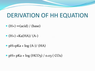
Henderson Hasselbalch equation: pH = 6.1 + log [HCO3-]
                                            0.03( Pco2)


Kassirer-Bleich equation: [H+] = 24 × PCO2 / [HCO3-]
DERIVATION OF HH EQUATION
 (H+) ∞(acid) / (base)


 (H+) =Ka(HA)/ (A-)


 pH=pKa + log (A-)/ (HA)


 pH= pKa + log (HCO3) / 0.03 ( CO2)
HYDROGEN ION CONC AT dif PH
           pH     [H+]

           7.7     20
            7.5     31
           7.4      40
           7.3      50
           7.1      80
           7.0     100
           6.8     160
APPROACH TO ABG
 Acidosis or alkalosis.
 Respiratory or metabolic.
 If respiratory – Acute or chronic.
 If metabolic acidosis – High AG or normal AG
 Is the compensation adequate
 Rule out mixed disorders
STEP 1
PH < 7.35 - ACIDOSIS
PH > 7.45 - ALKALOSIS
STEP 2
             ACID BASE CHANGES
Acid-Base Disorder      Primary Change   Compensatory
 Change

Respiratory acidosis    PCO2 up          HCO3 up
Respiratory alkalosis   PCO2 down        HCO3 down
Metabolic acidosis      HCO3 down        PCO2 down
Metabolic alkalosis     HCO3 up          PCO2 up
Compensation
Primary Disorder            Compensatory Mechanism

 Metabolic acidosis             Increased ventilation


Metabolic alkalosis             Decreased ventilation


Respiratory acidosis    Increased renal reabsorption of HCO3-
                                in the proximal tubule
                         Increased renal excretion of H in the
                                     distal tubule
Respiratory alkalosis   Decreased renal reabsorption of HCO3-
                               in the proximal tubule
                        Decreased renal excretion of H+ in the
                                    distal tubule
The Boston formulae*

State                          Rule          Formula                                               Range

Metabolic acidosisfor acid-base derangementPCOsimply guesstimated using the Boston formulae: + 8
        1.Compensation
                           1.5+8           can be 2
                                                    (mmHg) = 1.5*bicarbonate                           2

Metabolic alkalosis            0.7+20        PCO 2 (mmHg) = 0.7*bicarbonate +20                        5


Acute respiratory                            bicarbonate (mmol/l) drops 2 mmol/l
                               2 for 10                                                            ?
alkalosis                                       for every 10 mmHg PCO 2 drop


Chronic respiratory
                               4 for 10      likewise, but 5 mmol/l                                ?
alkalosis
                                             bicarbonate (mmol/l) increases 1
Acute respiratory
                               1 for 10      mmol/l                                                ?
acidosis
                                                for every 10 mmHg
Chronic respiratory
                               4 for 10      likewise, but 4 mmol/l                                ?
acidosis
HARRISON
METABOLIC ACIDOSIS-

          HCO3 –   1.25 PCO2


METABOLIC ALKALOSIS:

          HCO3-    0.75 PCO2
STEP 4 ANION GAP
 The principle of electroneutrality
 (Na+ + K+) – (Cl- + HCO3- )
 Usually 12-16 mEq/l
 Difference is due to the unmeasured –
  ve charge on the proteins, and SO4- and
  PO4-
 Low albumin will reduce the ‘normal’
  gap
HIGH AG            NORMAL AG

•LACTIC ACIDOSIS   •DIARRHOEA

•UREMIC ACIDOSIS   •FITUL A

•KETO ACIDOSIS     •RTA

•SALISYLSTE        •ACETOZOLAMIDE

•ETHELENE GLYCOL   •MASSIVE NS INFUSION

•ETHANOL           •HYPERALIMENTATION
HAGMA
 SAG increased. (Na – Cl + HCo3)
 The added Acid is buffered by
 Hco3, Hco3 Falls and Anion Gap is increased.
Key Point : Increased Anion Gap means an acid has been
 added to the blood. HAGMA.
NAGMA
 SAG normal.( Na – Cl + HCo3)
 When Hco3 is lost, to maintain electro neutrality
  Cl is conserved by the kidney’s and so Anion Gap is
  normal.
 Key Point : Normal Gap acidosis denotes loss of
  Hco3. Also called hypercholeremic acidosis.
 NAGMA.
HIGH AG            NORMAL AG

•LACTIC ACIDOSIS   •DIARRHOEA

•UREMIC ACIDOSIS   •FITUL A

•KETO ACIDOSIS     •RTA

•SALISYLSTE        •ACETOZOLAMIDE

•ETHELENE GLYCOL   •MASSIVE NS INFUSION

•ETHANOL           •HYPERALIMENTATION
OSMOLAL GAP
 osmolal gap = MO - CO

 MO = Measured Osmolality

 CO = Calculated Osmolality.
  2 x Na + GLU/18 + UN/2.8

     Normal OG = -10 to +10

     An OG value greater than + 14 has traditionally been
    considered a critical value or cutoff.
Urine Anion Gap
 UAG = (UNa +UK) – UCl.
 Normal UAG = -10 to +10.
 If UAG is negative,more than -20
  it is due GI loss.
 If the UAG is positive, more than +10 then it is due
 to renal loss of Hco3.

  UAG is an indirect measure of NH3 secretion in
 the Distal Tubule.
STEP 6
 If the decrease in bicarbonate is more than the rise
 in the AG, concurrent with the AG metabolic
 acidosis there is also a second type of metabolic
 acidosis present, a non-AG metabolic acidosis.
           AG/ HCO3 < 1

 If the decrease in bicarbonate is less than the rise
  in AG, a metabolic alkalosis is concurrently
  present with the AG metabolic acidosis.
            AG/ HCO3 > 1
RESPIRATORY ACIDOSIS
 Upper airway obstruction
 Lower airway obstruction
 Cardiogenic or non-cardiogenic pulmonary edema
 Pneumonia
 Pulmonary emboli
 Fat emboli
 Central nervous system depression
 Neuromascular impairment
 Ventilatory restriction
RESPIRATORY ALKALOSIS
 Central nervous system stimulation: Fever, pain, fear,
    cerebrovascular accident, CNS infection, trauma,
    tumor.
   Hypoxia: High altitude, profound anemia, pulmonary
    disease.
   Stimulation of chest receptors: Pulmonary edema,
    pulmonary emboli, pneumonia, pneumothorax,
    pleural effusion.
   Drugs or hormones : Salicylates,
    medroxyprogesterone, catecholamines.
   Miscellaneous: Sepsis, pregnancy, liver disease,
    hyperthyroidism.
METABOLIC ALKALOSIS
CHLORIDE RESPONSIVE               CHLORIDE RESISTANT
URINE CL < 25                     URINE CL> 25
•DIURETICS                        •HYPERALDOOSTERONE STATE

•CORTICOSTEROIDS                  •CUSHING

•GI LOSS- DIARRHOEA, VILLOUS      •BARTTERS
ADENOMA
                                  •POTTASIUM DEPLETION
•VOMITING
                                  •MASSIVE BLOOD TRANSFUSION
•SUCTION
                                  •Rx – K+ REPLACEMENT

•Rx – 0.9% NS , K+ REPLENIHMENT
TIPS
 Do not interpret any blood gas data for acid-base
  diagnosis without closely examining the serum
  electrolytes: Na+, K+, Cl-,
 Single acid-base disorders do not lead to normal
 blood pH. Although pH can end up in the normal
 range (7.35 - 7.45) with a single mild acid-base
 disorder, a truly normal pH with distinctly
 abnormal HCO3- and PaCO2 invariably suggests
 two or more primary disorders. and CO2.
TIPS
 Simplified rules predict the pH and HCO3- for a
  given change in PaCO2. If the pH or HCO3- is
  higher or lower than expected for the change in
  PaCO2, the patient probably has a metabolic acid-
  base disorder as well.
 In maximally-compensated metabolic acidosis, the
  numerical value of PaCO2 should be the same (or
  close to) as the last two digits of arterial pH. This
  observation reflects the formula for expected
  respiratory compensation in metabolic acidosis:
   Expected PaCO2 = [1.5 x serum hco3] + (8 ± 2)
correction
 Ph< 7.1
 HCO3 < 10
 BICARB DEFICIT
    VOD=Body wt( 0.4 + 2.4 / 5 )
    Correction = Vod ( bicarb defict)
   400 mEq increases Hco3 by 12 mEq
 JAYARAMAN
    AKI
 LATHA
    MYASTHENIA GRAVIS
 LATHA
    IN VENTILLATOR
 SAMUNDESHWARI
 DIABETIC KETO ACIDOSIS
THANK YOU

ABG APPROACH

  • 2.
    CONTRAINDICATION FOR ARTERIALPUNCTURE :  INFECTION AT SITE.  ALLEN’S TEST NEGATIVE.  ON ANTICOAGULANT THERAPY.  SEVERE PERIPHERAL VASCULAR DISEASE.  DISTAL TO SURGICAL SHUNT.
  • 3.
    WHY TO ORDERAN ABG  Aids in establishing a diagnosis  Helps guide treatment plan  Aids in ventilator management  Improvement in acid/base management allows for optimal function of medications  Acid/base status may alter electrolyte levels critical to patient status/care  Follow up
  • 5.
    NORMAL VALUES  Ph-7.35-7.45  Po2- 95mmhg- 100mmhg  PCO2- 36mmhg -44mmhg  HCO3- 22 – 26mEq/L  AG – 8-12  OG- -10 - +10
  • 6.
    EQUATIONS Henderson Hasselbalch equation:pH = 6.1 + log [HCO3-] 0.03( Pco2) Kassirer-Bleich equation: [H+] = 24 × PCO2 / [HCO3-]
  • 7.
    DERIVATION OF HHEQUATION  (H+) ∞(acid) / (base)  (H+) =Ka(HA)/ (A-)  pH=pKa + log (A-)/ (HA)  pH= pKa + log (HCO3) / 0.03 ( CO2)
  • 8.
    HYDROGEN ION CONCAT dif PH pH [H+] 7.7 20 7.5 31 7.4 40 7.3 50 7.1 80 7.0 100 6.8 160
  • 9.
    APPROACH TO ABG Acidosis or alkalosis.  Respiratory or metabolic.  If respiratory – Acute or chronic.  If metabolic acidosis – High AG or normal AG  Is the compensation adequate  Rule out mixed disorders
  • 10.
    STEP 1 PH <7.35 - ACIDOSIS PH > 7.45 - ALKALOSIS
  • 11.
    STEP 2 ACID BASE CHANGES Acid-Base Disorder Primary Change Compensatory Change Respiratory acidosis PCO2 up HCO3 up Respiratory alkalosis PCO2 down HCO3 down Metabolic acidosis HCO3 down PCO2 down Metabolic alkalosis HCO3 up PCO2 up
  • 12.
    Compensation Primary Disorder Compensatory Mechanism Metabolic acidosis Increased ventilation Metabolic alkalosis Decreased ventilation Respiratory acidosis Increased renal reabsorption of HCO3- in the proximal tubule Increased renal excretion of H in the distal tubule Respiratory alkalosis Decreased renal reabsorption of HCO3- in the proximal tubule Decreased renal excretion of H+ in the distal tubule
  • 13.
    The Boston formulae* State Rule Formula Range Metabolic acidosisfor acid-base derangementPCOsimply guesstimated using the Boston formulae: + 8 1.Compensation 1.5+8 can be 2 (mmHg) = 1.5*bicarbonate 2 Metabolic alkalosis 0.7+20 PCO 2 (mmHg) = 0.7*bicarbonate +20 5 Acute respiratory bicarbonate (mmol/l) drops 2 mmol/l 2 for 10 ? alkalosis for every 10 mmHg PCO 2 drop Chronic respiratory 4 for 10 likewise, but 5 mmol/l ? alkalosis bicarbonate (mmol/l) increases 1 Acute respiratory 1 for 10 mmol/l ? acidosis for every 10 mmHg Chronic respiratory 4 for 10 likewise, but 4 mmol/l ? acidosis
  • 14.
    HARRISON METABOLIC ACIDOSIS- HCO3 – 1.25 PCO2 METABOLIC ALKALOSIS: HCO3- 0.75 PCO2
  • 15.
    STEP 4 ANIONGAP  The principle of electroneutrality  (Na+ + K+) – (Cl- + HCO3- )  Usually 12-16 mEq/l  Difference is due to the unmeasured – ve charge on the proteins, and SO4- and PO4-  Low albumin will reduce the ‘normal’ gap
  • 16.
    HIGH AG NORMAL AG •LACTIC ACIDOSIS •DIARRHOEA •UREMIC ACIDOSIS •FITUL A •KETO ACIDOSIS •RTA •SALISYLSTE •ACETOZOLAMIDE •ETHELENE GLYCOL •MASSIVE NS INFUSION •ETHANOL •HYPERALIMENTATION
  • 17.
    HAGMA  SAG increased.(Na – Cl + HCo3)  The added Acid is buffered by Hco3, Hco3 Falls and Anion Gap is increased. Key Point : Increased Anion Gap means an acid has been added to the blood. HAGMA.
  • 18.
    NAGMA  SAG normal.(Na – Cl + HCo3)  When Hco3 is lost, to maintain electro neutrality Cl is conserved by the kidney’s and so Anion Gap is normal.  Key Point : Normal Gap acidosis denotes loss of Hco3. Also called hypercholeremic acidosis.  NAGMA.
  • 19.
    HIGH AG NORMAL AG •LACTIC ACIDOSIS •DIARRHOEA •UREMIC ACIDOSIS •FITUL A •KETO ACIDOSIS •RTA •SALISYLSTE •ACETOZOLAMIDE •ETHELENE GLYCOL •MASSIVE NS INFUSION •ETHANOL •HYPERALIMENTATION
  • 20.
    OSMOLAL GAP  osmolalgap = MO - CO  MO = Measured Osmolality  CO = Calculated Osmolality.  2 x Na + GLU/18 + UN/2.8  Normal OG = -10 to +10  An OG value greater than + 14 has traditionally been considered a critical value or cutoff.
  • 21.
    Urine Anion Gap UAG = (UNa +UK) – UCl.  Normal UAG = -10 to +10.  If UAG is negative,more than -20 it is due GI loss. If the UAG is positive, more than +10 then it is due to renal loss of Hco3. UAG is an indirect measure of NH3 secretion in the Distal Tubule.
  • 22.
    STEP 6  Ifthe decrease in bicarbonate is more than the rise in the AG, concurrent with the AG metabolic acidosis there is also a second type of metabolic acidosis present, a non-AG metabolic acidosis. AG/ HCO3 < 1  If the decrease in bicarbonate is less than the rise in AG, a metabolic alkalosis is concurrently present with the AG metabolic acidosis. AG/ HCO3 > 1
  • 23.
    RESPIRATORY ACIDOSIS  Upperairway obstruction  Lower airway obstruction  Cardiogenic or non-cardiogenic pulmonary edema  Pneumonia  Pulmonary emboli  Fat emboli  Central nervous system depression  Neuromascular impairment  Ventilatory restriction
  • 24.
    RESPIRATORY ALKALOSIS  Centralnervous system stimulation: Fever, pain, fear, cerebrovascular accident, CNS infection, trauma, tumor.  Hypoxia: High altitude, profound anemia, pulmonary disease.  Stimulation of chest receptors: Pulmonary edema, pulmonary emboli, pneumonia, pneumothorax, pleural effusion.  Drugs or hormones : Salicylates, medroxyprogesterone, catecholamines.  Miscellaneous: Sepsis, pregnancy, liver disease, hyperthyroidism.
  • 25.
    METABOLIC ALKALOSIS CHLORIDE RESPONSIVE CHLORIDE RESISTANT URINE CL < 25 URINE CL> 25 •DIURETICS •HYPERALDOOSTERONE STATE •CORTICOSTEROIDS •CUSHING •GI LOSS- DIARRHOEA, VILLOUS •BARTTERS ADENOMA •POTTASIUM DEPLETION •VOMITING •MASSIVE BLOOD TRANSFUSION •SUCTION •Rx – K+ REPLACEMENT •Rx – 0.9% NS , K+ REPLENIHMENT
  • 26.
    TIPS  Do notinterpret any blood gas data for acid-base diagnosis without closely examining the serum electrolytes: Na+, K+, Cl-,  Single acid-base disorders do not lead to normal blood pH. Although pH can end up in the normal range (7.35 - 7.45) with a single mild acid-base disorder, a truly normal pH with distinctly abnormal HCO3- and PaCO2 invariably suggests two or more primary disorders. and CO2.
  • 27.
    TIPS  Simplified rulespredict the pH and HCO3- for a given change in PaCO2. If the pH or HCO3- is higher or lower than expected for the change in PaCO2, the patient probably has a metabolic acid- base disorder as well.  In maximally-compensated metabolic acidosis, the numerical value of PaCO2 should be the same (or close to) as the last two digits of arterial pH. This observation reflects the formula for expected respiratory compensation in metabolic acidosis: Expected PaCO2 = [1.5 x serum hco3] + (8 ± 2)
  • 28.
    correction  Ph< 7.1 HCO3 < 10  BICARB DEFICIT  VOD=Body wt( 0.4 + 2.4 / 5 )  Correction = Vod ( bicarb defict) 400 mEq increases Hco3 by 12 mEq
  • 29.
  • 30.
     LATHA  MYASTHENIA GRAVIS
  • 31.
     LATHA  IN VENTILLATOR
  • 32.
  • 33.
  • 34.