
Recommended
More Related Content
What's hot
What's hot (20)
Viewers also liked
Viewers also liked (20)
Similar to Arterial blood gas analysis
Similar to Arterial blood gas analysis (20)
Recently uploaded
Recently uploaded (20)
Arterial blood gas analysis
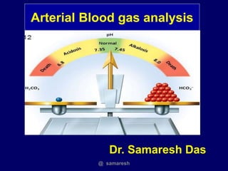
- 1. Arterial Blood gas analysis Dr. Samaresh Das @ samaresh
- 2. Alveolar Ventilation Alveolar Ventilation (V'A) : Gas exchange of lung during normal breathing. o High air exchange in functioning alveoli, higher alveolar ventilation, would bring in fresh oxygen-rich air and efflux carbon dioxide air rapidly o Healthy subjects the partial pressure of alveolar carbon dioxide (PACO2) is equivalent to the arterial carbon dioxide (PaCO2), we substitute the arterial variable for that of the alveolar value @ samaresh
- 3. Oxygenation Oxygenation : Process of adding oxygen to the body system. o clinical signs alone - Cyanosis, pallor and other physical findings are not reliable. o Saturation of peripheral oxygen (SpO2) levels measured with a pulse oximeter correlate highly with arterial oxygenation concentrations (PO2) @ samaresh
- 4. Acid Base Balance @ samaresh
- 5. Basic Terminology PH: Free H+ conc. , PH Inversely related to H+ Acid : Substance that can donate H+, lowers PH Base : Substance that can accept H+, raise PH Acidemia : PH <7.35 , raise H+ Alkalemia : PH >7.45, lower H+ Acidosis : Process/disease , ↓ PH , ↑ acid or ↓ alkali Alkalosis : Process /disease ,↑ PH , ↓ acid or ↑ alkali @ samaresh
- 6. Why Order an ABG? o Aids in establishing a diagnosis o Helps guide treatment plan o Aids in ventilator management o Improvement in acid/base management allows for optimal function of medications o Acid/base status may alter electrolyte levels critical to patient . @ samaresh
- 7. Logistics • Where to place -- the options – Radial – Femoral – Brachial – Dorsalis Pedis • When to Place an arterial line -- – Need for continuous BP monitoring – Need for multiple ABGs @ samaresh
- 8. Technical Errors Air bubbles o PO2 150 mmHg & PCO2 0 mm Hg in air bubble. o Discard sample if excessive air bubbles o Seal with cork/cap after taking sample Fever or Hypothermia o Most ABG analyzers report data at N body temp o If severe hyper/hypothermia, values of pH & PCO2 at 37 C can be significantly diff from pt’s actual values @ samaresh
- 9. Acid Base Status o Assessment via bicarbonate-carbon dioxide buffer system in blood. o H+ = 24 × ( Pco2 / HCO3) o PCO2 / HCo3 identifies the primary acid base disoders and secondary response @ samaresh
- 10. Sample collection o Only the person who collect the sample can tell if he has drawn a pulsating blood o Partly mixed sample- Difficult to recognize Arterial Venous PH 7.35 - 7.45 7.36- 7.39 Pco2 35- 45 44 - 48 Po2 80 - 100 38 - 42 Hco3 24- 26 20 - 24 Sao2 95- 100 % 75% @ samaresh
- 11. Primary Acid Base Disorders & secondary response Primary Disorder Primary change Secondary Response Resp. Acidosis ↑ PcO2 ↑ Hco3 Resp. Alkalosis ↓ Pco2 ↓ Hco3 Met. Acidosis ↓ Hco3 ↓ Pco2 Met. Alkalosis ↑ Hco3 ↑ Pco2 @ samaresh
- 12. During compensation HCO3¯ & PaCO2 move in the same direction @ samaresh
- 13. Compensation Respiratory compensation is always FAST …12-24 hrs Metabolic compensation • is always SLOW...2 - 3 days @ samaresh
- 15. Response to Metabolic Acidosis o Secondary response decrease Pco2 by increase ventilation o Appears in 30-120 minute & can take 12-24 hrs o Expected Paco2 = 40 - ( 1.2 × ∆ Hco3) EXAMPLE: Metabolic acidosis with a plasma HCO3 of 10 mEq/L, ∆ HCO3 is 24 – 10 = 14 mEq/L, so the expected PaCO2 is 40 – (1.2 × 14) = 23 mm Hg. o If the PaCO2 is >23 mm Hg, there is a secondary respiratory acidosis. o If the PaCO2 is <23 mmhg , there is secondary respiratory alkalosis @ samaresh
- 17. o Secondary response : Increase Pco2 by decrease ventilation o This response is not as vigorous as the response to metabolic acidosis o Expected Paco2 = 40 + ( 0.7 × ∆ Hco3 ) o EXAMPLE: Metabolic alkalosis with plasma HCO3 of 40 mEq/L, so ∆ HCO3 is 40 – 24 = 16 mEq/L, o So expected PaCO2 is 40 + ( 0.7 × 16) = 51 mm Hg. Response to Metabolic Alkalosis @ samaresh
- 19. o Secondary response of PaCO2 occurs in the kidneys, o HCO3 absorption in the proximal tubes o Response is relatively slow, take 2 or 3 days to reach completion. o Because of delay, respiratory acid-base disorders are separated into acute and chronic disorders. EXAMPLE: Acute increase in PaCO2 to 60 mm Hg, Expected HCO3 = 24+ (0.1 × ∆ co2 ) For an chronic respiratory acidosis o Expected HCO3 = 24+ (0.4 × ∆ co2 ) Response to Respiratory Acidosis @ samaresh
- 21. EXAMPLE: For an acute decrease in PaCO2 to 24 mm Hg, o Expected HCO3 = 24 - (0.2 × ∆ co2 ) For an chronic decrease in PaCO2 to 20 mm Hg, o Expected HCO3 = 24 - (0.4 × ∆ co2 ) Response to Respiratory Alkalosis @ samaresh
- 22. Stepwise approach to Acid Base analysis @ samaresh
- 23. @ samaresh
- 24. Stepwise approach to Acid Base analysis o Structured, rule-based approach to the diagnosis of primary, secondary, and mixed acid-base disorders using the relationships between the PH, PCO2, and HCO3 Normal Values : o pH = 7.35–7.45 o PCO2 = 35–45 mm Hg o HCO3 = 22–26 mEq/L @ samaresh
- 25. Acidemic pH < 7.35 Alkalemic pH > 7.45 Stepwise approach to Acid Base analysis @ samaresh
- 26. o Stage I: PaCO2 and pH are used to identify the primary acid- base disorder. o Rule 1: If PaCO2 and/or the pH is outside the normal range, there is an acid-base disorder. o Rule 2: If PaCO2 and pH both abnormal, compare the directional change. o 2a. If PaCO2 and pH change in same direction, there is primary metabolic acid-base disorder. o 2b. If PaCO2 and pH change in opposite directions, there is primary respiratory acid-base disorder. Stepwise approach to Acid Base analysis @ samaresh
- 27. EXAMPLE: o pH = 7.23 , PaCO2 = 23 mm Hg. o The pH and PaCO2 , both reduced (indicating a primary metabolic disorder) and the pH acidemic , so the diagnosis is primary metabolic acidosis. Stepwise approach to Acid Base analysis @ samaresh
- 28. Rule 3: If only pH or PaCO2 is abnormal, the condition is mixed metabolic and respiratory disorder o 3a. If PaCO2 is abnormal, the directional change in PaCO2 identifies the type of respiratory disorder (e.g., high PaCO2 indicates a respiratory acidosis), and the opposing metabolic disorder. o 3b. If the pH is abnormal, the directional change in pH identifies the type of metabolic disorder (e.g., low pH indicates a metabolic acidosis) and the opposing respiratory disorder Stepwise approach to Acid Base analysis @ samaresh
- 29. EXAMPLE: o pH = 7.38 and PaCO2 = 55 mm Hg. o Only PaCO2 is abnormal, so there is a mixed metabolic and respiratory disorder. o The PaCO2 is elevated, indicating a respiratory acidosis, so the metabolic disorder must be a metabolic alkalosis o So condition is a mixed respiratory acidosis and metabolic alkalosis. Both disorders are equivalent in severity because the pH is normal Stepwise approach to Acid Base analysis @ samaresh
- 30. Stage II: The goal in Stage II is to determine if there is an additional acid-base disorder. o Rule 4: For primary metabolic disorder, if the measured PaCO2 is higher than expected, there is a secondary respiratory acidosis, and if the measured PaCO2 is less than expected, there is a secondary respiratory alkalosis Stepwise approach to Acid Base analysis @ samaresh
- 31. EXAMPLE: o PaCO2 = 23 mm Hg, the pH = 7.32, and the HCO3 = 16 mEq/L. The pH and PCO2 change in the same direction, indicating a primary metabolic disorder, and the pH is acidemic, so the disorder is a primary metabolic acidosis. o Expected PaCO2 is 40 –1.2×(24 – 16) = 30 mm Hg. o The measured PaCO2 (23 mm Hg) , o so there is an additional respiratory alkalosis. Therefore, this condition is a primary metabolic acidosis with a secondary respiratory alkalosis Stepwise approach to Acid Base analysis @ samaresh
- 32. Rule 5: For a primary respiratory disorder, a normal or near- normal HCO3 indicates that the disorder is acute. Respiratory compensation is always FAST …12-24 hrs Metabolic compensation • is always SLOW...2 - 3 days Stepwise approach to Acid Base analysis @ samaresh
- 33. o Rule 6: Primary respiratory disorder where HCO3 is abnormal, determine the expected HCO3 for a chronic respiratory disorder. o 6a. For chronic respiratory acidosis, if the HCO3 is lower than expected, there is an incomplete renal response, and if the HCO3 is higher than expected, there is a secondary metabolic alkalosis. o 6b. For a chronic respiratory alkalosis, if the HCO3 is higher than expected, there is an incomplete renal response, and if the HCO3 is lower than expected, there is a secondary metabolic acidosis Stepwise approach to Acid Base analysis @ samaresh
- 34. EXAMPLE: PCO2 = 23 mm Hg, pH = 7.54, and the HCO3 = 32 mEq/L. o The PaCO2 and pH change in opposite directions, indicating a primary respiratory disorder, and the pH is alkaline, so the disorder is a primary respiratory alkalosis. o HCO3 is abnormal, indicating this is not an acute respiratory alkalosis. Stepwise approach to Acid Base analysis @ samaresh
- 35. PCO2 = 23 mm Hg, pH = 7.54, and the HCO3 = 37 mEq/L. Chr. Resp. Alk expected HCO3 = 24 + 0.4 ×(40 – 23) = 31 mEq/L. o If measured HCO3 < 31 mEq/L, condition would be a chr. Resp. alkalosis with incomplete renal response o If measured HCO3 is > 31 mEq/L, indicate secondary metabolic alkalosis Stepwise approach to Acid Base analysis @ samaresh
- 36. • ----- XXXX Diagnostics ------ • Blood Gas Report • Measured 37.0 o C • pH 7.523 • pCO2 30.1 mm Hg • pO2 105.3 mm Hg • Calculated Data • HCO3 act 22 mmol / L • O2 Sat 98.3 % • pO2 (A - a) 8 mm Hg D • pO2 (a / A) 0.93 • Entered Data • FiO2 21.0 % Case 1 30 year old female with sudden onset of dyspnea. No Cough or Chest Pain Vitals normal but RR 26, anxious. @ samaresh
- 37. • ----- XXXX Diagnostics ------ • Blood Gas Report • Measured 37.0 o C • pH 7.301 • pCO2 76.2 mm Hg • pO2 45.5 mm Hg • Calculated Data • HCO3 act 35.1 mmol / L • O2 Sat 78% • pO2 (A - a) 9.5 mm Hg D • pO2 (a / A) 0.83 • Entered Data • FiO2 21 % Case 2 60 year old male smoker with progressive respiratory distress and somnolence. @ samaresh
- 38. • ----- XXXX Diagnostics ------ • Blood Gas Report • Measured 37.0 o C • pH 7.23 • pCO2 23 mm Hg • pO2 110.5 mm Hg • Calculated Data • HCO3 act 14 mmol / L • O2 Sat % • pO2 (A - a) mm Hg D • pO2 (a / A) • Entered Data • FiO2 21.0% Case 3 28 year old diabetic with respiratory distress fatigue and loss of appetite. @ samaresh
- 39. @ samaresh
- 40. @ samaresh
- 41. 8) I shall practice gentle mechanical ventilation and not to try bring ABG to perfect normal. 9) I shall treat the patient, not the ABG report. 10) I shall always correlate ABG report clinically. @ samaresh
- 42. @ samaresh
- 43. @ samaresh