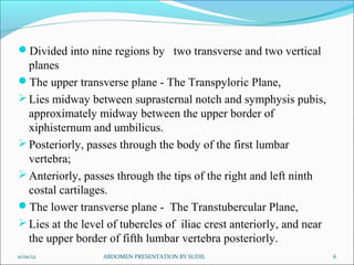
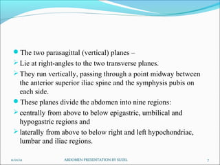
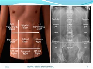
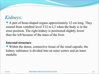
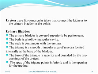
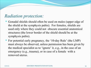
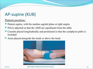
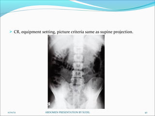
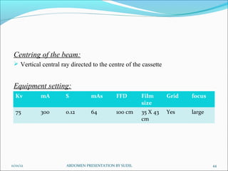
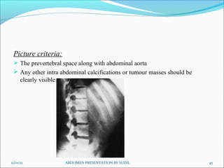
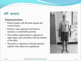
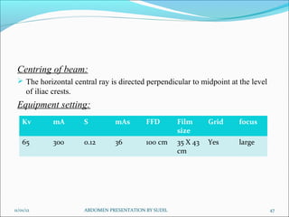
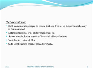
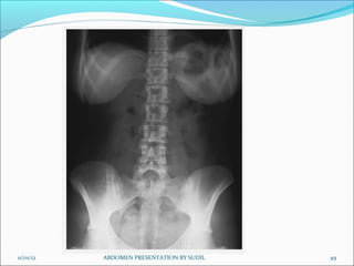
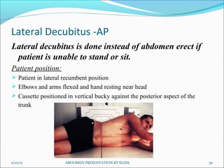
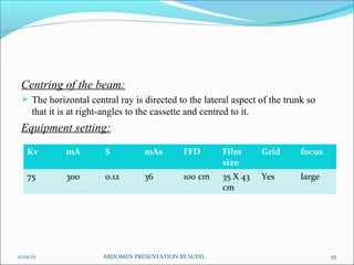
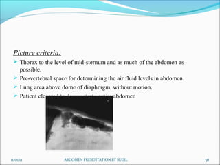
SlideShare a Scribd company logo
By:
Sudil Paudyal
B.Sc. MIT(51)
IOM,MMC
11/01/12 ABDOMEN PRESENTATION BY SUDIL 1
General Anatomy:
The largest cavity of the body,
Bounded
Anteriorly - by abdominal wall muscles
Posteriorly - by the vertebral column and posterior wall
muscles
Laterally - by lower ribs and parts of muscles of abdominal
wall
Superiorly - by the diaphragm
Inferiorly - by pelvic cavity
11/01/12 2
ABDOMEN PRESENTATION BY SUDIL
Abdominal walls:
 Bony support of the abdomen is minimal, consisting only of lumbar
vertebrae and portions of the pelvis (the ilium and the pubis).
 Muscles: Five pairs of muscles form anterior wall:
 Rectus abdominis
 External oblique
 Internal oblique
 Transversus abdominis
Three pairs form the posterior wall:
 Quadratus lumborum
 Psoas major
 Iliacus
Linea alba: A very strong midline tendinous cord, extends from xiphoid
process to symphysis pubis. Divides the anterior abdominal wall
longitudinally into two identical halves.
11/01/12 ABDOMEN PRESENTATION BY SUDIL 3
11/01/12 ABDOMEN PRESENTATION BY SUDIL 4
Fig: Anterior abdominal wall muscles Fig: Posterior abdominal wall muscles
Planes and regions:
Divided either into four quadrants or nine regions
Divided into four quadrants by a transverse and a mid sagittal
plane that intersect at the umbilicus.
Right Upper Quadrant (RUQ),
Right Lower Quadrant (RLQ),
Left Upper Quadrant (LUQ), and
Left Lower Quadrant (LLQ).
11/01/12 ABDOMEN PRESENTATION BY SUDIL 5
Divided into nine regions by two transverse and two vertical
planes
The upper transverse plane - The Transpyloric Plane,
Lies midway between suprasternal notch and symphysis pubis,
approximately midway between the upper border of
xiphisternum and umbilicus.
Posteriorly, passes through the body of the first lumbar
vertebra;
Anteriorly, passes through the tips of the right and left ninth
costal cartilages.
The lower transverse plane - The Transtubercular Plane,
Lies at the level of tubercles of iliac crest anteriorly, and near
the upper border of fifth lumbar vertebra posteriorly.
11/01/12 ABDOMEN PRESENTATION BY SUDIL 6
The two parasagittal (vertical) planes –
Lie at right-angles to the two transverse planes.
They run vertically, passing through a point midway between
the anterior superior iliac spine and the symphysis pubis on
each side.
These planes divide the abdomen into nine regions:
centrally from above to below epigastric, umbilical and
hypogastric regions and
laterally from above to below right and left hypochondriac,
lumbar and iliac regions.
11/01/12 7
ABDOMEN PRESENTATION BY SUDIL
11/01/12 ABDOMEN PRESENTATION BY SUDIL 8
Contents:
contains the greater part of the alimentary tract,
some of the accessory organs to digestion, viz. the liver,
pancreas and spleen,
some of the urinary organs i.e. the kidneys,upper part of the
ureters and the suprarenal glands.
Most of these structures, as well as the wall of the cavity are
more or less covered by an extensive and complicated serous
membrane, the peritoneum.
11/01/12 ABDOMEN PRESENTATION BY SUDIL 9
Fig: organs of anterior part of abdominal cavity
11/01/12 10
ABDOMEN PRESENTATION BY SUDIL
11/01/12 ABDOMEN PRESENTATION BY SUDIL 11
Fig: organs of posterior part of abdominal cavity
Peritoneum:
The serous membrane related to the viscera of the abdominal
cavity.
 Divided into two layers:
 Parietal Layer : Lines the body wall and covers the
retroperitoneal organs.
 Visceral Layer : Composed of two parts :
Covering of the surface of the peritoneal organs.
Mesentery-a double layer of peritoneum that suspends
part of the GI tract from the body wall.
 Peritoneal cavity : The potential space located between the
parietal and visceral layers.
11/01/12 ABDOMEN PRESENTATION BY SUDIL 12
Abdominal Viscera
Viscera are classified as:
Peritoneal organs - have a mesentery and are almost
completely
enclosed in peritoneum. These organs are mobile.
Retroperitoneal organs - are partially covered with peritoneum
and are immobile or fixed organs.
11/01/12 13
ABDOMEN PRESENTATION BY SUDIL
In a nutshell
Major Peritoneal organs: Stomach, Liver and gallbladder,
Spleen, Beginning of duodenum, Tail of pancreas, Jejunum,
Ileum, Appendix , Transverse colon, Sigmoid colon
Major Secondary Retroperitoneal organs: Most of duodenum,
Most of pancreas, Ascending colon ,Descending colon , Upper
rectum
Major Primary Retroperitoneal Organs: Kidney , Adrenal
gland, Ureter, Aorta, Inferior venacava, Lower rectum, Anal
canal
11/01/12 ABDOMEN PRESENTATION BY SUDIL 14
Liver:
Lies mostly in the right hypochondrium, and protected by rib
cage.
Divided into two lobes of unequal size by the falciform
ligament.
Fissures for the ligamentum teres and the ligamentum
venosum, the porta hepatis, and the fossa for the gallbladder
further subdivide the right lobe into the right lobe proper, the
quadrate lobe, and the caudate lobe.
Has a central hilus, or porta hepatis, which receives venous
blood from the portal vein and arterial blood from the hepatic
artery.
11/01/12 ABDOMEN PRESENTATION BY SUDIL 15
The central hilus also transmits the common bile duct, which
collects bile produced by the liver.
These structures, known collectively as the portal triad
The hepatic veins drain the liver by collecting blood from the
liver sinusoids and returning it to the inferior vena cava.
Gallbladder :
 lies in a fossa on the visceral surface of the liver to the right of
the quadrate lobe.
It stores and concentrates bile, which enters and leaves through
the cystic duct. The cystic duct joins the common hepatic duct
to form the common bile duct.
11/01/12 16
ABDOMEN PRESENTATION BY SUDIL
Fig: Liver, turned up to show posterior surface
11/01/12 17
ABDOMEN PRESENTATION BY SUDIL
Pancreas
Most of the pancreas is secondarily retroperitoneal, but the
distal part of the tail of the pancreas remains peritoneal . The
tip of the tail of the pancreas reaches the hilus of the spleen.
Both pancreatic ducts open into the second portion of the
duodenum.
Spleen
a peritoneal organ in the upper left quadrant that is related to
the left 9th, 10th, and 11th ribs. Fracture of these ribs may
lacerate the spleen.
11/01/12 ABDOMEN PRESENTATION BY SUDIL 18
Stomach:
has a lesser curvature, which is connected to the porta hepatis
of the liver by the lesser omentum, and a greater curvature
from which the greater omentum is suspended.
The cardiac region receives the esophagus.
The dome-shaped upper portion of the stomach, which is
normally filled with air, is the fundus.
The main center portion of the stomach is the body.
The pyloric portion of the stomach has a thick muscular wall
and narrow lumen that leads to the duodenum.
11/01/12 ABDOMEN PRESENTATION BY SUDIL 19
11/01/12 ABDOMEN PRESENTATION BY SUDIL 20
Fig: Abd. cavity showing greater and lesser
omentum
Fig:Longitudinal section of stomach
Kidneys and ureters:
Kidney's Relation to the Posterior Abdominal Wall
Both kidneys are in contact with the diaphragm, psoas major,
and quadratus lumborum .
Right kidney-contacts the above structures and the 12th rib.
Left kidney-contacts the above structures and the 11th and
12th ribs
Ureter's Relation to the Posterior Abdominal Wall
The ureter lies on the anterior surface of the psoas major.
11/01/12 ABDOMEN PRESENTATION BY SUDIL 21
11/01/12 ABDOMEN PRESENTATION BY SUDIL 22
Fig: Relation of kidneys and ureters to posterior abdominal wall
Kidneys:
A pair of bean-shaped organs approximately 12 cm long. They
extend from vertebral level T12 to L3 when the body is in the
erect position. The right kidney is positioned slightly lower
than the left because of the mass of the liver.
Internal structure
Within the dense, connective tissue of the renal capsule, the
kidney substance is divided into an outer cortex and an inner
medulla
11/01/12 ABDOMEN PRESENTATION BY SUDIL 23
Cortex-contains glomeruli, Bowman's capsules, and proximal
and distal convoluted tubules. It forms renal columns, which
extend between medullary pyramids.
Medulla--consists of 10 to 18 striated pyramids and contains
collecting ducts and loops of Henle. The apex of each pyramid
ends as a papilla where collecting ducts open.
Calyces-the minor calyces receive one or more papillae and
unite to form major calyces,of which there are two to three per
kidney.
Renal pelvis--the dilated upper portion of the ureter that
receives the major calyces.
11/01/12 24
ABDOMEN PRESENTATION BY SUDIL
11/01/12 ABDOMEN PRESENTATION BY SUDIL 25
Fig: cross section of a kidney
Ureters : are fibro-muscular tubes that connect the kidneys to
the urinary bladder in the pelvis.
Urinary Bladder:
The urinary bladder is covered superiorly by peritoneum.
The body is a hollow muscular cavity.
The neck is continuous with the urethra.
The trigone is a smooth triangular area of mucosa located
internally at the base of the bladder.
The base of the triangle is superior and bounded by the two
openings of the ureters.
 The apex of the trigone points inferiorly and is the opening
for the urethra.
11/01/12 ABDOMEN PRESENTATION BY SUDIL 26
Fig: Kidneys, Ureters and
Bladder
11/01/12 27
ABDOMEN PRESENTATION BY SUDIL
For any body habitus whether hypersthenic or asthenic,
abdominal viscera occupy a lower position:
in inspiration compared with expiration;
in the erect position compared with the recumbent position;
with age and the associated loss of muscle tone.
11/01/12 28
ABDOMEN PRESENTATION BY SUDIL
11/01/12 29
ABDOMEN PRESENTATION BY SUDIL
Radiography:
Preparation:
Careful preliminary patient preparation of the intestinal and
gastric contents is important for a clear view of all the
abdominal structures.
For non-acute conditions, patient preparation is as follows:
(1) Patient placed on a low-residue diet for (2 days) prior to x-
ray examination to prevent formation of gas due to excessive
fermentation of the intestinal contents
(2) Patient should be instructed to take some laxative the night
before the examination.
11/01/12 30
ABDOMEN PRESENTATION BY SUDIL
Exposure technique:
In examinations of the abdomen without a contrast medium, it
is necessary to obtain maximum soft tissue differentiation
throughout its different regions.
 Because of the wide range in thickness of the abdomen and the
delicate differences in physical density between the contained
viscera, it is necessary to use a more critical exposure
technique than is required to demonstrate the difference in
density between an opacified organ and the structures adjacent
to it.
The exposure factors should thus be adjusted to produce a
radiograph with moderate gray tones and less black and white
contrast.
11/01/12 31
ABDOMEN PRESENTATION BY SUDIL
A sharply demonstrated outline of the psoas muscles, lower
border of liver, kidneys ribs and spinous processes of the
lumbar vertebra are the best criteria for judging the quality of
an abdominal radiograph.
High mA and shorter exposure times must be used to freeze
voluntary and involuntary organ movements (breathing and
bowel peristalsis).
Exposure is taken on second full arrested expiration (to
displace diaphragm upward ) to give a better view of the
abdominal structures.
11/01/12 32
ABDOMEN PRESENTATION BY SUDIL
Immobilization:
One of the prime requisite in abdominal examinations is the
prevention of movement, both voluntary and involuntary.
To prevent muscle contraction, the patient must be adjusted
in a comfortable position so that he can relax.
A compression band may be applied across the abdomen for
immobilization but not compression.
The exposure should be made 1-2 sec after suspension of
respiration to allow involuntary movement of viscera to
subside.
11/01/12 33
ABDOMEN PRESENTATION BY SUDIL
Radiation protection:
 Gonadal shields should often be used on males (upper edge of
the shield at the symphysis pubis). For females, shields are
used only where they could not obscure essential anatomical
structures (the lower border of the shield should be at the
symphysis pubis).
 For potential early pregnancy, the ‘10-day Rule’ (the LMP)
must always be observed, unless permission has been given by
the medical specialist as to ‘ignore’ it, e.g., in the case of an
emergency (e.g., trauma), or in case of a female with a
removed uterus.
11/01/12 34
ABDOMEN PRESENTATION BY SUDIL
Radiographic projections:
Basic : Antero-posterior – supine (KUB) (so named because it
includes the kidneys, ureters and bladder).
Alternative: Postero-anterior – prone
Supplementary: Antero-posterior –erect
Anteroposterior – left lateral decubitus
Lateral
Lateral- dorsal decubitus
Anterior and posterior obliques ( for
contrast studies)
11/01/12 ABDOMEN PRESENTATION BY SUDIL 35
Indications:
Bowel obstruction
Perforation
Renal pathology
Acute abdomen
Foreign body localization
Toxic megacolon
Aortic aneurysm
Control or preliminary films for contrast studies
Detection of calcification or abnormal gas collection
11/01/12 ABDOMEN PRESENTATION BY SUDIL 36
AP-supine (KUB)
Patient position:
Patient supine, with the median sagittal plane at right angles
Pelvis adjusted so that the ASIS are equidistant from the table
Cassette placed longitudinally and positioned so that the symphysis pubis is
included
Arms placed alongside the trunk or above the head.
11/01/12 37
ABDOMEN PRESENTATION BY SUDIL
Centering of beam:
 Vertical central ray directed approx. at the level of a point 1 cm below the
line joining the iliac crests.
Equipment setting: ( for screen film combination)
11/01/12 38
ABDOMEN PRESENTATION BY SUDIL
Kv mA S mAs FFD Film
size
Grid focus
65 300 0.12 36 100 cm 35 X 43
cm
Yes large
Picture criteria:
 Whole of abdomen from upper abdomen to symphysis pubis.
 Lateral abdominal wall and the properitoneal fat layer.
 Psoas muscle, lower border of liver and the kidneys.
 Ribs and spinous processes of the lumbar vertebra.
 Whole of the urinary tract should be visualized.
 Bowel gas pattern with minimal unsharpness.
11/01/12 39
ABDOMEN PRESENTATION BY SUDIL
11/01/12 40
ABDOMEN PRESENTATION BY SUDIL
PA- prone
 When kidneys are not of primary interest, PA projection should be used.
 It reduces patient gonad dose compared to the AP projection
Patient position:
 Patient prone, with median sagittal plane at right angles to the table
 Arms up beside the head and both legs extended
11/01/12 ABDOMEN PRESENTATION BY SUDIL 41
11/01/12 ABDOMEN PRESENTATION BY SUDIL 42
 CR, equipment setting, picture criteria same as supine projection.
Lateral:
11/01/12 ABDOMEN PRESENTATION BY SUDIL 43
Position of patient:
Patient turned onto the side of examination, with hands resting near the
head. The hips and knees are flexed for stability.
With the MSP parallel to the table, the vertebral column( abt 8 cm anterior
to the posterior skin surface) positioned over the midline of the table
 Immobilization band applied across the pelvis.
Cassette centered at the level of iliac crests.
Centring of the beam:
 Vertical central ray directed to the centre of the cassette
Equipment setting:
11/01/12 ABDOMEN PRESENTATION BY SUDIL 44
Kv mA S mAs FFD Film
size
Grid focus
75 300 0.12 64 100 cm 35 X 43
cm
Yes large
Picture criteria:
 The prevertebral space along with abdominal aorta
 Any other intra abdominal calcifications or tumour masses should be
clearly visible.
11/01/12 ABDOMEN PRESENTATION BY SUDIL 45
AP -erect
Patient position:
 Patient stands with the back against the
vertical bucky.
 Patient’s legs separated well apart to
maintain a comfortable position.
 The median sagittal plane is adjusted at
right angles and coincident with the midline
of the table.
 The pelvis is adjusted so that the anterior
superior iliac spines are equidistant.
11/01/12 46
ABDOMEN PRESENTATION BY SUDIL
Centring of beam:
 The horizontal central ray is directed perpendicular to midpoint at the level
of iliac crests.
Equipment setting:
11/01/12 47
ABDOMEN PRESENTATION BY SUDIL
Kv mA S mAs FFD Film
size
Grid focus
65 300 0.12 36 100 cm 35 X 43
cm
Yes large
Picture criteria:
 Both domes of diaphragm to ensure that any free air in the peritoneal cavity
is demonstrated.
 Lateral abdominal wall and properitoneal fat
 Psoas muscle, lower border of liver and kidney shadows
 Vertebra in center of film.
 Side identification marker placed properly.
11/01/12 48
ABDOMEN PRESENTATION BY SUDIL
11/01/12 49
ABDOMEN PRESENTATION BY SUDIL
Lateral Decubitus -AP
Lateral decubitus is done instead of abdomen erect if
patient is unable to stand or sit.
Patient position:
 Patient in lateral recumbent position
 Elbows and arms flexed and hand resting near head
 Cassette positioned in vertical bucky against the posterior aspect of the
trunk
11/01/12 50
ABDOMEN PRESENTATION BY SUDIL
Centring of beam:
 The central ray is directed perpendicular to midpoint at the level of iliac
crest with x-ray tube horizontally.
Equipment setting:
 Note: Patient should be placed in lateral decubitus position for 5-10 mins to
allow the free air to rise
11/01/12 51
ABDOMEN PRESENTATION BY SUDIL
Kv mA S mAs FFD Film
size
Grid focus
65 300 0.12 36 100 cm 35 X 43
cm
Yes large
Picture criteria:
 Air fluid levels when an erect abdomen cannot be obtained.
 Lung area above dome of diaphragm
 Lateral abdominal wall and properitoneal fat
 Psoas muscle, lower border of liver and kidney shadows
 No rotation
11/01/12 52
ABDOMEN PRESENTATION BY SUDIL
11/01/12 53
ABDOMEN PRESENTATION BY SUDIL
Lateral dorsal decubitus (supine):
Occasionally, the patient cannot sit or even be rolled on to the
side, in which case the patient remains supine and a lateral
projection is taken using a horizontal central ray.
Patient position:
 Patient supine
 Arms raised away from the abdomen and thorax.
 Cassette positioned vertically against patient’s side
11/01/12 54
ABDOMEN PRESENTATION BY SUDIL
Centring of the beam:
 The horizontal central ray is directed to the lateral aspect of the trunk so
that it is at right-angles to the cassette and centred to it.
Equipment setting:
11/01/12 55
ABDOMEN PRESENTATION BY SUDIL
Kv mA S mAs FFD Film
size
Grid focus
75 300 0.12 36 100 cm 35 X 43
cm
Yes large
Picture criteria:
 Thorax to the level of mid-sternum and as much of the abdomen as
possible.
 Pre-vertebral space for determining the air fluid levels in abdomen.
 Lung area above dome of diaphragm, without motion.
 Patient elevated to demonstrate entire abdomen
11/01/12 56
ABDOMEN PRESENTATION BY SUDIL
References:
Clark’s positioning in radiography, 12th
edition
Merrill’s atlas of radiographic positions and radiologic
procedures, 12th
edition
Different other books and websites
11/01/12 57
ABDOMEN PRESENTATION BY SUDIL
11/01/12 ABDOMEN PRESENTATION BY SUDIL 58
THANK YOU
THANK YOU

More Related Content

Similar to abdomenradiography-121101102111-phpapp02 (1).pdf

THE UPPER FLOOR OF ABDOMINAL CAVITY.pptx
THE UPPER FLOOR OF ABDOMINAL CAVITY.pptxTHE UPPER FLOOR OF ABDOMINAL CAVITY.pptx
THE UPPER FLOOR OF ABDOMINAL CAVITY.pptx
shahajipawale0
 
RENAL ANATOMY.pptx
RENAL ANATOMY.pptxRENAL ANATOMY.pptx
RENAL ANATOMY.pptx
Anees Puthawala
 
small &large intestines .ppsx
small &large intestines  .ppsxsmall &large intestines  .ppsx
small &large intestines .ppsx
ssuser3cccba
 
abdominal cavity and Accessory.pdf
abdominal cavity and Accessory.pdfabdominal cavity and Accessory.pdf
abdominal cavity and Accessory.pdf
BariraAbdulfattah
 
peritoneum
 peritoneum peritoneum
peritoneum
husen123h
 
Anatomy Of The Stomach
Anatomy Of The StomachAnatomy Of The Stomach
Anatomy Of The Stomach
Lisa Brewer
 
The Small & Large Intestine.pptx
The Small & Large Intestine.pptxThe Small & Large Intestine.pptx
The Small & Large Intestine.pptx
Dr Ndayisaba Corneille
 
RENAL ANATOMY2.pptx
RENAL ANATOMY2.pptxRENAL ANATOMY2.pptx
RENAL ANATOMY2.pptx
Anees Puthawala
 
LIVER.pptx
LIVER.pptxLIVER.pptx
LIVER.pptx
JovialLife
 
ERITONEU
ERITONEUERITONEU
ERITONEU
dhruvkathuria8
 
1 Stomach
1  Stomach1  Stomach
1 Stomach
MBBS IMS MSU
 
Kidney
KidneyKidney
Abdomen & pelvis part I
Abdomen & pelvis part IAbdomen & pelvis part I
Abdomen & pelvis part I
SaruGosain
 
Kidney
KidneyKidney
Spleen
SpleenSpleen
Spleen
DibuDivesh1
 
Embryology alimentary system 2
Embryology   alimentary system 2Embryology   alimentary system 2
Embryology alimentary system 2
MBBS IMS MSU
 
Anatomy of the Ruminant Stomach
Anatomy of the Ruminant StomachAnatomy of the Ruminant Stomach
Anatomy of the Ruminant Stomach
Abdelmohaimen Mostafa Saleh
 
12. git[1]
12. git[1]12. git[1]
12. git[1]
MBBS IMS MSU
 
Isha zulfiqar
Isha zulfiqarIsha zulfiqar
Isha zulfiqar
ISHAZULFIQAR1
 
3. digestive system special procedure
3. digestive system special procedure3. digestive system special procedure
3. digestive system special procedure
CHERUDUGASE
 

Similar to abdomenradiography-121101102111-phpapp02 (1).pdf (20)

THE UPPER FLOOR OF ABDOMINAL CAVITY.pptx
THE UPPER FLOOR OF ABDOMINAL CAVITY.pptxTHE UPPER FLOOR OF ABDOMINAL CAVITY.pptx
THE UPPER FLOOR OF ABDOMINAL CAVITY.pptx
 
RENAL ANATOMY.pptx
RENAL ANATOMY.pptxRENAL ANATOMY.pptx
RENAL ANATOMY.pptx
 
small &large intestines .ppsx
small &large intestines  .ppsxsmall &large intestines  .ppsx
small &large intestines .ppsx
 
abdominal cavity and Accessory.pdf
abdominal cavity and Accessory.pdfabdominal cavity and Accessory.pdf
abdominal cavity and Accessory.pdf
 
peritoneum
 peritoneum peritoneum
peritoneum
 
Anatomy Of The Stomach
Anatomy Of The StomachAnatomy Of The Stomach
Anatomy Of The Stomach
 
The Small & Large Intestine.pptx
The Small & Large Intestine.pptxThe Small & Large Intestine.pptx
The Small & Large Intestine.pptx
 
RENAL ANATOMY2.pptx
RENAL ANATOMY2.pptxRENAL ANATOMY2.pptx
RENAL ANATOMY2.pptx
 
LIVER.pptx
LIVER.pptxLIVER.pptx
LIVER.pptx
 
ERITONEU
ERITONEUERITONEU
ERITONEU
 
1 Stomach
1  Stomach1  Stomach
1 Stomach
 
Kidney
KidneyKidney
Kidney
 
Abdomen & pelvis part I
Abdomen & pelvis part IAbdomen & pelvis part I
Abdomen & pelvis part I
 
Kidney
KidneyKidney
Kidney
 
Spleen
SpleenSpleen
Spleen
 
Embryology alimentary system 2
Embryology   alimentary system 2Embryology   alimentary system 2
Embryology alimentary system 2
 
Anatomy of the Ruminant Stomach
Anatomy of the Ruminant StomachAnatomy of the Ruminant Stomach
Anatomy of the Ruminant Stomach
 
12. git[1]
12. git[1]12. git[1]
12. git[1]
 
Isha zulfiqar
Isha zulfiqarIsha zulfiqar
Isha zulfiqar
 
3. digestive system special procedure
3. digestive system special procedure3. digestive system special procedure
3. digestive system special procedure
 

More from VanshikaGarg76

CT COMPONENTS
CT COMPONENTSCT COMPONENTS
CT COMPONENTS
VanshikaGarg76
 
upper limb
upper limbupper limb
upper limb
VanshikaGarg76
 
Imaging During Pregnancy
Imaging During Pregnancy Imaging During Pregnancy
Imaging During Pregnancy
VanshikaGarg76
 
macronutrients and micronutrients
macronutrients and micronutrientsmacronutrients and micronutrients
macronutrients and micronutrients
VanshikaGarg76
 
rdg4011stsem-dentalradiography-180219135013.pdf
rdg4011stsem-dentalradiography-180219135013.pdfrdg4011stsem-dentalradiography-180219135013.pdf
rdg4011stsem-dentalradiography-180219135013.pdf
VanshikaGarg76
 
OPERATION THEATRE RADIOGRAPHY (3)-1.pptx
OPERATION THEATRE RADIOGRAPHY (3)-1.pptxOPERATION THEATRE RADIOGRAPHY (3)-1.pptx
OPERATION THEATRE RADIOGRAPHY (3)-1.pptx
VanshikaGarg76
 
pleuraleffusion-160424141916 (1) (2)-1.pdf
pleuraleffusion-160424141916 (1) (2)-1.pdfpleuraleffusion-160424141916 (1) (2)-1.pdf
pleuraleffusion-160424141916 (1) (2)-1.pdf
VanshikaGarg76
 
pres-131112084302-phpapp01.pdf
pres-131112084302-phpapp01.pdfpres-131112084302-phpapp01.pdf
pres-131112084302-phpapp01.pdf
VanshikaGarg76
 
radiationunits-140511045254-phpapp02.pdf
radiationunits-140511045254-phpapp02.pdfradiationunits-140511045254-phpapp02.pdf
radiationunits-140511045254-phpapp02.pdf
VanshikaGarg76
 
Unit IV - Radio activity-1.pptx
Unit IV - Radio activity-1.pptxUnit IV - Radio activity-1.pptx
Unit IV - Radio activity-1.pptx
VanshikaGarg76
 
mri safety the dos and do nots-1.pptx
mri safety the dos and do nots-1.pptxmri safety the dos and do nots-1.pptx
mri safety the dos and do nots-1.pptx
VanshikaGarg76
 
socialissuesandenvironment-161129185634 (1).pdf
socialissuesandenvironment-161129185634 (1).pdfsocialissuesandenvironment-161129185634 (1).pdf
socialissuesandenvironment-161129185634 (1).pdf
VanshikaGarg76
 
Magnetic_Resonance_(MR)_spectroscopy-1.pptx
Magnetic_Resonance_(MR)_spectroscopy-1.pptxMagnetic_Resonance_(MR)_spectroscopy-1.pptx
Magnetic_Resonance_(MR)_spectroscopy-1.pptx
VanshikaGarg76
 
ultrasonography.pptx
ultrasonography.pptxultrasonography.pptx
ultrasonography.pptx
VanshikaGarg76
 
mammography
mammography mammography
mammography
VanshikaGarg76
 
PET CT-1.pptx
PET CT-1.pptxPET CT-1.pptx
PET CT-1.pptx
VanshikaGarg76
 
mr angiography.pptx
mr angiography.pptxmr angiography.pptx
mr angiography.pptx
VanshikaGarg76
 
GRIDS_TYPES.pptx
GRIDS_TYPES.pptxGRIDS_TYPES.pptx
GRIDS_TYPES.pptx
VanshikaGarg76
 
BASIC PRINCIPLE OF MRI.pptx
BASIC PRINCIPLE OF MRI.pptxBASIC PRINCIPLE OF MRI.pptx
BASIC PRINCIPLE OF MRI.pptx
VanshikaGarg76
 
RADIOGRAPHY CONTRAST MEDIA.pdf
RADIOGRAPHY CONTRAST MEDIA.pdfRADIOGRAPHY CONTRAST MEDIA.pdf
RADIOGRAPHY CONTRAST MEDIA.pdf
VanshikaGarg76
 

More from VanshikaGarg76 (20)

CT COMPONENTS
CT COMPONENTSCT COMPONENTS
CT COMPONENTS
 
upper limb
upper limbupper limb
upper limb
 
Imaging During Pregnancy
Imaging During Pregnancy Imaging During Pregnancy
Imaging During Pregnancy
 
macronutrients and micronutrients
macronutrients and micronutrientsmacronutrients and micronutrients
macronutrients and micronutrients
 
rdg4011stsem-dentalradiography-180219135013.pdf
rdg4011stsem-dentalradiography-180219135013.pdfrdg4011stsem-dentalradiography-180219135013.pdf
rdg4011stsem-dentalradiography-180219135013.pdf
 
OPERATION THEATRE RADIOGRAPHY (3)-1.pptx
OPERATION THEATRE RADIOGRAPHY (3)-1.pptxOPERATION THEATRE RADIOGRAPHY (3)-1.pptx
OPERATION THEATRE RADIOGRAPHY (3)-1.pptx
 
pleuraleffusion-160424141916 (1) (2)-1.pdf
pleuraleffusion-160424141916 (1) (2)-1.pdfpleuraleffusion-160424141916 (1) (2)-1.pdf
pleuraleffusion-160424141916 (1) (2)-1.pdf
 
pres-131112084302-phpapp01.pdf
pres-131112084302-phpapp01.pdfpres-131112084302-phpapp01.pdf
pres-131112084302-phpapp01.pdf
 
radiationunits-140511045254-phpapp02.pdf
radiationunits-140511045254-phpapp02.pdfradiationunits-140511045254-phpapp02.pdf
radiationunits-140511045254-phpapp02.pdf
 
Unit IV - Radio activity-1.pptx
Unit IV - Radio activity-1.pptxUnit IV - Radio activity-1.pptx
Unit IV - Radio activity-1.pptx
 
mri safety the dos and do nots-1.pptx
mri safety the dos and do nots-1.pptxmri safety the dos and do nots-1.pptx
mri safety the dos and do nots-1.pptx
 
socialissuesandenvironment-161129185634 (1).pdf
socialissuesandenvironment-161129185634 (1).pdfsocialissuesandenvironment-161129185634 (1).pdf
socialissuesandenvironment-161129185634 (1).pdf
 
Magnetic_Resonance_(MR)_spectroscopy-1.pptx
Magnetic_Resonance_(MR)_spectroscopy-1.pptxMagnetic_Resonance_(MR)_spectroscopy-1.pptx
Magnetic_Resonance_(MR)_spectroscopy-1.pptx
 
ultrasonography.pptx
ultrasonography.pptxultrasonography.pptx
ultrasonography.pptx
 
mammography
mammography mammography
mammography
 
PET CT-1.pptx
PET CT-1.pptxPET CT-1.pptx
PET CT-1.pptx
 
mr angiography.pptx
mr angiography.pptxmr angiography.pptx
mr angiography.pptx
 
GRIDS_TYPES.pptx
GRIDS_TYPES.pptxGRIDS_TYPES.pptx
GRIDS_TYPES.pptx
 
BASIC PRINCIPLE OF MRI.pptx
BASIC PRINCIPLE OF MRI.pptxBASIC PRINCIPLE OF MRI.pptx
BASIC PRINCIPLE OF MRI.pptx
 
RADIOGRAPHY CONTRAST MEDIA.pdf
RADIOGRAPHY CONTRAST MEDIA.pdfRADIOGRAPHY CONTRAST MEDIA.pdf
RADIOGRAPHY CONTRAST MEDIA.pdf
 

Recently uploaded

Communications Mining Series - Zero to Hero - Session 1
Communications Mining Series - Zero to Hero - Session 1Communications Mining Series - Zero to Hero - Session 1
Communications Mining Series - Zero to Hero - Session 1
DianaGray10
 
Best 20 SEO Techniques To Improve Website Visibility In SERP
Best 20 SEO Techniques To Improve Website Visibility In SERPBest 20 SEO Techniques To Improve Website Visibility In SERP
Best 20 SEO Techniques To Improve Website Visibility In SERP
Pixlogix Infotech
 
GraphSummit Singapore | The Future of Agility: Supercharging Digital Transfor...
GraphSummit Singapore | The Future of Agility: Supercharging Digital Transfor...GraphSummit Singapore | The Future of Agility: Supercharging Digital Transfor...
GraphSummit Singapore | The Future of Agility: Supercharging Digital Transfor...
Neo4j
 
Cosa hanno in comune un mattoncino Lego e la backdoor XZ?
Cosa hanno in comune un mattoncino Lego e la backdoor XZ?Cosa hanno in comune un mattoncino Lego e la backdoor XZ?
Cosa hanno in comune un mattoncino Lego e la backdoor XZ?
Speck&Tech
 
Introduction to CHERI technology - Cybersecurity
Introduction to CHERI technology - CybersecurityIntroduction to CHERI technology - Cybersecurity
Introduction to CHERI technology - Cybersecurity
mikeeftimakis1
 
Uni Systems Copilot event_05062024_C.Vlachos.pdf
Uni Systems Copilot event_05062024_C.Vlachos.pdfUni Systems Copilot event_05062024_C.Vlachos.pdf
Uni Systems Copilot event_05062024_C.Vlachos.pdf
Uni Systems S.M.S.A.
 
GenAI Pilot Implementation in the organizations
GenAI Pilot Implementation in the organizationsGenAI Pilot Implementation in the organizations
GenAI Pilot Implementation in the organizations
kumardaparthi1024
 
HCL Notes and Domino License Cost Reduction in the World of DLAU
HCL Notes and Domino License Cost Reduction in the World of DLAUHCL Notes and Domino License Cost Reduction in the World of DLAU
HCL Notes and Domino License Cost Reduction in the World of DLAU
panagenda
 
Mariano G Tinti - Decoding SpaceX
Mariano G Tinti - Decoding SpaceXMariano G Tinti - Decoding SpaceX
Mariano G Tinti - Decoding SpaceX
Mariano Tinti
 
Why You Should Replace Windows 11 with Nitrux Linux 3.5.0 for enhanced perfor...
Why You Should Replace Windows 11 with Nitrux Linux 3.5.0 for enhanced perfor...Why You Should Replace Windows 11 with Nitrux Linux 3.5.0 for enhanced perfor...
Why You Should Replace Windows 11 with Nitrux Linux 3.5.0 for enhanced perfor...
SOFTTECHHUB
 
GraphRAG for Life Science to increase LLM accuracy
GraphRAG for Life Science to increase LLM accuracyGraphRAG for Life Science to increase LLM accuracy
GraphRAG for Life Science to increase LLM accuracy
Tomaz Bratanic
 
“I’m still / I’m still / Chaining from the Block”
“I’m still / I’m still / Chaining from the Block”“I’m still / I’m still / Chaining from the Block”
“I’m still / I’m still / Chaining from the Block”
Claudio Di Ciccio
 
Microsoft - Power Platform_G.Aspiotis.pdf
Microsoft - Power Platform_G.Aspiotis.pdfMicrosoft - Power Platform_G.Aspiotis.pdf
Microsoft - Power Platform_G.Aspiotis.pdf
Uni Systems S.M.S.A.
 
GraphSummit Singapore | Enhancing Changi Airport Group's Passenger Experience...
GraphSummit Singapore | Enhancing Changi Airport Group's Passenger Experience...GraphSummit Singapore | Enhancing Changi Airport Group's Passenger Experience...
GraphSummit Singapore | Enhancing Changi Airport Group's Passenger Experience...
Neo4j
 
TrustArc Webinar - 2024 Global Privacy Survey
TrustArc Webinar - 2024 Global Privacy SurveyTrustArc Webinar - 2024 Global Privacy Survey
TrustArc Webinar - 2024 Global Privacy Survey
TrustArc
 
Unlock the Future of Search with MongoDB Atlas_ Vector Search Unleashed.pdf
Unlock the Future of Search with MongoDB Atlas_ Vector Search Unleashed.pdfUnlock the Future of Search with MongoDB Atlas_ Vector Search Unleashed.pdf
Unlock the Future of Search with MongoDB Atlas_ Vector Search Unleashed.pdf
Malak Abu Hammad
 
Climate Impact of Software Testing at Nordic Testing Days
Climate Impact of Software Testing at Nordic Testing DaysClimate Impact of Software Testing at Nordic Testing Days
Climate Impact of Software Testing at Nordic Testing Days
Kari Kakkonen
 
Presentation of the OECD Artificial Intelligence Review of Germany
Presentation of the OECD Artificial Intelligence Review of GermanyPresentation of the OECD Artificial Intelligence Review of Germany
Presentation of the OECD Artificial Intelligence Review of Germany
innovationoecd
 
20240607 QFM018 Elixir Reading List May 2024
20240607 QFM018 Elixir Reading List May 202420240607 QFM018 Elixir Reading List May 2024
20240607 QFM018 Elixir Reading List May 2024
Matthew Sinclair
 
Full-RAG: A modern architecture for hyper-personalization
Full-RAG: A modern architecture for hyper-personalizationFull-RAG: A modern architecture for hyper-personalization
Full-RAG: A modern architecture for hyper-personalization
Zilliz
 

Recently uploaded (20)

Communications Mining Series - Zero to Hero - Session 1
Communications Mining Series - Zero to Hero - Session 1Communications Mining Series - Zero to Hero - Session 1
Communications Mining Series - Zero to Hero - Session 1
 
Best 20 SEO Techniques To Improve Website Visibility In SERP
Best 20 SEO Techniques To Improve Website Visibility In SERPBest 20 SEO Techniques To Improve Website Visibility In SERP
Best 20 SEO Techniques To Improve Website Visibility In SERP
 
GraphSummit Singapore | The Future of Agility: Supercharging Digital Transfor...
GraphSummit Singapore | The Future of Agility: Supercharging Digital Transfor...GraphSummit Singapore | The Future of Agility: Supercharging Digital Transfor...
GraphSummit Singapore | The Future of Agility: Supercharging Digital Transfor...
 
Cosa hanno in comune un mattoncino Lego e la backdoor XZ?
Cosa hanno in comune un mattoncino Lego e la backdoor XZ?Cosa hanno in comune un mattoncino Lego e la backdoor XZ?
Cosa hanno in comune un mattoncino Lego e la backdoor XZ?
 
Introduction to CHERI technology - Cybersecurity
Introduction to CHERI technology - CybersecurityIntroduction to CHERI technology - Cybersecurity
Introduction to CHERI technology - Cybersecurity
 
Uni Systems Copilot event_05062024_C.Vlachos.pdf
Uni Systems Copilot event_05062024_C.Vlachos.pdfUni Systems Copilot event_05062024_C.Vlachos.pdf
Uni Systems Copilot event_05062024_C.Vlachos.pdf
 
GenAI Pilot Implementation in the organizations
GenAI Pilot Implementation in the organizationsGenAI Pilot Implementation in the organizations
GenAI Pilot Implementation in the organizations
 
HCL Notes and Domino License Cost Reduction in the World of DLAU
HCL Notes and Domino License Cost Reduction in the World of DLAUHCL Notes and Domino License Cost Reduction in the World of DLAU
HCL Notes and Domino License Cost Reduction in the World of DLAU
 
Mariano G Tinti - Decoding SpaceX
Mariano G Tinti - Decoding SpaceXMariano G Tinti - Decoding SpaceX
Mariano G Tinti - Decoding SpaceX
 
Why You Should Replace Windows 11 with Nitrux Linux 3.5.0 for enhanced perfor...
Why You Should Replace Windows 11 with Nitrux Linux 3.5.0 for enhanced perfor...Why You Should Replace Windows 11 with Nitrux Linux 3.5.0 for enhanced perfor...
Why You Should Replace Windows 11 with Nitrux Linux 3.5.0 for enhanced perfor...
 
GraphRAG for Life Science to increase LLM accuracy
GraphRAG for Life Science to increase LLM accuracyGraphRAG for Life Science to increase LLM accuracy
GraphRAG for Life Science to increase LLM accuracy
 
“I’m still / I’m still / Chaining from the Block”
“I’m still / I’m still / Chaining from the Block”“I’m still / I’m still / Chaining from the Block”
“I’m still / I’m still / Chaining from the Block”
 
Microsoft - Power Platform_G.Aspiotis.pdf
Microsoft - Power Platform_G.Aspiotis.pdfMicrosoft - Power Platform_G.Aspiotis.pdf
Microsoft - Power Platform_G.Aspiotis.pdf
 
GraphSummit Singapore | Enhancing Changi Airport Group's Passenger Experience...
GraphSummit Singapore | Enhancing Changi Airport Group's Passenger Experience...GraphSummit Singapore | Enhancing Changi Airport Group's Passenger Experience...
GraphSummit Singapore | Enhancing Changi Airport Group's Passenger Experience...
 
TrustArc Webinar - 2024 Global Privacy Survey
TrustArc Webinar - 2024 Global Privacy SurveyTrustArc Webinar - 2024 Global Privacy Survey
TrustArc Webinar - 2024 Global Privacy Survey
 
Unlock the Future of Search with MongoDB Atlas_ Vector Search Unleashed.pdf
Unlock the Future of Search with MongoDB Atlas_ Vector Search Unleashed.pdfUnlock the Future of Search with MongoDB Atlas_ Vector Search Unleashed.pdf
Unlock the Future of Search with MongoDB Atlas_ Vector Search Unleashed.pdf
 
Climate Impact of Software Testing at Nordic Testing Days
Climate Impact of Software Testing at Nordic Testing DaysClimate Impact of Software Testing at Nordic Testing Days
Climate Impact of Software Testing at Nordic Testing Days
 
Presentation of the OECD Artificial Intelligence Review of Germany
Presentation of the OECD Artificial Intelligence Review of GermanyPresentation of the OECD Artificial Intelligence Review of Germany
Presentation of the OECD Artificial Intelligence Review of Germany
 
20240607 QFM018 Elixir Reading List May 2024
20240607 QFM018 Elixir Reading List May 202420240607 QFM018 Elixir Reading List May 2024
20240607 QFM018 Elixir Reading List May 2024
 
Full-RAG: A modern architecture for hyper-personalization
Full-RAG: A modern architecture for hyper-personalizationFull-RAG: A modern architecture for hyper-personalization
Full-RAG: A modern architecture for hyper-personalization
 

abdomenradiography-121101102111-phpapp02 (1).pdf

  • 1. By: Sudil Paudyal B.Sc. MIT(51) IOM,MMC 11/01/12 ABDOMEN PRESENTATION BY SUDIL 1
  • 2. General Anatomy: The largest cavity of the body, Bounded Anteriorly - by abdominal wall muscles Posteriorly - by the vertebral column and posterior wall muscles Laterally - by lower ribs and parts of muscles of abdominal wall Superiorly - by the diaphragm Inferiorly - by pelvic cavity 11/01/12 2 ABDOMEN PRESENTATION BY SUDIL
  • 3. Abdominal walls:  Bony support of the abdomen is minimal, consisting only of lumbar vertebrae and portions of the pelvis (the ilium and the pubis).  Muscles: Five pairs of muscles form anterior wall:  Rectus abdominis  External oblique  Internal oblique  Transversus abdominis Three pairs form the posterior wall:  Quadratus lumborum  Psoas major  Iliacus Linea alba: A very strong midline tendinous cord, extends from xiphoid process to symphysis pubis. Divides the anterior abdominal wall longitudinally into two identical halves. 11/01/12 ABDOMEN PRESENTATION BY SUDIL 3
  • 4. 11/01/12 ABDOMEN PRESENTATION BY SUDIL 4 Fig: Anterior abdominal wall muscles Fig: Posterior abdominal wall muscles
  • 5. Planes and regions: Divided either into four quadrants or nine regions Divided into four quadrants by a transverse and a mid sagittal plane that intersect at the umbilicus. Right Upper Quadrant (RUQ), Right Lower Quadrant (RLQ), Left Upper Quadrant (LUQ), and Left Lower Quadrant (LLQ). 11/01/12 ABDOMEN PRESENTATION BY SUDIL 5
  • 6. Divided into nine regions by two transverse and two vertical planes The upper transverse plane - The Transpyloric Plane, Lies midway between suprasternal notch and symphysis pubis, approximately midway between the upper border of xiphisternum and umbilicus. Posteriorly, passes through the body of the first lumbar vertebra; Anteriorly, passes through the tips of the right and left ninth costal cartilages. The lower transverse plane - The Transtubercular Plane, Lies at the level of tubercles of iliac crest anteriorly, and near the upper border of fifth lumbar vertebra posteriorly. 11/01/12 ABDOMEN PRESENTATION BY SUDIL 6
  • 7. The two parasagittal (vertical) planes – Lie at right-angles to the two transverse planes. They run vertically, passing through a point midway between the anterior superior iliac spine and the symphysis pubis on each side. These planes divide the abdomen into nine regions: centrally from above to below epigastric, umbilical and hypogastric regions and laterally from above to below right and left hypochondriac, lumbar and iliac regions. 11/01/12 7 ABDOMEN PRESENTATION BY SUDIL
  • 9. Contents: contains the greater part of the alimentary tract, some of the accessory organs to digestion, viz. the liver, pancreas and spleen, some of the urinary organs i.e. the kidneys,upper part of the ureters and the suprarenal glands. Most of these structures, as well as the wall of the cavity are more or less covered by an extensive and complicated serous membrane, the peritoneum. 11/01/12 ABDOMEN PRESENTATION BY SUDIL 9
  • 10. Fig: organs of anterior part of abdominal cavity 11/01/12 10 ABDOMEN PRESENTATION BY SUDIL
  • 11. 11/01/12 ABDOMEN PRESENTATION BY SUDIL 11 Fig: organs of posterior part of abdominal cavity
  • 12. Peritoneum: The serous membrane related to the viscera of the abdominal cavity.  Divided into two layers:  Parietal Layer : Lines the body wall and covers the retroperitoneal organs.  Visceral Layer : Composed of two parts : Covering of the surface of the peritoneal organs. Mesentery-a double layer of peritoneum that suspends part of the GI tract from the body wall.  Peritoneal cavity : The potential space located between the parietal and visceral layers. 11/01/12 ABDOMEN PRESENTATION BY SUDIL 12
  • 13. Abdominal Viscera Viscera are classified as: Peritoneal organs - have a mesentery and are almost completely enclosed in peritoneum. These organs are mobile. Retroperitoneal organs - are partially covered with peritoneum and are immobile or fixed organs. 11/01/12 13 ABDOMEN PRESENTATION BY SUDIL
  • 14. In a nutshell Major Peritoneal organs: Stomach, Liver and gallbladder, Spleen, Beginning of duodenum, Tail of pancreas, Jejunum, Ileum, Appendix , Transverse colon, Sigmoid colon Major Secondary Retroperitoneal organs: Most of duodenum, Most of pancreas, Ascending colon ,Descending colon , Upper rectum Major Primary Retroperitoneal Organs: Kidney , Adrenal gland, Ureter, Aorta, Inferior venacava, Lower rectum, Anal canal 11/01/12 ABDOMEN PRESENTATION BY SUDIL 14
  • 15. Liver: Lies mostly in the right hypochondrium, and protected by rib cage. Divided into two lobes of unequal size by the falciform ligament. Fissures for the ligamentum teres and the ligamentum venosum, the porta hepatis, and the fossa for the gallbladder further subdivide the right lobe into the right lobe proper, the quadrate lobe, and the caudate lobe. Has a central hilus, or porta hepatis, which receives venous blood from the portal vein and arterial blood from the hepatic artery. 11/01/12 ABDOMEN PRESENTATION BY SUDIL 15
  • 16. The central hilus also transmits the common bile duct, which collects bile produced by the liver. These structures, known collectively as the portal triad The hepatic veins drain the liver by collecting blood from the liver sinusoids and returning it to the inferior vena cava. Gallbladder :  lies in a fossa on the visceral surface of the liver to the right of the quadrate lobe. It stores and concentrates bile, which enters and leaves through the cystic duct. The cystic duct joins the common hepatic duct to form the common bile duct. 11/01/12 16 ABDOMEN PRESENTATION BY SUDIL
  • 17. Fig: Liver, turned up to show posterior surface 11/01/12 17 ABDOMEN PRESENTATION BY SUDIL
  • 18. Pancreas Most of the pancreas is secondarily retroperitoneal, but the distal part of the tail of the pancreas remains peritoneal . The tip of the tail of the pancreas reaches the hilus of the spleen. Both pancreatic ducts open into the second portion of the duodenum. Spleen a peritoneal organ in the upper left quadrant that is related to the left 9th, 10th, and 11th ribs. Fracture of these ribs may lacerate the spleen. 11/01/12 ABDOMEN PRESENTATION BY SUDIL 18
  • 19. Stomach: has a lesser curvature, which is connected to the porta hepatis of the liver by the lesser omentum, and a greater curvature from which the greater omentum is suspended. The cardiac region receives the esophagus. The dome-shaped upper portion of the stomach, which is normally filled with air, is the fundus. The main center portion of the stomach is the body. The pyloric portion of the stomach has a thick muscular wall and narrow lumen that leads to the duodenum. 11/01/12 ABDOMEN PRESENTATION BY SUDIL 19
  • 20. 11/01/12 ABDOMEN PRESENTATION BY SUDIL 20 Fig: Abd. cavity showing greater and lesser omentum Fig:Longitudinal section of stomach
  • 21. Kidneys and ureters: Kidney's Relation to the Posterior Abdominal Wall Both kidneys are in contact with the diaphragm, psoas major, and quadratus lumborum . Right kidney-contacts the above structures and the 12th rib. Left kidney-contacts the above structures and the 11th and 12th ribs Ureter's Relation to the Posterior Abdominal Wall The ureter lies on the anterior surface of the psoas major. 11/01/12 ABDOMEN PRESENTATION BY SUDIL 21
  • 22. 11/01/12 ABDOMEN PRESENTATION BY SUDIL 22 Fig: Relation of kidneys and ureters to posterior abdominal wall
  • 23. Kidneys: A pair of bean-shaped organs approximately 12 cm long. They extend from vertebral level T12 to L3 when the body is in the erect position. The right kidney is positioned slightly lower than the left because of the mass of the liver. Internal structure Within the dense, connective tissue of the renal capsule, the kidney substance is divided into an outer cortex and an inner medulla 11/01/12 ABDOMEN PRESENTATION BY SUDIL 23
  • 24. Cortex-contains glomeruli, Bowman's capsules, and proximal and distal convoluted tubules. It forms renal columns, which extend between medullary pyramids. Medulla--consists of 10 to 18 striated pyramids and contains collecting ducts and loops of Henle. The apex of each pyramid ends as a papilla where collecting ducts open. Calyces-the minor calyces receive one or more papillae and unite to form major calyces,of which there are two to three per kidney. Renal pelvis--the dilated upper portion of the ureter that receives the major calyces. 11/01/12 24 ABDOMEN PRESENTATION BY SUDIL
  • 25. 11/01/12 ABDOMEN PRESENTATION BY SUDIL 25 Fig: cross section of a kidney
  • 26. Ureters : are fibro-muscular tubes that connect the kidneys to the urinary bladder in the pelvis. Urinary Bladder: The urinary bladder is covered superiorly by peritoneum. The body is a hollow muscular cavity. The neck is continuous with the urethra. The trigone is a smooth triangular area of mucosa located internally at the base of the bladder. The base of the triangle is superior and bounded by the two openings of the ureters.  The apex of the trigone points inferiorly and is the opening for the urethra. 11/01/12 ABDOMEN PRESENTATION BY SUDIL 26
  • 27. Fig: Kidneys, Ureters and Bladder 11/01/12 27 ABDOMEN PRESENTATION BY SUDIL
  • 28. For any body habitus whether hypersthenic or asthenic, abdominal viscera occupy a lower position: in inspiration compared with expiration; in the erect position compared with the recumbent position; with age and the associated loss of muscle tone. 11/01/12 28 ABDOMEN PRESENTATION BY SUDIL
  • 30. Radiography: Preparation: Careful preliminary patient preparation of the intestinal and gastric contents is important for a clear view of all the abdominal structures. For non-acute conditions, patient preparation is as follows: (1) Patient placed on a low-residue diet for (2 days) prior to x- ray examination to prevent formation of gas due to excessive fermentation of the intestinal contents (2) Patient should be instructed to take some laxative the night before the examination. 11/01/12 30 ABDOMEN PRESENTATION BY SUDIL
  • 31. Exposure technique: In examinations of the abdomen without a contrast medium, it is necessary to obtain maximum soft tissue differentiation throughout its different regions.  Because of the wide range in thickness of the abdomen and the delicate differences in physical density between the contained viscera, it is necessary to use a more critical exposure technique than is required to demonstrate the difference in density between an opacified organ and the structures adjacent to it. The exposure factors should thus be adjusted to produce a radiograph with moderate gray tones and less black and white contrast. 11/01/12 31 ABDOMEN PRESENTATION BY SUDIL
  • 32. A sharply demonstrated outline of the psoas muscles, lower border of liver, kidneys ribs and spinous processes of the lumbar vertebra are the best criteria for judging the quality of an abdominal radiograph. High mA and shorter exposure times must be used to freeze voluntary and involuntary organ movements (breathing and bowel peristalsis). Exposure is taken on second full arrested expiration (to displace diaphragm upward ) to give a better view of the abdominal structures. 11/01/12 32 ABDOMEN PRESENTATION BY SUDIL
  • 33. Immobilization: One of the prime requisite in abdominal examinations is the prevention of movement, both voluntary and involuntary. To prevent muscle contraction, the patient must be adjusted in a comfortable position so that he can relax. A compression band may be applied across the abdomen for immobilization but not compression. The exposure should be made 1-2 sec after suspension of respiration to allow involuntary movement of viscera to subside. 11/01/12 33 ABDOMEN PRESENTATION BY SUDIL
  • 34. Radiation protection:  Gonadal shields should often be used on males (upper edge of the shield at the symphysis pubis). For females, shields are used only where they could not obscure essential anatomical structures (the lower border of the shield should be at the symphysis pubis).  For potential early pregnancy, the ‘10-day Rule’ (the LMP) must always be observed, unless permission has been given by the medical specialist as to ‘ignore’ it, e.g., in the case of an emergency (e.g., trauma), or in case of a female with a removed uterus. 11/01/12 34 ABDOMEN PRESENTATION BY SUDIL
  • 35. Radiographic projections: Basic : Antero-posterior – supine (KUB) (so named because it includes the kidneys, ureters and bladder). Alternative: Postero-anterior – prone Supplementary: Antero-posterior –erect Anteroposterior – left lateral decubitus Lateral Lateral- dorsal decubitus Anterior and posterior obliques ( for contrast studies) 11/01/12 ABDOMEN PRESENTATION BY SUDIL 35
  • 36. Indications: Bowel obstruction Perforation Renal pathology Acute abdomen Foreign body localization Toxic megacolon Aortic aneurysm Control or preliminary films for contrast studies Detection of calcification or abnormal gas collection 11/01/12 ABDOMEN PRESENTATION BY SUDIL 36
  • 37. AP-supine (KUB) Patient position: Patient supine, with the median sagittal plane at right angles Pelvis adjusted so that the ASIS are equidistant from the table Cassette placed longitudinally and positioned so that the symphysis pubis is included Arms placed alongside the trunk or above the head. 11/01/12 37 ABDOMEN PRESENTATION BY SUDIL
  • 38. Centering of beam:  Vertical central ray directed approx. at the level of a point 1 cm below the line joining the iliac crests. Equipment setting: ( for screen film combination) 11/01/12 38 ABDOMEN PRESENTATION BY SUDIL Kv mA S mAs FFD Film size Grid focus 65 300 0.12 36 100 cm 35 X 43 cm Yes large
  • 39. Picture criteria:  Whole of abdomen from upper abdomen to symphysis pubis.  Lateral abdominal wall and the properitoneal fat layer.  Psoas muscle, lower border of liver and the kidneys.  Ribs and spinous processes of the lumbar vertebra.  Whole of the urinary tract should be visualized.  Bowel gas pattern with minimal unsharpness. 11/01/12 39 ABDOMEN PRESENTATION BY SUDIL
  • 41. PA- prone  When kidneys are not of primary interest, PA projection should be used.  It reduces patient gonad dose compared to the AP projection Patient position:  Patient prone, with median sagittal plane at right angles to the table  Arms up beside the head and both legs extended 11/01/12 ABDOMEN PRESENTATION BY SUDIL 41
  • 42. 11/01/12 ABDOMEN PRESENTATION BY SUDIL 42  CR, equipment setting, picture criteria same as supine projection.
  • 43. Lateral: 11/01/12 ABDOMEN PRESENTATION BY SUDIL 43 Position of patient: Patient turned onto the side of examination, with hands resting near the head. The hips and knees are flexed for stability. With the MSP parallel to the table, the vertebral column( abt 8 cm anterior to the posterior skin surface) positioned over the midline of the table  Immobilization band applied across the pelvis. Cassette centered at the level of iliac crests.
  • 44. Centring of the beam:  Vertical central ray directed to the centre of the cassette Equipment setting: 11/01/12 ABDOMEN PRESENTATION BY SUDIL 44 Kv mA S mAs FFD Film size Grid focus 75 300 0.12 64 100 cm 35 X 43 cm Yes large
  • 45. Picture criteria:  The prevertebral space along with abdominal aorta  Any other intra abdominal calcifications or tumour masses should be clearly visible. 11/01/12 ABDOMEN PRESENTATION BY SUDIL 45
  • 46. AP -erect Patient position:  Patient stands with the back against the vertical bucky.  Patient’s legs separated well apart to maintain a comfortable position.  The median sagittal plane is adjusted at right angles and coincident with the midline of the table.  The pelvis is adjusted so that the anterior superior iliac spines are equidistant. 11/01/12 46 ABDOMEN PRESENTATION BY SUDIL
  • 47. Centring of beam:  The horizontal central ray is directed perpendicular to midpoint at the level of iliac crests. Equipment setting: 11/01/12 47 ABDOMEN PRESENTATION BY SUDIL Kv mA S mAs FFD Film size Grid focus 65 300 0.12 36 100 cm 35 X 43 cm Yes large
  • 48. Picture criteria:  Both domes of diaphragm to ensure that any free air in the peritoneal cavity is demonstrated.  Lateral abdominal wall and properitoneal fat  Psoas muscle, lower border of liver and kidney shadows  Vertebra in center of film.  Side identification marker placed properly. 11/01/12 48 ABDOMEN PRESENTATION BY SUDIL
  • 50. Lateral Decubitus -AP Lateral decubitus is done instead of abdomen erect if patient is unable to stand or sit. Patient position:  Patient in lateral recumbent position  Elbows and arms flexed and hand resting near head  Cassette positioned in vertical bucky against the posterior aspect of the trunk 11/01/12 50 ABDOMEN PRESENTATION BY SUDIL
  • 51. Centring of beam:  The central ray is directed perpendicular to midpoint at the level of iliac crest with x-ray tube horizontally. Equipment setting:  Note: Patient should be placed in lateral decubitus position for 5-10 mins to allow the free air to rise 11/01/12 51 ABDOMEN PRESENTATION BY SUDIL Kv mA S mAs FFD Film size Grid focus 65 300 0.12 36 100 cm 35 X 43 cm Yes large
  • 52. Picture criteria:  Air fluid levels when an erect abdomen cannot be obtained.  Lung area above dome of diaphragm  Lateral abdominal wall and properitoneal fat  Psoas muscle, lower border of liver and kidney shadows  No rotation 11/01/12 52 ABDOMEN PRESENTATION BY SUDIL
  • 54. Lateral dorsal decubitus (supine): Occasionally, the patient cannot sit or even be rolled on to the side, in which case the patient remains supine and a lateral projection is taken using a horizontal central ray. Patient position:  Patient supine  Arms raised away from the abdomen and thorax.  Cassette positioned vertically against patient’s side 11/01/12 54 ABDOMEN PRESENTATION BY SUDIL
  • 55. Centring of the beam:  The horizontal central ray is directed to the lateral aspect of the trunk so that it is at right-angles to the cassette and centred to it. Equipment setting: 11/01/12 55 ABDOMEN PRESENTATION BY SUDIL Kv mA S mAs FFD Film size Grid focus 75 300 0.12 36 100 cm 35 X 43 cm Yes large
  • 56. Picture criteria:  Thorax to the level of mid-sternum and as much of the abdomen as possible.  Pre-vertebral space for determining the air fluid levels in abdomen.  Lung area above dome of diaphragm, without motion.  Patient elevated to demonstrate entire abdomen 11/01/12 56 ABDOMEN PRESENTATION BY SUDIL
  • 57. References: Clark’s positioning in radiography, 12th edition Merrill’s atlas of radiographic positions and radiologic procedures, 12th edition Different other books and websites 11/01/12 57 ABDOMEN PRESENTATION BY SUDIL
  • 58. 11/01/12 ABDOMEN PRESENTATION BY SUDIL 58 THANK YOU THANK YOU