SlideShare a Scribd company logo
1 of 54
Topic:
General Abdomen
Prepared by:
Safi. Khan
Bowel
Sonography can be performed following ingestion of water to
improve evaluation of the stomach and proximal small bowel.
Retrograde infusion of water can also be used to distend the
colon (hydrocolonic sonography) and evaluate colonic
lesions.
The normal bowel has five layers that can be seen
sonographically.
The inner hyperechoic layer arises from the interface reflection
between the lumen and the surface of the mucosa.
The second layer is hypoechoic and arises from the combined
mucosa and the muscularis mucosa.
The third layer is hyperechoic and arises from the submucosa.
The fourth layer is hypoechoic and arises from the muscularis
propria. The final outer layer is hyperechoic and arises from
the interface reflection between the muscularis propria and the
serosa or adventitia (plus peri-intestinal fat).
These five layers are routinely seen on endoscopic ultrasound
and are intermittently seen on transcutaneous scans.
Normal bowel layers. Long axis views of the stomach with the lumen
filled with water (A) and with the lumen collapsed (B) show the
following: 1, The interface from the mucosal surface is hyperechoic. 2,
The deep mucosa and muscularis mucosa are hypoechoic. 3, The
submucosa is hyperechoic. 4, The muscularis propria is hypoechoic.
5, The interface between the muscularis propria and the serosa is
hyperechoic.
Note that the same layers labeled on one wall of the antrum are repeated
on the opposite wall.
On many transabdominal scans when the lumen is collapsed, the
mucosa, the muscularis mucosa, and the submucosa cannot be
distinguished into separate layers. This produces a central
echogenic region that is surrounded by the hypoechoic
muscularis propria, resulting in the typical bull’seye Appearance.
Normal colon. Transverse view of the colon shows the typical
bull’s-eye appearance of collapsed bowel (arrows).
In other cases the mucosa and the muscularis mucosa combine to
form a central hypoechoic region, followed by the hyperechoic
submucosa and then the hypoechoic muscularis propria (Fig. 9-3).
The normal intestinal wall should be less than 5 mm in thickness.
Normal stomach. A, Transverse view of the collapsed stomach shows
the combined mucosa and muscularis mucosa as a hypoechoic
central region surrounded by the echogenic submucosa and the
hypoechoic muscularis propria. B, Similar view in the distended
state shows all five layers.
Distended, fluid-filled, peristalsing loops are the hallmark of bowel
obstruction scanning in the lateral flanks where dependent fluid-
filled loops are located. When a bowel obstruction is detected, a
search for the cause should be performed. Hernias, intrinsic bowel
wall lesions, abdominal masses, and abdominal fluid collections
can all be detected with sonography.
Small bowel obstruction. Static view of the left upper quadrant shows
a dilated fluid-filled loop of small bowel (cursors) measuring greater
than 3 cm in diameter. An adjacent fluid collection is seen.
Small bowel lymphoma in different patients. A, Longitudinal view
shows a focal, hypoechoic, solid mass (cursors) along the
mesenteric wall of the small bowel. Gas in the lumen (arrows)
obscures most of the adjacent mesenteric wall of the bowel. The
antimesenteric wall of the bowel (arrowheads) is thickened, as
are the small bowel folds.
B, Short axis view shows marked concentric thickening of the small
bowel wall (cursors). Gas in the lumen (arrow) produces a dirty
acoustic shadow (S).
Gastrointestinal stromal tumors in different patients. A, Transverse
view of the mid abdomen shows a hypoechoic mass (cursors)
with central areas of liquefaction consistent with necrosis.
B, Longitudinal view of the lower abdomen shows a complex mass
(cursors) with a large area of central liquefaction. A small faintly
shadowing calcification (arrow) is also identified. C, View of the
fluid-filled stomach (S) shows a hypoechoic, solid mass (cursors)
arising from the posterior gastric wall.
D, Transverse view of the fluid-filled stomach (S) shows a lobulated
solid mass (cursors) with a small intraluminal component and a
larger exophytic component. Longitudinal sonogram (E) and
endoscopic (F) views of the stomach show a solid, hypoechoic
mass (cursors) arising from the collapsed stomach (S). The
retroflexed endoscopic view shows the submucosal portion of the
mass (cursors) adjacent to the endoscope (E).
Gastric lipoma. A, Longitudinal sonogram shows a hyperechoic
lesion (arrow) projecting into the lumen of the stomach.
Colitis in different patients. Transverse (A) and longitudinal (B)
views of the left colon show diffuse concentric thickening.
Longitudinal sonogram (C) show diffuse thickening of the
transverse colon due to colitis caused by Clostridium difficile.
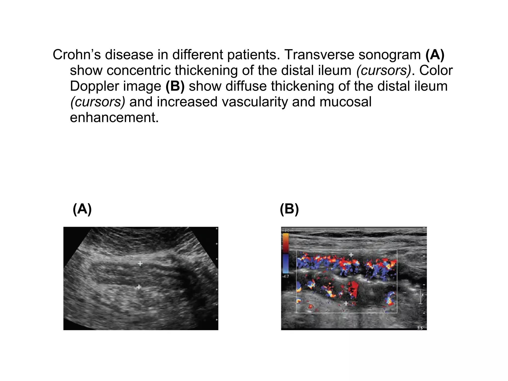
Crohn’s disease in different patients. Transverse sonogram (A)
show concentric thickening of the distal ileum (cursors). Color
Doppler image (B) show diffuse thickening of the distal ileum
(cursors) and increased vascularity and mucosal
enhancement.
(A) (B)
Peri-intestinal abscess. A, Short axis view of a small bowel loop
(cursors) shows concentric thickening of the small bowel with an
adjacent irregular fluid collection due to an abscess (A) in a
patient with Crohn’s disease.
B, Short axis view of the sigmoid colon (S) shows concentric colonic
wall thickening with extraluminal inflammatory changes and fluid
(asterisks) in a patient with diverticulitis. The urinary bladder (B) is
seen at the edge of the image.
Intussusception in different patients. A, Short axis view of a loop of
the small bowel shows multiple concentric rings of increased and
decreased echogenicity and thickening of the outer small bowel
layer typical of an intussusception.
B, Longitudinal static view shows a focally thickened small bowel
loop (cursors) with multiple layers. The leading edge of the
intussusceptum is seen (arrows).
Appendicitis is a common cause of acute abdominal pain and is the
most common condition requiring urgent abdominal surgery. CT is
the primary imaging modality. A high-resolution probe (usually a
linear array or a curved array operating at 5 MHz or higher) should
be used. Patient can point to a specific area of pain and this usually
corresponds closely to the site of the abnormal appendix.
The primary criterion for the diagnosis of appendicitis is an appendiceal
diameter greater than 6 mm. In some patients an intraluminal
appendicolith can be detected.
Appendicitis in different patients. A, Long axis view of the appendix
shows wall thickening with an appendiceal diameter of
9.0 mm. B, Long axis view of the appendix (cursors) shows increased
diameter as well as an echogenic shadowing appendicolith (arrow).
Tip appendicitis. A, CT scan shows a normal diameter appendix
with an enhancing wall. B, Long axis view of the base of the
appendix shows a normal diameter of 5.6 mm. A shadowing
appendicolith (arrow) is present. C, Short axis view of the tip of
the appendix shows mild thickening with a diameter of 6.5 mm.
the normal appendix can be seen only by experienced
sonographers, and usually only in thin patients (Fig. 9-18). It is
not necessary to see a normal appendix to exclude appendicitis.
Normal appendix. Longitudinal view shows a normal appendix
(cursors) measuring 5 mm arising from the cecum (C).
PERITONEUM
The most common abnormality of the peritoneal cavity is Ascites.
Sonography is quite sensitive at detecting ascites as well as
guiding aspiration of ascites. Small amounts of ascites are seen
as anechoic collections most often in the pelvic cul-desac and in
the right upper quadrant between the liver and the abdominal
wall or in the hepatorenal fossa (Morison’s pouch) Ascites. A,
Longitudinal view shows a trace amount of ascites (asterisk) in
the hepatorenal fossa. B, Oblique view shows a trace amount of
ascites (asterisk) between a cirrhotic liver and the abdominal
wall.
C, Transverse view shows anechoic ascites (asterisks) floating
around loops of the small bowel (B) and small bowel mesentery
(arrowheads) without compression or distortion of these
structures.
D, Longitudinal view shows complex ascites (asterisks) with diffuse
low-level echoes in the perihepatic space. This is the typical
appearance of hemorrhagic fluid.
E, Longitudinal view shows echogenic fluid (asterisk) in the
hepatorenal fossa several minutes following a liver biopsy. This
is typical of acute hemoperitoneum. F, Transverse view of the
right lower quadrant shows a complex multiseptated collection
of ascites (asterisks) between the abdominal wall and
compressed loops of the bowel (B).
Mesothelioma, a rare primary malignancy of the peritoneum, can
closely mimic peritoneal carcinomatosis
Mesothelioma in different patients. A, View of the right flank
abdominal wall shows a small, solid peritoneal nodule (cursors).
Larger nodules were seen elsewhere. B, Transverse color Doppler
view shows a large sheet of solid, hypoechoic, hypervascular
tissue (cursors) between the abdominal wall and the liver (L). C,
Transverse view shows a solid, hyperechoic peritoneal mass
(cursors) between the liver (L) and the kidney (K).
Splenosis is also a cause of solid peritoneal masses that can
simulate peritoneal metastases. They are more homogeneous
than most metastatic lesions but are otherwise Indistinguishable
Splenosis. Gray-scale (A) and power Doppler (B) views of the right
lower quadrant show a well-defined, hypoechoic, solid nodule
with readily detectable blood flow.
Fluid- and fat-containing hernia. Longitudinal view shows a fluid-
filled hernia sac (cursors) communicating with ascites in the
abdominal cavity through a small defect. compression shows
fluid and fat passing in and out of the hernia sac.
General abdomen ultrasound sonography

More Related Content

What's hot

Presentation1.pptx, ultrasound examination of the liver and gall bladder.
Presentation1.pptx, ultrasound examination of the liver and gall bladder.Presentation1.pptx, ultrasound examination of the liver and gall bladder.
Presentation1.pptx, ultrasound examination of the liver and gall bladder.Abdellah Nazeer
 
RADIOLOGIC ANATOMY OF SMALL INTESTINE AND INTRODUCTION TO SMALL BOWEL OBSTRUC...
RADIOLOGIC ANATOMY OF SMALL INTESTINE AND INTRODUCTION TO SMALL BOWEL OBSTRUC...RADIOLOGIC ANATOMY OF SMALL INTESTINE AND INTRODUCTION TO SMALL BOWEL OBSTRUC...
RADIOLOGIC ANATOMY OF SMALL INTESTINE AND INTRODUCTION TO SMALL BOWEL OBSTRUC...Mohammad Naufal
 
Liver Ultrasound
Liver UltrasoundLiver Ultrasound
Liver UltrasoundSafi. Khan
 
Acute Abdomen-Radiology
Acute Abdomen-RadiologyAcute Abdomen-Radiology
Acute Abdomen-RadiologyParvathy Nair
 
Intestinal Ultrasound
Intestinal UltrasoundIntestinal Ultrasound
Intestinal UltrasoundJoann Vargas
 
Presentation1.pptx, ultrasound study of the spleen and pancreas.
Presentation1.pptx, ultrasound study of the spleen and pancreas.Presentation1.pptx, ultrasound study of the spleen and pancreas.
Presentation1.pptx, ultrasound study of the spleen and pancreas.Abdellah Nazeer
 
Spleen Ultrasound anatomy structure scanning techniques and pathologies
Spleen Ultrasound anatomy structure scanning techniques and pathologies Spleen Ultrasound anatomy structure scanning techniques and pathologies
Spleen Ultrasound anatomy structure scanning techniques and pathologies Safi. Khan
 
Imaging of Bowel Obstruction
Imaging of Bowel ObstructionImaging of Bowel Obstruction
Imaging of Bowel ObstructionRathachai Kaewlai
 
Imaging of the abdomen & the gastrointestinal
Imaging of the abdomen & the gastrointestinalImaging of the abdomen & the gastrointestinal
Imaging of the abdomen & the gastrointestinalHidayat Shariff
 
Ultrasound of the urinary tract - Renal infections
Ultrasound of the urinary tract - Renal infectionsUltrasound of the urinary tract - Renal infections
Ultrasound of the urinary tract - Renal infectionsSamir Haffar
 
small intestine imaging
small intestine imagingsmall intestine imaging
small intestine imagingSumer Yadav
 
Gall bladder Ultrasound
Gall bladder UltrasoundGall bladder Ultrasound
Gall bladder UltrasoundSafi. Khan
 
Ultrasound of pancrease in Radiology
Ultrasound of pancrease in RadiologyUltrasound of pancrease in Radiology
Ultrasound of pancrease in RadiologyMahesh Kumar
 
Presentation1, radiological imaging of barium studies.
Presentation1, radiological imaging of barium studies.Presentation1, radiological imaging of barium studies.
Presentation1, radiological imaging of barium studies.Abdellah Nazeer
 
The Radiology of Malrotation
The Radiology of MalrotationThe Radiology of Malrotation
The Radiology of Malrotationtboulden
 
Abdominal Sonography
Abdominal SonographyAbdominal Sonography
Abdominal Sonographyu.surgery
 

What's hot (20)

Presentation1.pptx, ultrasound examination of the liver and gall bladder.
Presentation1.pptx, ultrasound examination of the liver and gall bladder.Presentation1.pptx, ultrasound examination of the liver and gall bladder.
Presentation1.pptx, ultrasound examination of the liver and gall bladder.
 
RADIOLOGIC ANATOMY OF SMALL INTESTINE AND INTRODUCTION TO SMALL BOWEL OBSTRUC...
RADIOLOGIC ANATOMY OF SMALL INTESTINE AND INTRODUCTION TO SMALL BOWEL OBSTRUC...RADIOLOGIC ANATOMY OF SMALL INTESTINE AND INTRODUCTION TO SMALL BOWEL OBSTRUC...
RADIOLOGIC ANATOMY OF SMALL INTESTINE AND INTRODUCTION TO SMALL BOWEL OBSTRUC...
 
Liver Ultrasound
Liver UltrasoundLiver Ultrasound
Liver Ultrasound
 
Acute Abdomen-Radiology
Acute Abdomen-RadiologyAcute Abdomen-Radiology
Acute Abdomen-Radiology
 
Intestinal Ultrasound
Intestinal UltrasoundIntestinal Ultrasound
Intestinal Ultrasound
 
Presentation1.pptx, ultrasound study of the spleen and pancreas.
Presentation1.pptx, ultrasound study of the spleen and pancreas.Presentation1.pptx, ultrasound study of the spleen and pancreas.
Presentation1.pptx, ultrasound study of the spleen and pancreas.
 
Spleen Ultrasound anatomy structure scanning techniques and pathologies
Spleen Ultrasound anatomy structure scanning techniques and pathologies Spleen Ultrasound anatomy structure scanning techniques and pathologies
Spleen Ultrasound anatomy structure scanning techniques and pathologies
 
Imaging of Bowel Obstruction
Imaging of Bowel ObstructionImaging of Bowel Obstruction
Imaging of Bowel Obstruction
 
Imaging of the abdomen & the gastrointestinal
Imaging of the abdomen & the gastrointestinalImaging of the abdomen & the gastrointestinal
Imaging of the abdomen & the gastrointestinal
 
Ultrasound Normal Anatomy of Major Organs
Ultrasound Normal Anatomy of Major OrgansUltrasound Normal Anatomy of Major Organs
Ultrasound Normal Anatomy of Major Organs
 
Ultrasound of the urinary tract - Renal infections
Ultrasound of the urinary tract - Renal infectionsUltrasound of the urinary tract - Renal infections
Ultrasound of the urinary tract - Renal infections
 
Usg artifacts
Usg artifactsUsg artifacts
Usg artifacts
 
small intestine imaging
small intestine imagingsmall intestine imaging
small intestine imaging
 
Gall bladder Ultrasound
Gall bladder UltrasoundGall bladder Ultrasound
Gall bladder Ultrasound
 
Ultrasound of pancrease in Radiology
Ultrasound of pancrease in RadiologyUltrasound of pancrease in Radiology
Ultrasound of pancrease in Radiology
 
Presentation1, radiological imaging of barium studies.
Presentation1, radiological imaging of barium studies.Presentation1, radiological imaging of barium studies.
Presentation1, radiological imaging of barium studies.
 
Imaging of Large Bowel Polyp
Imaging of Large Bowel PolypImaging of Large Bowel Polyp
Imaging of Large Bowel Polyp
 
The Radiology of Malrotation
The Radiology of MalrotationThe Radiology of Malrotation
The Radiology of Malrotation
 
Abdominal Sonography
Abdominal SonographyAbdominal Sonography
Abdominal Sonography
 
Abdominal Imaging Lecture
Abdominal Imaging LectureAbdominal Imaging Lecture
Abdominal Imaging Lecture
 

Similar to General abdomen ultrasound sonography

Presentation1.pptx, ultrasound examination of the appendix.
Presentation1.pptx, ultrasound examination of the appendix.Presentation1.pptx, ultrasound examination of the appendix.
Presentation1.pptx, ultrasound examination of the appendix.Abdellah Nazeer
 
Presentation1, Ultrasound of the bowel loops and the lymph nodes..pptx
Presentation1, Ultrasound of the bowel loops and the lymph nodes..pptxPresentation1, Ultrasound of the bowel loops and the lymph nodes..pptx
Presentation1, Ultrasound of the bowel loops and the lymph nodes..pptxAbdellah Nazeer
 
Presentation1, ultrasound of the bowel loops and the lymph nodes.
Presentation1, ultrasound of the bowel loops and the lymph nodes.Presentation1, ultrasound of the bowel loops and the lymph nodes.
Presentation1, ultrasound of the bowel loops and the lymph nodes.AbdullahNazeerYassin
 
Presentation1, ultrasound of the bowel loops and the lymph nodes.
Presentation1, ultrasound of the bowel loops and the lymph nodes.Presentation1, ultrasound of the bowel loops and the lymph nodes.
Presentation1, ultrasound of the bowel loops and the lymph nodes.AbdullahNazeerYassin
 
Presentation1, ultrasound examination of the chest.
Presentation1, ultrasound examination of the chest.Presentation1, ultrasound examination of the chest.
Presentation1, ultrasound examination of the chest.Abdellah Nazeer
 
Diagnostic modalities for Gastric diseases.pptx
Diagnostic modalities for Gastric diseases.pptxDiagnostic modalities for Gastric diseases.pptx
Diagnostic modalities for Gastric diseases.pptxPushpa Lal Bhadel
 
X-ray signs of gastrointestinal pathology.ppt
X-ray signs of gastrointestinal pathology.pptX-ray signs of gastrointestinal pathology.ppt
X-ray signs of gastrointestinal pathology.pptmalkeet011999
 
Imaging of small bowel pathology
Imaging of small bowel pathologyImaging of small bowel pathology
Imaging of small bowel pathologySunil Kumar
 
Presentation1, radiological imaging of internal abdominal hernia.
Presentation1, radiological imaging of internal abdominal hernia.Presentation1, radiological imaging of internal abdominal hernia.
Presentation1, radiological imaging of internal abdominal hernia.Abdellah Nazeer
 
GI Classic Sign.ppt
GI Classic Sign.pptGI Classic Sign.ppt
GI Classic Sign.pptssuser86266b
 
Imaging of fetal GIT anomalies
Imaging of fetal GIT anomaliesImaging of fetal GIT anomalies
Imaging of fetal GIT anomaliesDeepak Garg
 
Lectures on diagnostic_ultrasound
Lectures on diagnostic_ultrasoundLectures on diagnostic_ultrasound
Lectures on diagnostic_ultrasoundusgaile
 
Cystic Duct Normal Anatomy and Anatomic Variants.pptx
Cystic Duct Normal Anatomy and Anatomic Variants.pptxCystic Duct Normal Anatomy and Anatomic Variants.pptx
Cystic Duct Normal Anatomy and Anatomic Variants.pptxSaharGhosn
 
Spontaneous Esophageal Rupture 修改后
Spontaneous Esophageal Rupture  修改后Spontaneous Esophageal Rupture  修改后
Spontaneous Esophageal Rupture 修改后Deep Deep
 
Abdomen Gastrointestinal tract.pptx
Abdomen Gastrointestinal tract.pptxAbdomen Gastrointestinal tract.pptx
Abdomen Gastrointestinal tract.pptxPushkarBhure1
 

Similar to General abdomen ultrasound sonography (20)

Presentation1.pptx, ultrasound examination of the appendix.
Presentation1.pptx, ultrasound examination of the appendix.Presentation1.pptx, ultrasound examination of the appendix.
Presentation1.pptx, ultrasound examination of the appendix.
 
Presentation1, Ultrasound of the bowel loops and the lymph nodes..pptx
Presentation1, Ultrasound of the bowel loops and the lymph nodes..pptxPresentation1, Ultrasound of the bowel loops and the lymph nodes..pptx
Presentation1, Ultrasound of the bowel loops and the lymph nodes..pptx
 
Presentation1, ultrasound of the bowel loops and the lymph nodes.
Presentation1, ultrasound of the bowel loops and the lymph nodes.Presentation1, ultrasound of the bowel loops and the lymph nodes.
Presentation1, ultrasound of the bowel loops and the lymph nodes.
 
Presentation1, ultrasound of the bowel loops and the lymph nodes.
Presentation1, ultrasound of the bowel loops and the lymph nodes.Presentation1, ultrasound of the bowel loops and the lymph nodes.
Presentation1, ultrasound of the bowel loops and the lymph nodes.
 
Learn ultrasonography
Learn ultrasonographyLearn ultrasonography
Learn ultrasonography
 
Presentation1, ultrasound examination of the chest.
Presentation1, ultrasound examination of the chest.Presentation1, ultrasound examination of the chest.
Presentation1, ultrasound examination of the chest.
 
Abdominal Plain.pptx
Abdominal Plain.pptxAbdominal Plain.pptx
Abdominal Plain.pptx
 
Abdominal Plain.pptx
Abdominal Plain.pptxAbdominal Plain.pptx
Abdominal Plain.pptx
 
Diagnostic modalities for Gastric diseases.pptx
Diagnostic modalities for Gastric diseases.pptxDiagnostic modalities for Gastric diseases.pptx
Diagnostic modalities for Gastric diseases.pptx
 
X-ray signs of gastrointestinal pathology.ppt
X-ray signs of gastrointestinal pathology.pptX-ray signs of gastrointestinal pathology.ppt
X-ray signs of gastrointestinal pathology.ppt
 
Imaging of small bowel pathology
Imaging of small bowel pathologyImaging of small bowel pathology
Imaging of small bowel pathology
 
Presentation1, radiological imaging of internal abdominal hernia.
Presentation1, radiological imaging of internal abdominal hernia.Presentation1, radiological imaging of internal abdominal hernia.
Presentation1, radiological imaging of internal abdominal hernia.
 
Plain abdomen
Plain abdomenPlain abdomen
Plain abdomen
 
GI Classic Sign.ppt
GI Classic Sign.pptGI Classic Sign.ppt
GI Classic Sign.ppt
 
Imaging of fetal GIT anomalies
Imaging of fetal GIT anomaliesImaging of fetal GIT anomalies
Imaging of fetal GIT anomalies
 
Lectures on diagnostic_ultrasound
Lectures on diagnostic_ultrasoundLectures on diagnostic_ultrasound
Lectures on diagnostic_ultrasound
 
Cystic Duct Normal Anatomy and Anatomic Variants.pptx
Cystic Duct Normal Anatomy and Anatomic Variants.pptxCystic Duct Normal Anatomy and Anatomic Variants.pptx
Cystic Duct Normal Anatomy and Anatomic Variants.pptx
 
Spontaneous Esophageal Rupture 修改后
Spontaneous Esophageal Rupture  修改后Spontaneous Esophageal Rupture  修改后
Spontaneous Esophageal Rupture 修改后
 
Abdomen Gastrointestinal tract.pptx
Abdomen Gastrointestinal tract.pptxAbdomen Gastrointestinal tract.pptx
Abdomen Gastrointestinal tract.pptx
 
Imaging in Appendicitis
Imaging in AppendicitisImaging in Appendicitis
Imaging in Appendicitis
 

Recently uploaded

Russian Call Girls in Pune Tanvi 9907093804 Short 1500 Night 6000 Best call g...
Russian Call Girls in Pune Tanvi 9907093804 Short 1500 Night 6000 Best call g...Russian Call Girls in Pune Tanvi 9907093804 Short 1500 Night 6000 Best call g...
Russian Call Girls in Pune Tanvi 9907093804 Short 1500 Night 6000 Best call g...Miss joya
 
Kesar Bagh Call Girl Price 9548273370 , Lucknow Call Girls Service
Kesar Bagh Call Girl Price 9548273370 , Lucknow Call Girls ServiceKesar Bagh Call Girl Price 9548273370 , Lucknow Call Girls Service
Kesar Bagh Call Girl Price 9548273370 , Lucknow Call Girls Servicemakika9823
 
College Call Girls Pune Mira 9907093804 Short 1500 Night 6000 Best call girls...
College Call Girls Pune Mira 9907093804 Short 1500 Night 6000 Best call girls...College Call Girls Pune Mira 9907093804 Short 1500 Night 6000 Best call girls...
College Call Girls Pune Mira 9907093804 Short 1500 Night 6000 Best call girls...Miss joya
 
Call Girls Service Navi Mumbai Samaira 8617697112 Independent Escort Service ...
Call Girls Service Navi Mumbai Samaira 8617697112 Independent Escort Service ...Call Girls Service Navi Mumbai Samaira 8617697112 Independent Escort Service ...
Call Girls Service Navi Mumbai Samaira 8617697112 Independent Escort Service ...Call girls in Ahmedabad High profile
 
Bangalore Call Girls Nelamangala Number 7001035870 Meetin With Bangalore Esc...
Bangalore Call Girls Nelamangala Number 7001035870  Meetin With Bangalore Esc...Bangalore Call Girls Nelamangala Number 7001035870  Meetin With Bangalore Esc...
Bangalore Call Girls Nelamangala Number 7001035870 Meetin With Bangalore Esc...narwatsonia7
 
Call Girls Colaba Mumbai ❤️ 9920874524 👈 Cash on Delivery
Call Girls Colaba Mumbai ❤️ 9920874524 👈 Cash on DeliveryCall Girls Colaba Mumbai ❤️ 9920874524 👈 Cash on Delivery
Call Girls Colaba Mumbai ❤️ 9920874524 👈 Cash on Deliverynehamumbai
 
VIP Call Girls Indore Kirti 💚😋 9256729539 🚀 Indore Escorts
VIP Call Girls Indore Kirti 💚😋  9256729539 🚀 Indore EscortsVIP Call Girls Indore Kirti 💚😋  9256729539 🚀 Indore Escorts
VIP Call Girls Indore Kirti 💚😋 9256729539 🚀 Indore Escortsaditipandeya
 
Call Girls Service Jaipur Grishma WhatsApp ❤8445551418 VIP Call Girls Jaipur
Call Girls Service Jaipur Grishma WhatsApp ❤8445551418 VIP Call Girls JaipurCall Girls Service Jaipur Grishma WhatsApp ❤8445551418 VIP Call Girls Jaipur
Call Girls Service Jaipur Grishma WhatsApp ❤8445551418 VIP Call Girls Jaipurparulsinha
 
(👑VVIP ISHAAN ) Russian Call Girls Service Navi Mumbai🖕9920874524🖕Independent...
(👑VVIP ISHAAN ) Russian Call Girls Service Navi Mumbai🖕9920874524🖕Independent...(👑VVIP ISHAAN ) Russian Call Girls Service Navi Mumbai🖕9920874524🖕Independent...
(👑VVIP ISHAAN ) Russian Call Girls Service Navi Mumbai🖕9920874524🖕Independent...Taniya Sharma
 
💎VVIP Kolkata Call Girls Parganas🩱7001035870🩱Independent Girl ( Ac Rooms Avai...
💎VVIP Kolkata Call Girls Parganas🩱7001035870🩱Independent Girl ( Ac Rooms Avai...💎VVIP Kolkata Call Girls Parganas🩱7001035870🩱Independent Girl ( Ac Rooms Avai...
💎VVIP Kolkata Call Girls Parganas🩱7001035870🩱Independent Girl ( Ac Rooms Avai...Taniya Sharma
 
VIP Call Girls Pune Vrinda 9907093804 Short 1500 Night 6000 Best call girls S...
VIP Call Girls Pune Vrinda 9907093804 Short 1500 Night 6000 Best call girls S...VIP Call Girls Pune Vrinda 9907093804 Short 1500 Night 6000 Best call girls S...
VIP Call Girls Pune Vrinda 9907093804 Short 1500 Night 6000 Best call girls S...Miss joya
 
Vip Call Girls Anna Salai Chennai 👉 8250192130 ❣️💯 Top Class Girls Available
Vip Call Girls Anna Salai Chennai 👉 8250192130 ❣️💯 Top Class Girls AvailableVip Call Girls Anna Salai Chennai 👉 8250192130 ❣️💯 Top Class Girls Available
Vip Call Girls Anna Salai Chennai 👉 8250192130 ❣️💯 Top Class Girls AvailableNehru place Escorts
 
Artifacts in Nuclear Medicine with Identifying and resolving artifacts.
Artifacts in Nuclear Medicine with Identifying and resolving artifacts.Artifacts in Nuclear Medicine with Identifying and resolving artifacts.
Artifacts in Nuclear Medicine with Identifying and resolving artifacts.MiadAlsulami
 
CALL ON ➥9907093804 🔝 Call Girls Hadapsar ( Pune) Girls Service
CALL ON ➥9907093804 🔝 Call Girls Hadapsar ( Pune)  Girls ServiceCALL ON ➥9907093804 🔝 Call Girls Hadapsar ( Pune)  Girls Service
CALL ON ➥9907093804 🔝 Call Girls Hadapsar ( Pune) Girls ServiceMiss joya
 
VIP Mumbai Call Girls Hiranandani Gardens Just Call 9920874524 with A/C Room ...
VIP Mumbai Call Girls Hiranandani Gardens Just Call 9920874524 with A/C Room ...VIP Mumbai Call Girls Hiranandani Gardens Just Call 9920874524 with A/C Room ...
VIP Mumbai Call Girls Hiranandani Gardens Just Call 9920874524 with A/C Room ...Garima Khatri
 
VIP Call Girls Tirunelveli Aaradhya 8250192130 Independent Escort Service Tir...
VIP Call Girls Tirunelveli Aaradhya 8250192130 Independent Escort Service Tir...VIP Call Girls Tirunelveli Aaradhya 8250192130 Independent Escort Service Tir...
VIP Call Girls Tirunelveli Aaradhya 8250192130 Independent Escort Service Tir...narwatsonia7
 
♛VVIP Hyderabad Call Girls Chintalkunta🖕7001035870🖕Riya Kappor Top Call Girl ...
♛VVIP Hyderabad Call Girls Chintalkunta🖕7001035870🖕Riya Kappor Top Call Girl ...♛VVIP Hyderabad Call Girls Chintalkunta🖕7001035870🖕Riya Kappor Top Call Girl ...
♛VVIP Hyderabad Call Girls Chintalkunta🖕7001035870🖕Riya Kappor Top Call Girl ...astropune
 
Call Girls Horamavu WhatsApp Number 7001035870 Meeting With Bangalore Escorts
Call Girls Horamavu WhatsApp Number 7001035870 Meeting With Bangalore EscortsCall Girls Horamavu WhatsApp Number 7001035870 Meeting With Bangalore Escorts
Call Girls Horamavu WhatsApp Number 7001035870 Meeting With Bangalore Escortsvidya singh
 

Recently uploaded (20)

Russian Call Girls in Pune Tanvi 9907093804 Short 1500 Night 6000 Best call g...
Russian Call Girls in Pune Tanvi 9907093804 Short 1500 Night 6000 Best call g...Russian Call Girls in Pune Tanvi 9907093804 Short 1500 Night 6000 Best call g...
Russian Call Girls in Pune Tanvi 9907093804 Short 1500 Night 6000 Best call g...
 
Kesar Bagh Call Girl Price 9548273370 , Lucknow Call Girls Service
Kesar Bagh Call Girl Price 9548273370 , Lucknow Call Girls ServiceKesar Bagh Call Girl Price 9548273370 , Lucknow Call Girls Service
Kesar Bagh Call Girl Price 9548273370 , Lucknow Call Girls Service
 
College Call Girls Pune Mira 9907093804 Short 1500 Night 6000 Best call girls...
College Call Girls Pune Mira 9907093804 Short 1500 Night 6000 Best call girls...College Call Girls Pune Mira 9907093804 Short 1500 Night 6000 Best call girls...
College Call Girls Pune Mira 9907093804 Short 1500 Night 6000 Best call girls...
 
Call Girls Service Navi Mumbai Samaira 8617697112 Independent Escort Service ...
Call Girls Service Navi Mumbai Samaira 8617697112 Independent Escort Service ...Call Girls Service Navi Mumbai Samaira 8617697112 Independent Escort Service ...
Call Girls Service Navi Mumbai Samaira 8617697112 Independent Escort Service ...
 
Escort Service Call Girls In Sarita Vihar,, 99530°56974 Delhi NCR
Escort Service Call Girls In Sarita Vihar,, 99530°56974 Delhi NCREscort Service Call Girls In Sarita Vihar,, 99530°56974 Delhi NCR
Escort Service Call Girls In Sarita Vihar,, 99530°56974 Delhi NCR
 
Bangalore Call Girls Nelamangala Number 7001035870 Meetin With Bangalore Esc...
Bangalore Call Girls Nelamangala Number 7001035870  Meetin With Bangalore Esc...Bangalore Call Girls Nelamangala Number 7001035870  Meetin With Bangalore Esc...
Bangalore Call Girls Nelamangala Number 7001035870 Meetin With Bangalore Esc...
 
Call Girls Colaba Mumbai ❤️ 9920874524 👈 Cash on Delivery
Call Girls Colaba Mumbai ❤️ 9920874524 👈 Cash on DeliveryCall Girls Colaba Mumbai ❤️ 9920874524 👈 Cash on Delivery
Call Girls Colaba Mumbai ❤️ 9920874524 👈 Cash on Delivery
 
VIP Call Girls Indore Kirti 💚😋 9256729539 🚀 Indore Escorts
VIP Call Girls Indore Kirti 💚😋  9256729539 🚀 Indore EscortsVIP Call Girls Indore Kirti 💚😋  9256729539 🚀 Indore Escorts
VIP Call Girls Indore Kirti 💚😋 9256729539 🚀 Indore Escorts
 
Call Girls Service Jaipur Grishma WhatsApp ❤8445551418 VIP Call Girls Jaipur
Call Girls Service Jaipur Grishma WhatsApp ❤8445551418 VIP Call Girls JaipurCall Girls Service Jaipur Grishma WhatsApp ❤8445551418 VIP Call Girls Jaipur
Call Girls Service Jaipur Grishma WhatsApp ❤8445551418 VIP Call Girls Jaipur
 
(👑VVIP ISHAAN ) Russian Call Girls Service Navi Mumbai🖕9920874524🖕Independent...
(👑VVIP ISHAAN ) Russian Call Girls Service Navi Mumbai🖕9920874524🖕Independent...(👑VVIP ISHAAN ) Russian Call Girls Service Navi Mumbai🖕9920874524🖕Independent...
(👑VVIP ISHAAN ) Russian Call Girls Service Navi Mumbai🖕9920874524🖕Independent...
 
💎VVIP Kolkata Call Girls Parganas🩱7001035870🩱Independent Girl ( Ac Rooms Avai...
💎VVIP Kolkata Call Girls Parganas🩱7001035870🩱Independent Girl ( Ac Rooms Avai...💎VVIP Kolkata Call Girls Parganas🩱7001035870🩱Independent Girl ( Ac Rooms Avai...
💎VVIP Kolkata Call Girls Parganas🩱7001035870🩱Independent Girl ( Ac Rooms Avai...
 
VIP Call Girls Pune Vrinda 9907093804 Short 1500 Night 6000 Best call girls S...
VIP Call Girls Pune Vrinda 9907093804 Short 1500 Night 6000 Best call girls S...VIP Call Girls Pune Vrinda 9907093804 Short 1500 Night 6000 Best call girls S...
VIP Call Girls Pune Vrinda 9907093804 Short 1500 Night 6000 Best call girls S...
 
Vip Call Girls Anna Salai Chennai 👉 8250192130 ❣️💯 Top Class Girls Available
Vip Call Girls Anna Salai Chennai 👉 8250192130 ❣️💯 Top Class Girls AvailableVip Call Girls Anna Salai Chennai 👉 8250192130 ❣️💯 Top Class Girls Available
Vip Call Girls Anna Salai Chennai 👉 8250192130 ❣️💯 Top Class Girls Available
 
sauth delhi call girls in Bhajanpura 🔝 9953056974 🔝 escort Service
sauth delhi call girls in Bhajanpura 🔝 9953056974 🔝 escort Servicesauth delhi call girls in Bhajanpura 🔝 9953056974 🔝 escort Service
sauth delhi call girls in Bhajanpura 🔝 9953056974 🔝 escort Service
 
Artifacts in Nuclear Medicine with Identifying and resolving artifacts.
Artifacts in Nuclear Medicine with Identifying and resolving artifacts.Artifacts in Nuclear Medicine with Identifying and resolving artifacts.
Artifacts in Nuclear Medicine with Identifying and resolving artifacts.
 
CALL ON ➥9907093804 🔝 Call Girls Hadapsar ( Pune) Girls Service
CALL ON ➥9907093804 🔝 Call Girls Hadapsar ( Pune)  Girls ServiceCALL ON ➥9907093804 🔝 Call Girls Hadapsar ( Pune)  Girls Service
CALL ON ➥9907093804 🔝 Call Girls Hadapsar ( Pune) Girls Service
 
VIP Mumbai Call Girls Hiranandani Gardens Just Call 9920874524 with A/C Room ...
VIP Mumbai Call Girls Hiranandani Gardens Just Call 9920874524 with A/C Room ...VIP Mumbai Call Girls Hiranandani Gardens Just Call 9920874524 with A/C Room ...
VIP Mumbai Call Girls Hiranandani Gardens Just Call 9920874524 with A/C Room ...
 
VIP Call Girls Tirunelveli Aaradhya 8250192130 Independent Escort Service Tir...
VIP Call Girls Tirunelveli Aaradhya 8250192130 Independent Escort Service Tir...VIP Call Girls Tirunelveli Aaradhya 8250192130 Independent Escort Service Tir...
VIP Call Girls Tirunelveli Aaradhya 8250192130 Independent Escort Service Tir...
 
♛VVIP Hyderabad Call Girls Chintalkunta🖕7001035870🖕Riya Kappor Top Call Girl ...
♛VVIP Hyderabad Call Girls Chintalkunta🖕7001035870🖕Riya Kappor Top Call Girl ...♛VVIP Hyderabad Call Girls Chintalkunta🖕7001035870🖕Riya Kappor Top Call Girl ...
♛VVIP Hyderabad Call Girls Chintalkunta🖕7001035870🖕Riya Kappor Top Call Girl ...
 
Call Girls Horamavu WhatsApp Number 7001035870 Meeting With Bangalore Escorts
Call Girls Horamavu WhatsApp Number 7001035870 Meeting With Bangalore EscortsCall Girls Horamavu WhatsApp Number 7001035870 Meeting With Bangalore Escorts
Call Girls Horamavu WhatsApp Number 7001035870 Meeting With Bangalore Escorts
 

General abdomen ultrasound sonography

  • 2. Bowel Sonography can be performed following ingestion of water to improve evaluation of the stomach and proximal small bowel. Retrograde infusion of water can also be used to distend the colon (hydrocolonic sonography) and evaluate colonic lesions.
  • 3.
  • 4. The normal bowel has five layers that can be seen sonographically. The inner hyperechoic layer arises from the interface reflection between the lumen and the surface of the mucosa. The second layer is hypoechoic and arises from the combined mucosa and the muscularis mucosa. The third layer is hyperechoic and arises from the submucosa. The fourth layer is hypoechoic and arises from the muscularis propria. The final outer layer is hyperechoic and arises from the interface reflection between the muscularis propria and the serosa or adventitia (plus peri-intestinal fat). These five layers are routinely seen on endoscopic ultrasound and are intermittently seen on transcutaneous scans.
  • 5.
  • 6.
  • 7. Normal bowel layers. Long axis views of the stomach with the lumen filled with water (A) and with the lumen collapsed (B) show the following: 1, The interface from the mucosal surface is hyperechoic. 2, The deep mucosa and muscularis mucosa are hypoechoic. 3, The submucosa is hyperechoic. 4, The muscularis propria is hypoechoic. 5, The interface between the muscularis propria and the serosa is hyperechoic. Note that the same layers labeled on one wall of the antrum are repeated on the opposite wall.
  • 8. On many transabdominal scans when the lumen is collapsed, the mucosa, the muscularis mucosa, and the submucosa cannot be distinguished into separate layers. This produces a central echogenic region that is surrounded by the hypoechoic muscularis propria, resulting in the typical bull’seye Appearance. Normal colon. Transverse view of the colon shows the typical bull’s-eye appearance of collapsed bowel (arrows).
  • 9. In other cases the mucosa and the muscularis mucosa combine to form a central hypoechoic region, followed by the hyperechoic submucosa and then the hypoechoic muscularis propria (Fig. 9-3). The normal intestinal wall should be less than 5 mm in thickness. Normal stomach. A, Transverse view of the collapsed stomach shows the combined mucosa and muscularis mucosa as a hypoechoic central region surrounded by the echogenic submucosa and the hypoechoic muscularis propria. B, Similar view in the distended state shows all five layers.
  • 10. Distended, fluid-filled, peristalsing loops are the hallmark of bowel obstruction scanning in the lateral flanks where dependent fluid- filled loops are located. When a bowel obstruction is detected, a search for the cause should be performed. Hernias, intrinsic bowel wall lesions, abdominal masses, and abdominal fluid collections can all be detected with sonography. Small bowel obstruction. Static view of the left upper quadrant shows a dilated fluid-filled loop of small bowel (cursors) measuring greater than 3 cm in diameter. An adjacent fluid collection is seen.
  • 11.
  • 12. Small bowel lymphoma in different patients. A, Longitudinal view shows a focal, hypoechoic, solid mass (cursors) along the mesenteric wall of the small bowel. Gas in the lumen (arrows) obscures most of the adjacent mesenteric wall of the bowel. The antimesenteric wall of the bowel (arrowheads) is thickened, as are the small bowel folds. B, Short axis view shows marked concentric thickening of the small bowel wall (cursors). Gas in the lumen (arrow) produces a dirty acoustic shadow (S).
  • 13.
  • 14. Gastrointestinal stromal tumors in different patients. A, Transverse view of the mid abdomen shows a hypoechoic mass (cursors) with central areas of liquefaction consistent with necrosis. B, Longitudinal view of the lower abdomen shows a complex mass (cursors) with a large area of central liquefaction. A small faintly shadowing calcification (arrow) is also identified. C, View of the fluid-filled stomach (S) shows a hypoechoic, solid mass (cursors) arising from the posterior gastric wall.
  • 15.
  • 16. D, Transverse view of the fluid-filled stomach (S) shows a lobulated solid mass (cursors) with a small intraluminal component and a larger exophytic component. Longitudinal sonogram (E) and endoscopic (F) views of the stomach show a solid, hypoechoic mass (cursors) arising from the collapsed stomach (S). The retroflexed endoscopic view shows the submucosal portion of the mass (cursors) adjacent to the endoscope (E).
  • 17. Gastric lipoma. A, Longitudinal sonogram shows a hyperechoic lesion (arrow) projecting into the lumen of the stomach.
  • 18.
  • 19. Colitis in different patients. Transverse (A) and longitudinal (B) views of the left colon show diffuse concentric thickening. Longitudinal sonogram (C) show diffuse thickening of the transverse colon due to colitis caused by Clostridium difficile.
  • 20.
  • 21. Crohn’s disease in different patients. Transverse sonogram (A) show concentric thickening of the distal ileum (cursors). Color Doppler image (B) show diffuse thickening of the distal ileum (cursors) and increased vascularity and mucosal enhancement. (A) (B)
  • 22.
  • 23.
  • 24. Peri-intestinal abscess. A, Short axis view of a small bowel loop (cursors) shows concentric thickening of the small bowel with an adjacent irregular fluid collection due to an abscess (A) in a patient with Crohn’s disease. B, Short axis view of the sigmoid colon (S) shows concentric colonic wall thickening with extraluminal inflammatory changes and fluid (asterisks) in a patient with diverticulitis. The urinary bladder (B) is seen at the edge of the image.
  • 25. Intussusception in different patients. A, Short axis view of a loop of the small bowel shows multiple concentric rings of increased and decreased echogenicity and thickening of the outer small bowel layer typical of an intussusception. B, Longitudinal static view shows a focally thickened small bowel loop (cursors) with multiple layers. The leading edge of the intussusceptum is seen (arrows).
  • 26.
  • 27. Appendicitis is a common cause of acute abdominal pain and is the most common condition requiring urgent abdominal surgery. CT is the primary imaging modality. A high-resolution probe (usually a linear array or a curved array operating at 5 MHz or higher) should be used. Patient can point to a specific area of pain and this usually corresponds closely to the site of the abnormal appendix. The primary criterion for the diagnosis of appendicitis is an appendiceal diameter greater than 6 mm. In some patients an intraluminal appendicolith can be detected. Appendicitis in different patients. A, Long axis view of the appendix shows wall thickening with an appendiceal diameter of 9.0 mm. B, Long axis view of the appendix (cursors) shows increased diameter as well as an echogenic shadowing appendicolith (arrow).
  • 28.
  • 29.
  • 30.
  • 31.
  • 32.
  • 33.
  • 34.
  • 35. Tip appendicitis. A, CT scan shows a normal diameter appendix with an enhancing wall. B, Long axis view of the base of the appendix shows a normal diameter of 5.6 mm. A shadowing appendicolith (arrow) is present. C, Short axis view of the tip of the appendix shows mild thickening with a diameter of 6.5 mm.
  • 36. the normal appendix can be seen only by experienced sonographers, and usually only in thin patients (Fig. 9-18). It is not necessary to see a normal appendix to exclude appendicitis. Normal appendix. Longitudinal view shows a normal appendix (cursors) measuring 5 mm arising from the cecum (C).
  • 37.
  • 38. PERITONEUM The most common abnormality of the peritoneal cavity is Ascites. Sonography is quite sensitive at detecting ascites as well as guiding aspiration of ascites. Small amounts of ascites are seen as anechoic collections most often in the pelvic cul-desac and in the right upper quadrant between the liver and the abdominal wall or in the hepatorenal fossa (Morison’s pouch) Ascites. A, Longitudinal view shows a trace amount of ascites (asterisk) in the hepatorenal fossa. B, Oblique view shows a trace amount of ascites (asterisk) between a cirrhotic liver and the abdominal wall.
  • 39.
  • 40.
  • 41. C, Transverse view shows anechoic ascites (asterisks) floating around loops of the small bowel (B) and small bowel mesentery (arrowheads) without compression or distortion of these structures. D, Longitudinal view shows complex ascites (asterisks) with diffuse low-level echoes in the perihepatic space. This is the typical appearance of hemorrhagic fluid.
  • 42. E, Longitudinal view shows echogenic fluid (asterisk) in the hepatorenal fossa several minutes following a liver biopsy. This is typical of acute hemoperitoneum. F, Transverse view of the right lower quadrant shows a complex multiseptated collection of ascites (asterisks) between the abdominal wall and compressed loops of the bowel (B).
  • 43. Mesothelioma, a rare primary malignancy of the peritoneum, can closely mimic peritoneal carcinomatosis Mesothelioma in different patients. A, View of the right flank abdominal wall shows a small, solid peritoneal nodule (cursors). Larger nodules were seen elsewhere. B, Transverse color Doppler view shows a large sheet of solid, hypoechoic, hypervascular tissue (cursors) between the abdominal wall and the liver (L). C, Transverse view shows a solid, hyperechoic peritoneal mass (cursors) between the liver (L) and the kidney (K).
  • 44.
  • 45.
  • 46.
  • 47.
  • 48. Splenosis is also a cause of solid peritoneal masses that can simulate peritoneal metastases. They are more homogeneous than most metastatic lesions but are otherwise Indistinguishable Splenosis. Gray-scale (A) and power Doppler (B) views of the right lower quadrant show a well-defined, hypoechoic, solid nodule with readily detectable blood flow.
  • 49.
  • 50. Fluid- and fat-containing hernia. Longitudinal view shows a fluid- filled hernia sac (cursors) communicating with ascites in the abdominal cavity through a small defect. compression shows fluid and fat passing in and out of the hernia sac.