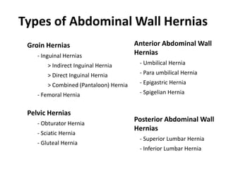
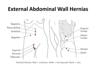
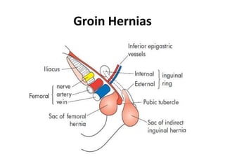
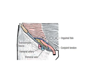
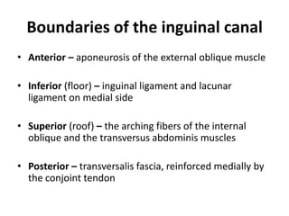
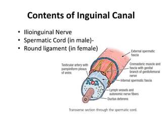
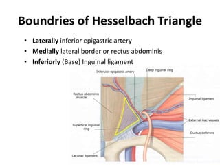
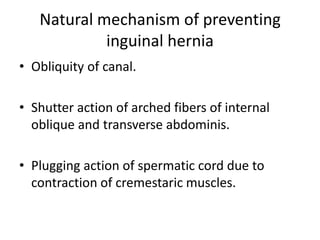
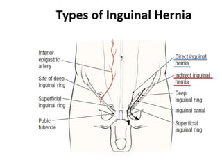
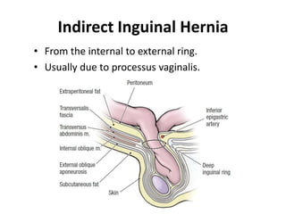
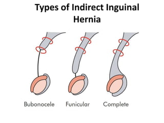
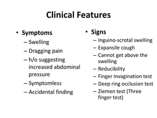
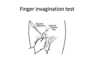
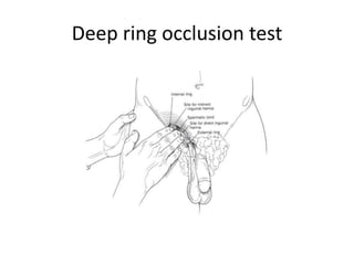
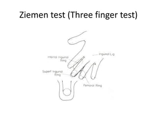
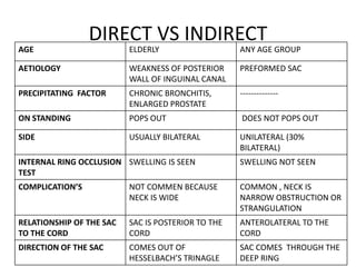
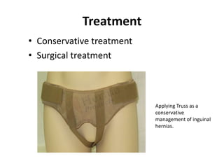
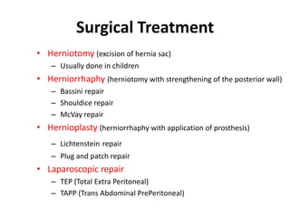
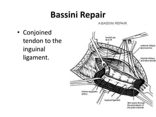
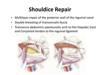
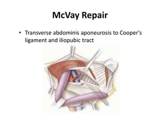
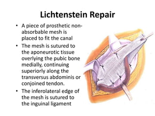
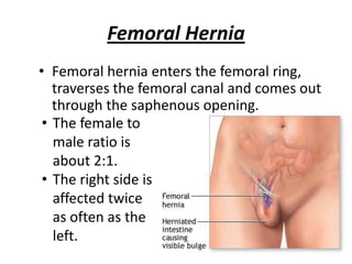
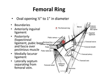
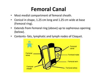
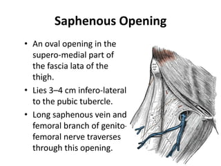
Abdominal wall hernia (INDIRECT,
    DIRECT AND FEMORAL )


          BY DR NITIN KEYAL
               INTERN
       DEPARTMENT OF SURGERY
     KATHMANDU MODEL HOSPITAL .
CONTENT’S
• DEFINITION
• TYPES OF HERNIA AND ETIOLOGY
• COMPOSITION OF HERNIA
• CLASSIFICATION OF HERNIA AND CONTENTS OF
  HERNIA .
• ANATOMY OF HERNIA AND CLINICAL FEATURES
• DIFFERENT KINDS OF TEST
• MANAGEMENT.
Types of Abdominal Wall Hernias
 Groin Hernias                          Anterior Abdominal Wall
    - Inguinal Hernias                  Hernias
        > Indirect Inguinal Hernia       - Umbilical Hernia
        > Direct Inguinal Hernia         - Para umbilical Hernia
        > Combined (Pantaloon) Hernia    - Epigastric Hernia
    - Femoral Hernia                     - Spigelian Hernia


 Pelvic Hernias
    - Obturator Hernia
                                        Posterior Abdominal Wall
                                        Hernias
    - Sciatic Hernia
                                         - Superior Lumbar Hernia
    - Gluteal Hernia
                                         - Inferior Lumbar Hernia
External Abdominal Wall Hernias
Etiologies
• Increased abdominal pressure
   – Cough, urinary trouble, constipation, straining, ascites,
     intra abdominal malignancies, pregnancy

• Weakness of abdominal wall
   – Congenital
      • Patent processes vaginalis, patent canal of nuck in females
   – Acquired
      •   Excess fat (obesity)
      •   Post Pregnancy
      •   Surgical incisions
      •   Connective tissue disorders like Marfan’s syndrome
Composition of a hernia
• A hernia consists of three parts –
   – the sac
   – the coverings of the sac
   – the contents of the sac
• The sac is a diverticulum of peritoneum, consisting of
   –   Mouth
   –   Neck
   –   Body
   –   Fundus
• The coverings are derived from the layers of the
  abdominal wall through which the sac passes.
Contents
• omentum = omentocele;
• intestine = enterocele; more commonly small
  bowel but may be large intestine or appendix;
• a portion of the circumference of the intestine =
  Richter’s hernia;
• a portion of the bladder (or a diverticulum);
• ovary with or without the corresponding fallopian
  tube;
• a Meckel’s diverticulum = a Littre’s hernia;
• fluid, as part of ascites or peritoneal fluid.
Classification of Hernia
• Reducible – if contents can be returned to abdomen
• Irreducible – if contents cannot be returned but there
  are no other complications
• Obstructed – if bowel in the hernia has good blood
  supply but bowel is obstructed
• Strangulated – if blood supply of bowel is obstructed
• Inflamed – if contents of sac have become inflamed
• Incarcerated – if the portion of the colon occupying a
  hernial sac is blocked with faeces
Groin Hernias
Nyhus Classification of Groin Hernia
• Type I: Indirect inguinal hernia with internal inguinal ring normal
• Type II: Indirect inguinal hernia with internal inguinal ring dilated
   but posterior wall intact, inferior epigastric vessels not displaced
• Type III: Posterior wall defect
    A: Direct inguinal hernia
    B: Indirect inguinal hernia with internal inguinal ring dilated,
    medially encroaching on or destroying the transversalis fascia of
    hesselbach’s triangle.
    C: Femoral hernia
• Type IV: Recurrent hernia
    A: Direct   B: Indirect       C: Femoral       D: Combined
Anatomy of inguinal canal
• 4 cm in length passing downward and medially
  from deep to superficial ring

• Deep/internal ring is ‘U’ shaped in fascia
  transversalis which lies 1.25 cm above the mid
  inguinal point.

• Superficial/External ring is in external oblique
  aponeurosis situated just above and lateral to the
  pubic crest.
Boundaries of the inguinal canal
• Anterior – aponeurosis of the external oblique muscle

• Inferior (floor) – inguinal ligament and lacunar
  ligament on medial side

• Superior (roof) – the arching fibers of the internal
  oblique and the transversus abdominis muscles

• Posterior – transversalis fascia, reinforced medially by
  the conjoint tendon
Contents of Inguinal Canal
• Ilioinguinal Nerve
• Spermatic Cord (in male)-
• Round ligament (in female)
Boundries of Hesselbach Triangle
 • Laterally inferior epigastric artery
 • Medially lateral border or rectus abdominis
 • Inferiorly (Base) Inguinal ligament
Natural mechanism of preventing
            inguinal hernia
• Obliquity of canal.

• Shutter action of arched fibers of internal
  oblique and transverse abdominis.

• Plugging action of spermatic cord due to
  contraction of cremestaric muscles.
Types of Inguinal Hernia
Indirect Inguinal Hernia
• From the internal to external ring.
• Usually due to processus vaginalis.
Types of Indirect Inguinal
          Hernia
Direct Inguinal Hernia
• A direct inguinal hernia is always acquired.
• The sac passes through a weakness or defect of the
  transversalis fascia in the posterior wall of the
  inguinal canal.
Clinical Features
• Symptoms                • Signs
  – Swelling                – Inguino-scrotal swelling
                            – Expansile cough
  – Dragging pain
                            – Cannot get above the
  – h/o suggesting            swelling
    increased abdominal     – Reducibility
    pressure                – Finger invagination test
  – Symptomless             – Deep ring occlusion test
  – Accidental finding      – Ziemen test (Three
                              finger test)
Finger invagination test
Deep ring occlusion test
Ziemen test (Three finger test)
DIRECT VS INDIRECT
AGE                       ELDERLY                   ANY AGE GROUP

AETIOLOGY                 WEAKNESS OF POSTERIOR     PREFORMED SAC
                          WALL OF INGUINAL CANAL
PRECIPITATING FACTOR      CHRONIC BRONCHITIS,       --------------
                          ENLARGED PROSTATE
ON STANDING               POPS OUT                  DOES NOT POPS OUT

SIDE                      USUALLY BILATERAL         UNILATERAL (30%
                                                    BILATERAL)
INTERNAL RING OCCLUSION SWELLING IS SEEN            SWELLING NOT SEEN
TEST
COMPLICATION’S            NOT COMMEN BECAUSE        COMMON , NECK IS
                          NECK IS WIDE              NARROW OBSTRUCTION OR
                                                    STRANGULATION
RELATIONSHIP OF THE SAC   SAC IS POSTERIOR TO THE   ANTEROLATERAL TO THE
TO THE CORD               CORD                      CORD
DIRECTION OF THE SAC      COMES OUT OF              SAC COMES THROUGH THE
                          HESSELBACH’S TRINAGLE     DEEP RING
Treatment
• Conservative treatment
• Surgical treatment


                           Applying Truss as a
                           conservative
                           management of inguinal
                           hernias.
Surgical Treatment
• Herniotomy (excision of hernia sac)
    – Usually done in children
• Herniorrhaphy (herniotomy with strengthening of the posterior wall)
    – Bassini repair
    – Shouldice repair
    – McVay repair
• Hernioplasty (herniorrhaphy with application of prosthesis)
    – Lichtenstein repair
    – Plug and patch repair
• Laparoscopic repair
    – TEP (Total Extra Peritoneal)
    – TAPP (Trans Abdominal PrePeritoneal)
Bassini Repair
• Conjoined
  tendon to the
  inguinal
  ligament.
Shouldice Repair
• Multilayer repair of the posterior wall of the inguinal canal
• Double breasting of transversalis fascia
• Transverse abdominis aponeurotic arch to the iliopubic tract
  and Conjoined tendon to the inguinal ligament
McVay Repair
• Transverse abdominis aponeurosis to Cooper's
  ligament and iliopubic tract
Lichtenstein Repair
• A piece of prosthetic non-
  absorbable mesh is
  placed to fit the canal
• The mesh is sutured to
  the aponeurotic tissue
  overlying the pubic bone
  medially, continuing
  superiorly along the
  transversus abdominis or
  conjoined tendon.
• The inferolateral edge of
  the mesh is sutured to
  the inguinal ligament
Femoral Hernia
• Femoral hernia enters the femoral ring,
   traverses the femoral canal and comes out
   through the saphenous opening.
 • The female to
   male ratio is
   about 2:1.
 • The right side is
   affected twice
   as often as the
   left.
Femoral Ring
• Oval opening ½” to 1” in diameter
• Boundaries
• Anteriorly inguinal
  ligament
• Posteriorly
  iliopectineal
  ligament, pubic bone
  and fascia over
  pectinious muscle
• Medially lacunar
  ligament
• Laterally septum
  separating from
  femoral vein.
Femoral Canal
• Most medial compartment of femoral sheath.
• Conical in shape, 1.25 cm long and 1.25 cm wide at base
  (Femoral ring).
• Extends from Femoral ring (above) up to saphenous opening
  (below).
• Contents- fats, lymphatic and lymph nodes of Cloquet.
Saphenous Opening
• An oval opening in the
  supero-medial part of
  the fascia lata of the
  thigh.
• Lies 3–4 cm infero-lateral
  to the pubic tubercle.
• Long saphenous vein and
  femoral branch of genito-
  femoral nerve traverses
  through this opening.
Surgical Treatment
• The low (Lockwood) operation- the inguinal
  ligament to the iliopectineal ligament.
• The high (McEvedy) operation- the conjoint
  tendon to the iliopectineal ligament.
• Lotheissen’s operation- the conjoint tendon
  or inguinal ligament to the iliopectineal
  ligament through inguinal canal
REFERENCE’S

•   BALLEY AND LOVE 25TH EDITION
•   TEXTBOOK OF MANIPAL 3RD EDITION
•   MEDSCAPE.COM
•   GOOGLE.COM/IMAGES .
Abdominal hernias by dr. nitin

Abdominal hernias by dr. nitin

  • 1.
    Abdominal wall hernia(INDIRECT, DIRECT AND FEMORAL ) BY DR NITIN KEYAL INTERN DEPARTMENT OF SURGERY KATHMANDU MODEL HOSPITAL .
  • 2.
    CONTENT’S • DEFINITION • TYPESOF HERNIA AND ETIOLOGY • COMPOSITION OF HERNIA • CLASSIFICATION OF HERNIA AND CONTENTS OF HERNIA . • ANATOMY OF HERNIA AND CLINICAL FEATURES • DIFFERENT KINDS OF TEST • MANAGEMENT.
  • 3.
    Types of AbdominalWall Hernias Groin Hernias Anterior Abdominal Wall - Inguinal Hernias Hernias > Indirect Inguinal Hernia - Umbilical Hernia > Direct Inguinal Hernia - Para umbilical Hernia > Combined (Pantaloon) Hernia - Epigastric Hernia - Femoral Hernia - Spigelian Hernia Pelvic Hernias - Obturator Hernia Posterior Abdominal Wall Hernias - Sciatic Hernia - Superior Lumbar Hernia - Gluteal Hernia - Inferior Lumbar Hernia
  • 4.
  • 5.
    Etiologies • Increased abdominalpressure – Cough, urinary trouble, constipation, straining, ascites, intra abdominal malignancies, pregnancy • Weakness of abdominal wall – Congenital • Patent processes vaginalis, patent canal of nuck in females – Acquired • Excess fat (obesity) • Post Pregnancy • Surgical incisions • Connective tissue disorders like Marfan’s syndrome
  • 6.
    Composition of ahernia • A hernia consists of three parts – – the sac – the coverings of the sac – the contents of the sac • The sac is a diverticulum of peritoneum, consisting of – Mouth – Neck – Body – Fundus • The coverings are derived from the layers of the abdominal wall through which the sac passes.
  • 7.
    Contents • omentum =omentocele; • intestine = enterocele; more commonly small bowel but may be large intestine or appendix; • a portion of the circumference of the intestine = Richter’s hernia; • a portion of the bladder (or a diverticulum); • ovary with or without the corresponding fallopian tube; • a Meckel’s diverticulum = a Littre’s hernia; • fluid, as part of ascites or peritoneal fluid.
  • 8.
    Classification of Hernia •Reducible – if contents can be returned to abdomen • Irreducible – if contents cannot be returned but there are no other complications • Obstructed – if bowel in the hernia has good blood supply but bowel is obstructed • Strangulated – if blood supply of bowel is obstructed • Inflamed – if contents of sac have become inflamed • Incarcerated – if the portion of the colon occupying a hernial sac is blocked with faeces
  • 9.
  • 10.
    Nyhus Classification ofGroin Hernia • Type I: Indirect inguinal hernia with internal inguinal ring normal • Type II: Indirect inguinal hernia with internal inguinal ring dilated but posterior wall intact, inferior epigastric vessels not displaced • Type III: Posterior wall defect A: Direct inguinal hernia B: Indirect inguinal hernia with internal inguinal ring dilated, medially encroaching on or destroying the transversalis fascia of hesselbach’s triangle. C: Femoral hernia • Type IV: Recurrent hernia A: Direct B: Indirect C: Femoral D: Combined
  • 11.
    Anatomy of inguinalcanal • 4 cm in length passing downward and medially from deep to superficial ring • Deep/internal ring is ‘U’ shaped in fascia transversalis which lies 1.25 cm above the mid inguinal point. • Superficial/External ring is in external oblique aponeurosis situated just above and lateral to the pubic crest.
  • 16.
    Boundaries of theinguinal canal • Anterior – aponeurosis of the external oblique muscle • Inferior (floor) – inguinal ligament and lacunar ligament on medial side • Superior (roof) – the arching fibers of the internal oblique and the transversus abdominis muscles • Posterior – transversalis fascia, reinforced medially by the conjoint tendon
  • 17.
    Contents of InguinalCanal • Ilioinguinal Nerve • Spermatic Cord (in male)- • Round ligament (in female)
  • 18.
    Boundries of HesselbachTriangle • Laterally inferior epigastric artery • Medially lateral border or rectus abdominis • Inferiorly (Base) Inguinal ligament
  • 19.
    Natural mechanism ofpreventing inguinal hernia • Obliquity of canal. • Shutter action of arched fibers of internal oblique and transverse abdominis. • Plugging action of spermatic cord due to contraction of cremestaric muscles.
  • 20.
  • 21.
    Indirect Inguinal Hernia •From the internal to external ring. • Usually due to processus vaginalis.
  • 22.
    Types of IndirectInguinal Hernia
  • 23.
    Direct Inguinal Hernia •A direct inguinal hernia is always acquired. • The sac passes through a weakness or defect of the transversalis fascia in the posterior wall of the inguinal canal.
  • 24.
    Clinical Features • Symptoms • Signs – Swelling – Inguino-scrotal swelling – Expansile cough – Dragging pain – Cannot get above the – h/o suggesting swelling increased abdominal – Reducibility pressure – Finger invagination test – Symptomless – Deep ring occlusion test – Accidental finding – Ziemen test (Three finger test)
  • 25.
  • 26.
  • 27.
    Ziemen test (Threefinger test)
  • 28.
    DIRECT VS INDIRECT AGE ELDERLY ANY AGE GROUP AETIOLOGY WEAKNESS OF POSTERIOR PREFORMED SAC WALL OF INGUINAL CANAL PRECIPITATING FACTOR CHRONIC BRONCHITIS, -------------- ENLARGED PROSTATE ON STANDING POPS OUT DOES NOT POPS OUT SIDE USUALLY BILATERAL UNILATERAL (30% BILATERAL) INTERNAL RING OCCLUSION SWELLING IS SEEN SWELLING NOT SEEN TEST COMPLICATION’S NOT COMMEN BECAUSE COMMON , NECK IS NECK IS WIDE NARROW OBSTRUCTION OR STRANGULATION RELATIONSHIP OF THE SAC SAC IS POSTERIOR TO THE ANTEROLATERAL TO THE TO THE CORD CORD CORD DIRECTION OF THE SAC COMES OUT OF SAC COMES THROUGH THE HESSELBACH’S TRINAGLE DEEP RING
  • 29.
    Treatment • Conservative treatment •Surgical treatment Applying Truss as a conservative management of inguinal hernias.
  • 30.
    Surgical Treatment • Herniotomy(excision of hernia sac) – Usually done in children • Herniorrhaphy (herniotomy with strengthening of the posterior wall) – Bassini repair – Shouldice repair – McVay repair • Hernioplasty (herniorrhaphy with application of prosthesis) – Lichtenstein repair – Plug and patch repair • Laparoscopic repair – TEP (Total Extra Peritoneal) – TAPP (Trans Abdominal PrePeritoneal)
  • 31.
    Bassini Repair • Conjoined tendon to the inguinal ligament.
  • 32.
    Shouldice Repair • Multilayerrepair of the posterior wall of the inguinal canal • Double breasting of transversalis fascia • Transverse abdominis aponeurotic arch to the iliopubic tract and Conjoined tendon to the inguinal ligament
  • 33.
    McVay Repair • Transverseabdominis aponeurosis to Cooper's ligament and iliopubic tract
  • 34.
    Lichtenstein Repair • Apiece of prosthetic non- absorbable mesh is placed to fit the canal • The mesh is sutured to the aponeurotic tissue overlying the pubic bone medially, continuing superiorly along the transversus abdominis or conjoined tendon. • The inferolateral edge of the mesh is sutured to the inguinal ligament
  • 35.
    Femoral Hernia • Femoralhernia enters the femoral ring, traverses the femoral canal and comes out through the saphenous opening. • The female to male ratio is about 2:1. • The right side is affected twice as often as the left.
  • 36.
    Femoral Ring • Ovalopening ½” to 1” in diameter • Boundaries • Anteriorly inguinal ligament • Posteriorly iliopectineal ligament, pubic bone and fascia over pectinious muscle • Medially lacunar ligament • Laterally septum separating from femoral vein.
  • 37.
    Femoral Canal • Mostmedial compartment of femoral sheath. • Conical in shape, 1.25 cm long and 1.25 cm wide at base (Femoral ring). • Extends from Femoral ring (above) up to saphenous opening (below). • Contents- fats, lymphatic and lymph nodes of Cloquet.
  • 38.
    Saphenous Opening • Anoval opening in the supero-medial part of the fascia lata of the thigh. • Lies 3–4 cm infero-lateral to the pubic tubercle. • Long saphenous vein and femoral branch of genito- femoral nerve traverses through this opening.
  • 39.
    Surgical Treatment • Thelow (Lockwood) operation- the inguinal ligament to the iliopectineal ligament. • The high (McEvedy) operation- the conjoint tendon to the iliopectineal ligament. • Lotheissen’s operation- the conjoint tendon or inguinal ligament to the iliopectineal ligament through inguinal canal
  • 40.
    REFERENCE’S • BALLEY AND LOVE 25TH EDITION • TEXTBOOK OF MANIPAL 3RD EDITION • MEDSCAPE.COM • GOOGLE.COM/IMAGES .