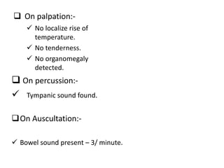
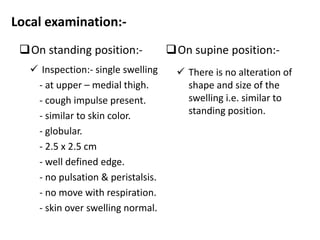
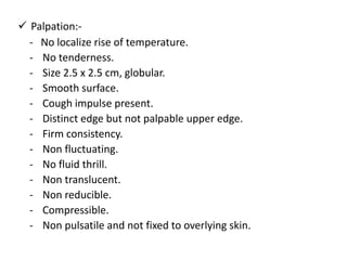
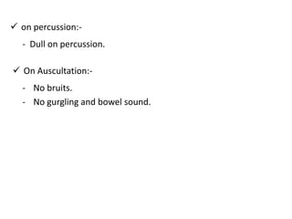
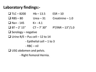
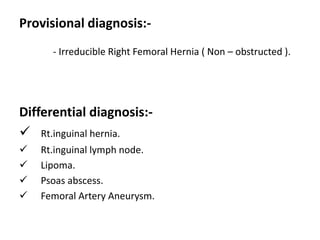
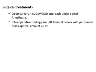
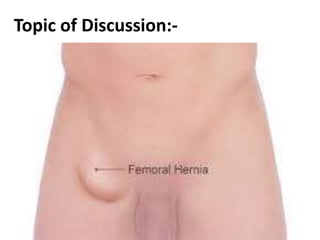
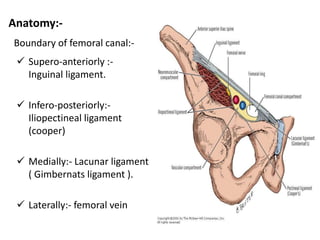
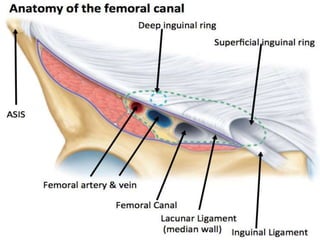
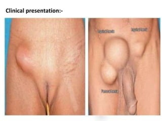
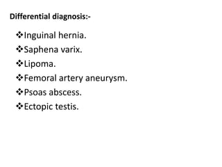
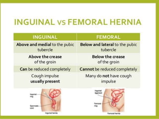
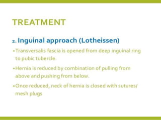
Case presentation:-
Intern :- Amrit Pokhrel
GMCTH,Pokhara
Personal details:-
A 49 yrs female residing on Baglung, Housewife by occupation
presented at Surgery-OPD with
Chief complaints:-
 Swelling on right inguinal region since 4 yrs.
 Pain for 15 days
History of present illness:-
Swelling on rt.inguinal region:-
 Since 4 yrs.
 Develops spontaneously.
 Associated with pain.
 Grows slowly.
 Not a/w fever, ulceration, loss of body weight and
swelling is single.
Pain:-
 At rt.inguinal region.
 Insidious onset.
 Pin pricking.
 Radiates toward thigh.
 Aggravated by standing & weight lifting.
 Relief by rest on supine position.
 Pain is mild.
Past history:-
 No significant past medical ( like:- chronic cough, constipation
and retention of urine ) and surgical history in past.
Personal history:-
 Patient is non-alcoholic, non-smoker, non-vegetarian diet.
Family history:-
 No significant family history.
 Eight member family with five kids.
General physical examination:-
 General condition – fair
 Jaundice
 Anemia
 Cyanosis
 Clubbing
 Lymph adenopathy
 Edema
 Well hydrated
Vital signs:-
 B.P. - 110/70 mm of Hg.
 Pulse – 76 beats/minute – regular.
 Temperature – 98.F
 RR – 18 breathe / minute
 Spo2 – 98 %
Not found
Systemic examination:-
Chest:- normal vesicular breathe sound present.
CVS :- S1 + S2 + Mo
CNS:- grossly intact.
Per abdomen:-
 Inspection:-
 Scaphoid shape.
 Umbilicus inverted and centrally located
 All quadrants moves equally with respiration.
 Visible pulsation and peristalsis are absent.
 Protrusion from right sided saphenous opening.
 On palpation:-
 On percussion:-
 Tympanic sound found.
On Auscultation:-
 Bowel sound present – 3/ minute.
 No localize rise of
temperature.
 No tenderness.
 No organomegaly
detected.
Local examination:-
On standing position:-
 Inspection:- single swelling
- at upper – medial thigh.
- cough impulse present.
- similar to skin color.
- globular.
- 2.5 x 2.5 cm
- well defined edge.
- no pulsation & peristalsis.
- no move with respiration.
- skin over swelling normal.
On supine position:-
 There is no alteration of
shape and size of the
swelling i.e. similar to
standing position.
 Palpation:-
- No localize rise of temperature.
- No tenderness.
- Size 2.5 x 2.5 cm, globular.
- Smooth surface.
- Cough impulse present.
- Distinct edge but not palpable upper edge.
- Firm consistency.
- Non fluctuating.
- No fluid thrill.
- Non translucent.
- Non reducible.
- Compressible.
- Non pulsatile and not fixed to overlying skin.
 on percussion:-
- Dull on percussion.
 On Auscultation:-
- No bruits.
- No gurgling and bowel sound.
Laboratory findings:-
 TLC – 8200 Hb – 13.5 ESR – 10
 RBS – 80 Urea – 31 Creatinine – 1.0
 Na+ - 145 K+ - 4.1
 BT – 2’ 15” CT – 7’ 30” PT/INR – 13”/1.0
 Serology – negative
 Urine R/E – Pus cell – 12 to 14
- Epithelial cell – 1 to 3
- RBC – nil
 USG abdomen and pelvis.
- Right Femoral Hernia.
Provisional diagnosis:-
- Irreducible Right Femoral Hernia ( Non – obstructed ).
Differential diagnosis:-
 Rt.inguinal hernia.
 Rt.inguinal lymph node.
 Lipoma.
 Psoas abscess.
 Femoral Artery Aneurysm.
Surgical treatment:-
 Open surgery – LOCKWOOD approach under Spinal
Anesthesia.
 Intra operative findings are:- Rt.femoral hernia with peritoneal
fluids approx. amount 20 ml
Topic of Discussion:-
Anatomy:-
Boundary of femoral canal:-
 Supero-anteriorly :-
Inguinal ligament.
 Infero-posteriorly:-
Iliopectineal ligament
(cooper)
 Medially:- Lacunar ligament
( Gimbernats ligament ).
 Laterally:- femoral vein
Hernia:-
Femoral hernia :-
Clinical presentation:-
Clinical features:-
 Usually presents with pain or discomfort in groin.
 Groin lump.
 Usually not reducible.
 Mild pain exacerbated by bending or lifting.
 Mild tenderness.
In case of obstruction:-
 Colicky abdominal pain.
 Vomiting, constipation
 Abdominal distension.
 Lump irreducible and tender.
In case of strangulation:-
 Lump is very tender.
 Skin over the lump red and hot.
 Features of shock.
Diagnosis:-
History and Clinical examination.
Ultrasonography – abdomen and pelvis.
Differential diagnosis:-
Inguinal hernia.
Saphena varix.
Lipoma.
Femoral artery aneurysm.
Psoas abscess.
Ectopic testis.
Treatment:-
Surgery case presentation. femoral hernia.

Surgery case presentation. femoral hernia.

  • 1.
    Case presentation:- Intern :-Amrit Pokhrel GMCTH,Pokhara
  • 2.
    Personal details:- A 49yrs female residing on Baglung, Housewife by occupation presented at Surgery-OPD with Chief complaints:-  Swelling on right inguinal region since 4 yrs.  Pain for 15 days
  • 3.
    History of presentillness:- Swelling on rt.inguinal region:-  Since 4 yrs.  Develops spontaneously.  Associated with pain.  Grows slowly.  Not a/w fever, ulceration, loss of body weight and swelling is single.
  • 4.
    Pain:-  At rt.inguinalregion.  Insidious onset.  Pin pricking.  Radiates toward thigh.  Aggravated by standing & weight lifting.  Relief by rest on supine position.  Pain is mild.
  • 5.
    Past history:-  Nosignificant past medical ( like:- chronic cough, constipation and retention of urine ) and surgical history in past. Personal history:-  Patient is non-alcoholic, non-smoker, non-vegetarian diet. Family history:-  No significant family history.  Eight member family with five kids.
  • 6.
    General physical examination:- General condition – fair  Jaundice  Anemia  Cyanosis  Clubbing  Lymph adenopathy  Edema  Well hydrated Vital signs:-  B.P. - 110/70 mm of Hg.  Pulse – 76 beats/minute – regular.  Temperature – 98.F  RR – 18 breathe / minute  Spo2 – 98 % Not found
  • 7.
    Systemic examination:- Chest:- normalvesicular breathe sound present. CVS :- S1 + S2 + Mo CNS:- grossly intact. Per abdomen:-  Inspection:-  Scaphoid shape.  Umbilicus inverted and centrally located  All quadrants moves equally with respiration.  Visible pulsation and peristalsis are absent.  Protrusion from right sided saphenous opening.
  • 8.
     On palpation:- On percussion:-  Tympanic sound found. On Auscultation:-  Bowel sound present – 3/ minute.  No localize rise of temperature.  No tenderness.  No organomegaly detected.
  • 9.
    Local examination:- On standingposition:-  Inspection:- single swelling - at upper – medial thigh. - cough impulse present. - similar to skin color. - globular. - 2.5 x 2.5 cm - well defined edge. - no pulsation & peristalsis. - no move with respiration. - skin over swelling normal. On supine position:-  There is no alteration of shape and size of the swelling i.e. similar to standing position.
  • 10.
     Palpation:- - Nolocalize rise of temperature. - No tenderness. - Size 2.5 x 2.5 cm, globular. - Smooth surface. - Cough impulse present. - Distinct edge but not palpable upper edge. - Firm consistency. - Non fluctuating. - No fluid thrill. - Non translucent. - Non reducible. - Compressible. - Non pulsatile and not fixed to overlying skin.
  • 11.
     on percussion:- -Dull on percussion.  On Auscultation:- - No bruits. - No gurgling and bowel sound.
  • 12.
    Laboratory findings:-  TLC– 8200 Hb – 13.5 ESR – 10  RBS – 80 Urea – 31 Creatinine – 1.0  Na+ - 145 K+ - 4.1  BT – 2’ 15” CT – 7’ 30” PT/INR – 13”/1.0  Serology – negative  Urine R/E – Pus cell – 12 to 14 - Epithelial cell – 1 to 3 - RBC – nil  USG abdomen and pelvis. - Right Femoral Hernia.
  • 13.
    Provisional diagnosis:- - IrreducibleRight Femoral Hernia ( Non – obstructed ). Differential diagnosis:-  Rt.inguinal hernia.  Rt.inguinal lymph node.  Lipoma.  Psoas abscess.  Femoral Artery Aneurysm.
  • 14.
    Surgical treatment:-  Opensurgery – LOCKWOOD approach under Spinal Anesthesia.  Intra operative findings are:- Rt.femoral hernia with peritoneal fluids approx. amount 20 ml
  • 15.
  • 16.
    Anatomy:- Boundary of femoralcanal:-  Supero-anteriorly :- Inguinal ligament.  Infero-posteriorly:- Iliopectineal ligament (cooper)  Medially:- Lacunar ligament ( Gimbernats ligament ).  Laterally:- femoral vein
  • 18.
  • 19.
  • 21.
  • 22.
    Clinical features:-  Usuallypresents with pain or discomfort in groin.  Groin lump.  Usually not reducible.  Mild pain exacerbated by bending or lifting.  Mild tenderness. In case of obstruction:-  Colicky abdominal pain.  Vomiting, constipation  Abdominal distension.  Lump irreducible and tender.
  • 23.
    In case ofstrangulation:-  Lump is very tender.  Skin over the lump red and hot.  Features of shock.
  • 24.
    Diagnosis:- History and Clinicalexamination. Ultrasonography – abdomen and pelvis.
  • 25.
    Differential diagnosis:- Inguinal hernia. Saphenavarix. Lipoma. Femoral artery aneurysm. Psoas abscess. Ectopic testis.
  • 28.