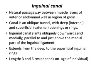
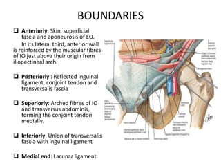
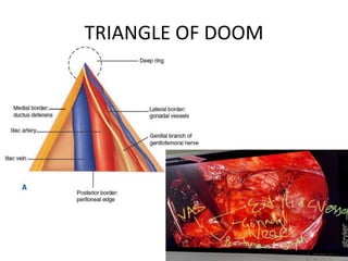
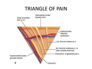
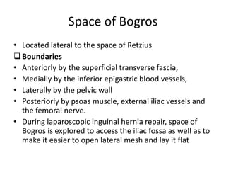
Hernia: Inguinal – Surgical anatomy,
presentation, treatment,
complications
Introduction
Abnormal protrusion of organ or tissue
through opening in the layer that normally
confines it.
Inguinal ligament
• Thick lower border of
aponeurosis of external
oblique
• Stretches between ASIS &
pubic tubercle.
• Lower border, it is fused
with fascia lata.
• Laterally fused with
iliopsoas fascia
• Medial end of inguinal
ligament, near its site of
attachment to pubic
tubercle, form triangular,
shelf-like lacunar ligament.
Inguinal canal
• Natural passageway between muscle layers of
anterior abdominal wall in region of groin
• Canal is an oblique tunnel, with deep (internal)
and superficial (external) openings or rings.
• Inguinal canal slants obliquely downwards and
medially, parallel to and just above the medial
part of the inguinal ligament.
• Extends from the deep to the superficial inguinal
rings
• Length: 3 and 6 cm(depends on age of individual)
BOUNDARIES
 Anteriorly: Skin, superficial
fascia and aponeurosis of EO.
In its lateral third, anterior wall
is reinforced by the muscular fibres
of IO just above their origin from
iliopectineal arch.
 Posteriorly : Reflected inguinal
ligament, conjoint tendon and
transversalis fascia
 Superiorly: Arched fibres of IO
and transversus abdominis,
forming the conjoint tendon
medially.
 Inferiorly: Union of transversalis
fascia with inguinal ligament
 Medial end: Lacunar ligament.
Contents
• Spermatic cord
Spermatic cord constitutes- vas deferens, testicular & cremastic
arteries , pampiniform plexus of veins, lymphatics
• Ilioinguinal nerve
• Genital branch of genitofemoral nerve
• Females – Round ligament is present instead of spermatic cord.
Superficial inguinal ring
• Hiatus in the aponeurosis of external oblique, Lies 1.25
cm above and lateral to pubic tubercle
• Triangular, with its apex pointing laterally towards
ASIS
• The ring is smaller in the female.
Deep inguinal ring
• U shaped condensation of the fascia
trasversalis
• Approximately midway between the anterior
superior iliac spine and the pubic symphysis,
and about 1.25 cm above inguinal ligament.
• Oval, with a roughly vertical long axis.
• Size varies between individuals but it is usually
1–2 cm wide in adults and larger in male.
• Inferior epigastric vessels run in medial border
of deep inguinal ring.
• Traction on fascial ring exerted by contraction
of internal oblique may narrow opening when
intra-abdominal pressure is increased.
Conjoint tendon
• Formed from lower fibres of IO and lower part of the aponeurosis
of transversus abdominis.
• Attached to pubic crest and extends to a variable extent along
pectineal line
• Descends behind superficial inguinal ring and acts to strengthen the
medial portion of posterior wall of inguinal canal.
• Medially, upper fibres of tendon fuse with anterior wall of rectus
sheath
Midinguinal point : Halfway between ASIS &
pubic symphysis.
• In adults, it is the approximate surface marking
of the femoral artery (just below the ligament)
Midpoint of inguinal ligament: Midpoint
between ASIS and pubic tubercle
• Lies just lateral to mid-inguinal point.
Hesselbach’s inguinal triangle
• Lateral border : Inferior
epigastric artery
• Inferior border: Inguinal
ligament
• Medial border: Lateral
margin of rectus
abdominis
TRIANGLE OF DOOM
TRIANGLE OF PAIN
Myopectineal orifice of Fruchaud
Lateral: Iliopsoas
Medial: Rectus Sheath & rectus abdominis
Superiorly: Arching fiber of transversus
abdominis and internal oblique
Inferior: Iliopectineal line and Cooper's
ligament and Pecten pubis
Pectineal ligament
• Known as the inguinal ligament of Cooper
• Extension of the lacunar ligament that runs
along the pectineal line of the pubis (also
known as the pecten pubis).
COOPERS LIGAMENT-LIGHT HOUSE
• 2 spaces are potential non-natural cavities
under the lower anterior abdominal wall, and
they lie in between the superficial transverse
fascia and the peritoneum
• Created by blunt separation when performing
a laparoscopic inguinal hernia repair.
• Space of Retzius: Space formed by the fold of
the tight fusion of deep transverse fascia and
peritoneum between bladder and the
peritoneum, which includes the bladder and is
filled with loose connective tissue
Space of Bogros
• Located lateral to the space of Retzius
Boundaries
• Anteriorly by the superficial transverse fascia,
• Medially by the inferior epigastric blood vessels,
• Laterally by the pelvic wall
• Posteriorly by psoas muscle, external iliac vessels and
the femoral nerve.
• During laparoscopic inguinal hernia repair, space of
Bogros is explored to access the iliac fossa as well as to
make it easier to open lateral mesh and lay it flat
RETZIUS SPACE AND SPACE OF
BOGROS
Corona mortis
• Abnormal arterial or venous anastomosis
between external iliac, Inferior epigastric
artery and obturator system of vessels
• May cause significant hemorrhage during
pelvi-acetabular fracture surgeries, hernia
repair and laparoscopic gynecological
procedures.
• 21 studies (n=2184 hemi-pelvises) were included in the
meta-analysis.
• Overall prevalence of the corona mortis in hemi-pelvises is
high (49.3%).
• A venous corona mortis is more prevalent than an arterial
corona mortis (41.7% vs. 17.0%). The corona mortis is more
common in Asia (59.3%) than in Europe (42.8%) and North
America (44.3%).
Defense mechanism of Inguinal canal
1. Obliquity of inguinal canal
two inguinal ring do not lie opposite to each other ,
therefore rise in intra-abdominal pressure
approximate anterior and posterior abdominal wall
called flap valve mechanism .
2. Ball and valve mechanism :
contraction of cremaster helps spermatic cord to
plug superficial inguinal ring
3 . Shutter mechanism of internal oblique : as
internal oblique contribute to anterior wall , roof
and posterior wall of canal
When it contracts the roof is approximated to
floor like a shutter
4. Slit valve mechanism – Contraction of external
oblique , approximate two crura of superficial
inguinal ring
Clinical types
• Reducible –contents can be returned into the abdominal
cavity.
• Irreducible – contents cannot be returned into the abdominal
cavity.
• Obstructed – irreducibilty + intestinal obstruction, but the
blood supply is not impaired.
• Strangulated- Arrest of the blood supply leading to ischemia
and infarction.
• Incarcerated: Hernia is not only irreducible but also potentially
developing strangulation.
Epidemiology
•Approximately 7% of all surgical outpatient.
•Accounts for 96% groin hernias (other 4% are femoral)
•Bilateral in 20% of cases
•Lifetime risk of inguinal hernia: 10%
•M:F 9:1
• Affects 1-3% of young children
• In men the incidence rises from 11 per 10,000 person years aged
16-24 years to 200 per 10,000 person years aged 75 years or above.
• Extremely common; represents the most frequent problem
requiring surgical intervention in the paediatric age group
• Much more common in boys (90% of cases) than girls
• Definite familial tendency,
• more frequent on the right side as a result of later descent of the
right testis and delayed obliteration of the right processus vaginalis.
Anatomical classification
• Indirect hernia – more common about 2/3 of
inguinal hernia .
• It is more common in young
• Direct hernia- more common in old
Indirect inguinal hernia
• Indirect/Lateral/oblique is most common of
two types of inguinal hernia (65% of inguinal
hernia , and 55% are right sided , 12% B/L)
• In most of all indirect hernia ,embryonic
processus vaginalis remains open or patent
considered congenital origin
• Protruding peritoneal sac enters inguinal canal
through deep inguinal ring
• Origin is lateral to inferior epigastric artery
• Bubonocele –Limited to inguinal canal
• Funicular – Just above testis
• Complete or Vaginal – Reached to scrotum
Direct inguinal hernia
• Peritoneal sac enters medial end of inguinal
canal directly through weakened posterior
wall
• Considered acquired , as it develops when
musculature has been weakened
• Medial to inferior epigastric vessels
Sliding hernia
• Third type of inguinal hernia ,
• Also an acquired hernia due to wearing of
abdominal wall , but occurs at deep inguinal
ring lateral to inferior epigastric vessels ,
• Retroperitoneal fatty tissue is pushed
downwards along inguinal canal
• However sac has formed secondarily ,
distinguishing it from classical indirect hernia ,
• Left- sigmoid colon may be pulled
• Right - caecum
Composition of hernia
• Sac
• Covering of sac
• Content of sac
Sac
• Diverticuliculum of peritoneum
• Mouth, neck , body , fundus
• Usually neck is well defined and its diameter is
important
• Covering –Derived from layers of abdominal wall
through which sac passes , in long standing cases
, covering may be atrophied
That makes anatomy indistinguishable
Contents
• Omentum – omentocele
• Intestine –enterocle
• Portion of the circumference of the bowel: Richter’s Hernia
• Portion of the urinary bladder.
• Appendix: Amyand's hernia
• Meckel’s diverticulum: Littre’s hernia
• Fallopian tubes
• Maydl's hernia:
 Rare type of incarcerated hernia popularly known as a hernia in “W”
which describes the orientation of the bowel in hernia sac
 vulnerability of central segment of bowel to undergo intra-abdominal
closed-loop strangulation which may go unnoticed.
• Fluid – part of ascites
Direct hernia
• Contents herniate directly through posterior wall of
inguinal canal through Hesselbach’s triangle
• Weakness in posterior wall of inguinal canal
• Bounded laterally -inferior epigastric artery,
medially – lateral border of rectus abdominus muscle
inferiorly – inguinal ligament
Presentation
• Often Asymptomatic
• Groin bulge
• Dull felling or heaviness in groin
• Focal pain/ burning sensation (suspicion of incarceration or strangulation)
• Pain
 Localized pain
 Referred pain
 Generalized pain
• Nausea and vomiting
• Constipation
• Urinary symptoms
Presentation
• At first appearance, it is easily reducible.
• With time it can no longer be reduced, it is irreducible or
incarcerated.
• Strangulation: when visceral contents of the hernia become
twisted or entrapped by the narrow opening.
Strangulation usually leads to bowel obstruction with sudden,
severe pain in the hernia, vomiting and irreducibility.
Nyhus Classification System
European hernia society (EHS) for
groin hernia
• PL2 – primary , indirect , inguinal hernia with 3cm
defect
Other
• Casting, Halverson and McVay , zollinger ,
Gilbert classifications
REFERENCE
• GREYS ANATOMY
• NETTER ATLAS
• SABISTON TEXTBOOK OF SURGERY
• BAILEY AND LOVE

hernia.pptx

  • 1.
    Hernia: Inguinal –Surgical anatomy, presentation, treatment, complications
  • 2.
    Introduction Abnormal protrusion oforgan or tissue through opening in the layer that normally confines it.
  • 3.
    Inguinal ligament • Thicklower border of aponeurosis of external oblique • Stretches between ASIS & pubic tubercle. • Lower border, it is fused with fascia lata. • Laterally fused with iliopsoas fascia • Medial end of inguinal ligament, near its site of attachment to pubic tubercle, form triangular, shelf-like lacunar ligament.
  • 4.
    Inguinal canal • Naturalpassageway between muscle layers of anterior abdominal wall in region of groin • Canal is an oblique tunnel, with deep (internal) and superficial (external) openings or rings. • Inguinal canal slants obliquely downwards and medially, parallel to and just above the medial part of the inguinal ligament. • Extends from the deep to the superficial inguinal rings • Length: 3 and 6 cm(depends on age of individual)
  • 5.
    BOUNDARIES  Anteriorly: Skin,superficial fascia and aponeurosis of EO. In its lateral third, anterior wall is reinforced by the muscular fibres of IO just above their origin from iliopectineal arch.  Posteriorly : Reflected inguinal ligament, conjoint tendon and transversalis fascia  Superiorly: Arched fibres of IO and transversus abdominis, forming the conjoint tendon medially.  Inferiorly: Union of transversalis fascia with inguinal ligament  Medial end: Lacunar ligament.
  • 6.
    Contents • Spermatic cord Spermaticcord constitutes- vas deferens, testicular & cremastic arteries , pampiniform plexus of veins, lymphatics • Ilioinguinal nerve • Genital branch of genitofemoral nerve • Females – Round ligament is present instead of spermatic cord.
  • 7.
    Superficial inguinal ring •Hiatus in the aponeurosis of external oblique, Lies 1.25 cm above and lateral to pubic tubercle • Triangular, with its apex pointing laterally towards ASIS • The ring is smaller in the female.
  • 8.
    Deep inguinal ring •U shaped condensation of the fascia trasversalis • Approximately midway between the anterior superior iliac spine and the pubic symphysis, and about 1.25 cm above inguinal ligament. • Oval, with a roughly vertical long axis. • Size varies between individuals but it is usually 1–2 cm wide in adults and larger in male.
  • 9.
    • Inferior epigastricvessels run in medial border of deep inguinal ring. • Traction on fascial ring exerted by contraction of internal oblique may narrow opening when intra-abdominal pressure is increased.
  • 10.
    Conjoint tendon • Formedfrom lower fibres of IO and lower part of the aponeurosis of transversus abdominis. • Attached to pubic crest and extends to a variable extent along pectineal line • Descends behind superficial inguinal ring and acts to strengthen the medial portion of posterior wall of inguinal canal. • Medially, upper fibres of tendon fuse with anterior wall of rectus sheath
  • 11.
    Midinguinal point :Halfway between ASIS & pubic symphysis. • In adults, it is the approximate surface marking of the femoral artery (just below the ligament) Midpoint of inguinal ligament: Midpoint between ASIS and pubic tubercle • Lies just lateral to mid-inguinal point.
  • 13.
    Hesselbach’s inguinal triangle •Lateral border : Inferior epigastric artery • Inferior border: Inguinal ligament • Medial border: Lateral margin of rectus abdominis
  • 14.
  • 15.
  • 18.
    Myopectineal orifice ofFruchaud Lateral: Iliopsoas Medial: Rectus Sheath & rectus abdominis Superiorly: Arching fiber of transversus abdominis and internal oblique Inferior: Iliopectineal line and Cooper's ligament and Pecten pubis
  • 20.
    Pectineal ligament • Knownas the inguinal ligament of Cooper • Extension of the lacunar ligament that runs along the pectineal line of the pubis (also known as the pecten pubis).
  • 21.
  • 22.
    • 2 spacesare potential non-natural cavities under the lower anterior abdominal wall, and they lie in between the superficial transverse fascia and the peritoneum • Created by blunt separation when performing a laparoscopic inguinal hernia repair.
  • 23.
    • Space ofRetzius: Space formed by the fold of the tight fusion of deep transverse fascia and peritoneum between bladder and the peritoneum, which includes the bladder and is filled with loose connective tissue
  • 24.
    Space of Bogros •Located lateral to the space of Retzius Boundaries • Anteriorly by the superficial transverse fascia, • Medially by the inferior epigastric blood vessels, • Laterally by the pelvic wall • Posteriorly by psoas muscle, external iliac vessels and the femoral nerve. • During laparoscopic inguinal hernia repair, space of Bogros is explored to access the iliac fossa as well as to make it easier to open lateral mesh and lay it flat
  • 25.
    RETZIUS SPACE ANDSPACE OF BOGROS
  • 26.
    Corona mortis • Abnormalarterial or venous anastomosis between external iliac, Inferior epigastric artery and obturator system of vessels • May cause significant hemorrhage during pelvi-acetabular fracture surgeries, hernia repair and laparoscopic gynecological procedures.
  • 28.
    • 21 studies(n=2184 hemi-pelvises) were included in the meta-analysis. • Overall prevalence of the corona mortis in hemi-pelvises is high (49.3%). • A venous corona mortis is more prevalent than an arterial corona mortis (41.7% vs. 17.0%). The corona mortis is more common in Asia (59.3%) than in Europe (42.8%) and North America (44.3%).
  • 29.
    Defense mechanism ofInguinal canal 1. Obliquity of inguinal canal two inguinal ring do not lie opposite to each other , therefore rise in intra-abdominal pressure approximate anterior and posterior abdominal wall called flap valve mechanism . 2. Ball and valve mechanism : contraction of cremaster helps spermatic cord to plug superficial inguinal ring
  • 30.
    3 . Shuttermechanism of internal oblique : as internal oblique contribute to anterior wall , roof and posterior wall of canal When it contracts the roof is approximated to floor like a shutter 4. Slit valve mechanism – Contraction of external oblique , approximate two crura of superficial inguinal ring
  • 31.
    Clinical types • Reducible–contents can be returned into the abdominal cavity. • Irreducible – contents cannot be returned into the abdominal cavity. • Obstructed – irreducibilty + intestinal obstruction, but the blood supply is not impaired. • Strangulated- Arrest of the blood supply leading to ischemia and infarction. • Incarcerated: Hernia is not only irreducible but also potentially developing strangulation.
  • 32.
    Epidemiology •Approximately 7% ofall surgical outpatient. •Accounts for 96% groin hernias (other 4% are femoral) •Bilateral in 20% of cases •Lifetime risk of inguinal hernia: 10% •M:F 9:1
  • 33.
    • Affects 1-3%of young children • In men the incidence rises from 11 per 10,000 person years aged 16-24 years to 200 per 10,000 person years aged 75 years or above. • Extremely common; represents the most frequent problem requiring surgical intervention in the paediatric age group • Much more common in boys (90% of cases) than girls • Definite familial tendency, • more frequent on the right side as a result of later descent of the right testis and delayed obliteration of the right processus vaginalis.
  • 35.
    Anatomical classification • Indirecthernia – more common about 2/3 of inguinal hernia . • It is more common in young • Direct hernia- more common in old
  • 36.
    Indirect inguinal hernia •Indirect/Lateral/oblique is most common of two types of inguinal hernia (65% of inguinal hernia , and 55% are right sided , 12% B/L) • In most of all indirect hernia ,embryonic processus vaginalis remains open or patent considered congenital origin • Protruding peritoneal sac enters inguinal canal through deep inguinal ring • Origin is lateral to inferior epigastric artery
  • 37.
    • Bubonocele –Limitedto inguinal canal • Funicular – Just above testis • Complete or Vaginal – Reached to scrotum
  • 38.
    Direct inguinal hernia •Peritoneal sac enters medial end of inguinal canal directly through weakened posterior wall • Considered acquired , as it develops when musculature has been weakened • Medial to inferior epigastric vessels
  • 40.
    Sliding hernia • Thirdtype of inguinal hernia , • Also an acquired hernia due to wearing of abdominal wall , but occurs at deep inguinal ring lateral to inferior epigastric vessels , • Retroperitoneal fatty tissue is pushed downwards along inguinal canal • However sac has formed secondarily , distinguishing it from classical indirect hernia , • Left- sigmoid colon may be pulled • Right - caecum
  • 41.
    Composition of hernia •Sac • Covering of sac • Content of sac
  • 42.
    Sac • Diverticuliculum ofperitoneum • Mouth, neck , body , fundus • Usually neck is well defined and its diameter is important • Covering –Derived from layers of abdominal wall through which sac passes , in long standing cases , covering may be atrophied That makes anatomy indistinguishable
  • 43.
    Contents • Omentum –omentocele • Intestine –enterocle • Portion of the circumference of the bowel: Richter’s Hernia • Portion of the urinary bladder. • Appendix: Amyand's hernia • Meckel’s diverticulum: Littre’s hernia • Fallopian tubes • Maydl's hernia:  Rare type of incarcerated hernia popularly known as a hernia in “W” which describes the orientation of the bowel in hernia sac  vulnerability of central segment of bowel to undergo intra-abdominal closed-loop strangulation which may go unnoticed. • Fluid – part of ascites
  • 44.
    Direct hernia • Contentsherniate directly through posterior wall of inguinal canal through Hesselbach’s triangle • Weakness in posterior wall of inguinal canal • Bounded laterally -inferior epigastric artery, medially – lateral border of rectus abdominus muscle inferiorly – inguinal ligament
  • 45.
    Presentation • Often Asymptomatic •Groin bulge • Dull felling or heaviness in groin • Focal pain/ burning sensation (suspicion of incarceration or strangulation) • Pain  Localized pain  Referred pain  Generalized pain • Nausea and vomiting • Constipation • Urinary symptoms
  • 46.
    Presentation • At firstappearance, it is easily reducible. • With time it can no longer be reduced, it is irreducible or incarcerated. • Strangulation: when visceral contents of the hernia become twisted or entrapped by the narrow opening. Strangulation usually leads to bowel obstruction with sudden, severe pain in the hernia, vomiting and irreducibility.
  • 47.
  • 48.
    European hernia society(EHS) for groin hernia • PL2 – primary , indirect , inguinal hernia with 3cm defect
  • 49.
    Other • Casting, Halversonand McVay , zollinger , Gilbert classifications
  • 50.
    REFERENCE • GREYS ANATOMY •NETTER ATLAS • SABISTON TEXTBOOK OF SURGERY • BAILEY AND LOVE