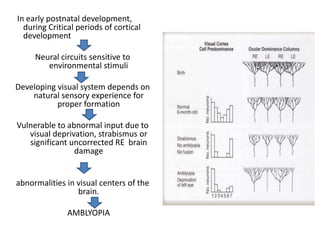
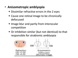
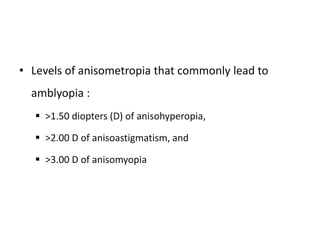
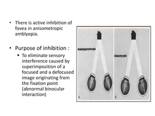
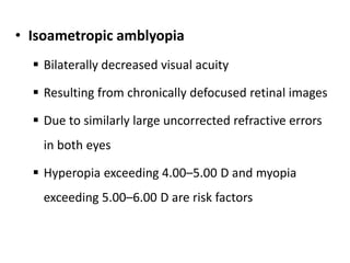
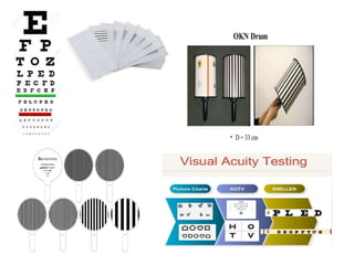
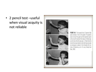
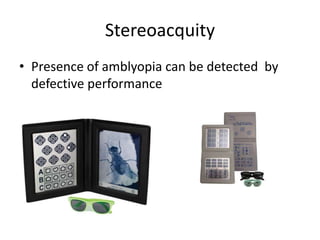
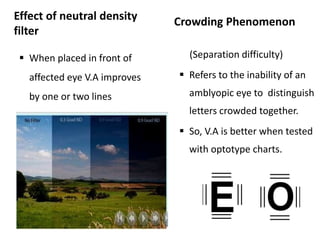
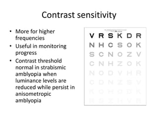
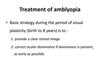
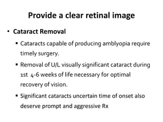
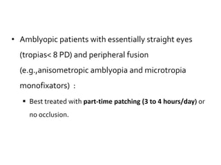
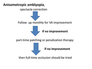
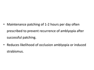
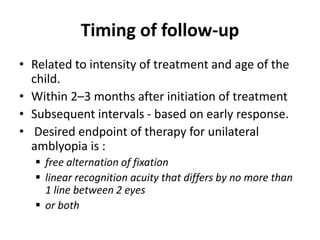
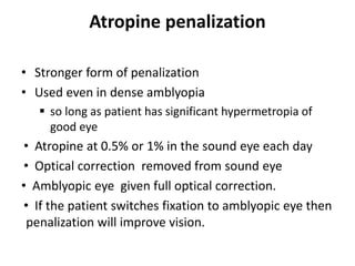
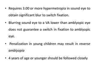
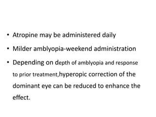
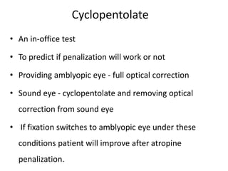
Amblyopia
Arika Poudel
1st year resident
MD Ophthalmology
Contents
Definition of Amblyopia
Pathophysiology
Classification
Clinical characteristics
Evaluation and diagnosis
Treatment
Prognosis
• Derived from Greek language
• Amblys – dull; ops – eye ‘‘dullness of vision’’
• U/L or ( less commonly) B/L reduction of Best
corrected visual acuity
• Cannot be attributed directly to any structural
abnormality of eye or visual pathways
• A spectrum of visual
loss, ranging from
missing a few letters on
6/6 line to HM
• At least 2 Snellen lines
difference in BCVA
between 2 eyes
Pathophysiology
• During fetal development:
 Differentiation and organization of the visual system are
likely guided by intrinsic control mechanisms
• At birth:
 Process is not complete and must continue to develop
throughout the first decade of life.
 Unlike the prenatal period, environmental factors and
visual experience influence the process in postnatal life.
For normal visual development
Both eyes must be presented
with equally clear and similar
images
This constant stimulation is
crucial to the development of
normal vision
Any process that interferes with
this process can lead to the
development of amblyopia
Sensitive or critical period
• The human visual system is
sensitive to the effects of
depriving vision.
• Extends roughly from birth
through the end of the first
decade of life.
• Vulnerability is greatest
during the first few months
of life and thereafter
gradually decreases.
• During these periods:
 Visual system’s plasticity allows greatest
opportunity for reversal of amblyopia.
• Abnormal visual experiences
 Cells of primary visual cortex can lose innate
ability to respond to stimulation of 1 or both eyes
 Cells that remain responsive can show significant
functional deficiencies.
• Abnormal visual experience during the critical period
can take one of two forms:
 A lack of exposure to the well-formed images necessary
for normal development interferes with the maturation of
form vision.
 A marked disparity in the quality or directionality of
inputs from the two eyes leads to abnormal competitive
binocular interaction.
• Either of these two mechanisms can contribute to
unilateral or bilateral amblyopia.
In early postnatal development,
during Critical periods of cortical
development
Neural circuits sensitive to
environmental stimuli
Developing visual system depends on
natural sensory experience for
proper formation
Vulnerable to abnormal input due to
visual deprivation, strabismus or
significant uncorrected RE brain
damage
abnormalities in visual centers of the
brain.
AMBLYOPIA
Classification
• Strabismic amblyopia
• Refractive amblyopia
• Visual deprivational amblyopia
• Special forms of amblyopia
• Occlusion amblyopia
• Idiopathic amblyopia
Strabismic amblyopia
• Occurs despite both eyes having clearly focused
retinal images
• Occur in those
 Have strong fixation preference for 1 eye
 Have U/l rather than an alternating fixation
pattern
 Constantly suppress cortical activity from deviated
eye
• Those with alternate fixation and alternate
suppression do not have amblyopia, but have
abnormal binocular function
• Competitive or inhibitory interaction between
neurons carrying nonfusible input from 2 eyes
Domination of cortical vision centers by input from
fixating eye
Reduced responsiveness to input from nonfixating
eye
• In young children with strabismus, suppression
develops rapidly.
Overlap of different foveal
images from retinas of
fixating eye and deviating
eye
transmitted to visual centers
Inhibition of visual input
from fovea of deviating
eye
(visual adaptation to avoid
diplopia and visual
confusion )
• Refractive Amblyopia
 Refractive amblyopia results from consistent
retinal defocus in 1 or both eyes.
 Anisometropia causes unilateral amblyopia
 Isoametropia causes bilateral amblyopia
• Anisometropic amblyopia
 Dissimilar refractive errors in the 2 eyes
 Cause one retinal image to be chronically
defocused
 Image blur and partly from interocular
competition
 Or inhibition similar (but not identical) to that
responsible for strabismic amblyopia
• Levels of anisometropia that commonly lead to
amblyopia :
 >1.50 diopters (D) of anisohyperopia,
 >2.00 D of anisoastigmatism, and
 >3.00 D of anisomyopia
• There is active inhibition of
fovea in anisometropic
amblyopia.
• Purpose of inhibition :
 To eliminate sensory
interference caused by
superimposition of a
focused and a defocused
image originating from
the fixation point
(abnormal binocular
interaction)
• Isoametropic amblyopia
 Bilaterally decreased visual acuity
 Resulting from chronically defocused retinal images
 Due to similarly large uncorrected refractive errors
in both eyes
 Hyperopia exceeding 4.00–5.00 D and myopia
exceeding 5.00–6.00 D are risk factors
• Meridional amblyopia
 Bilateral high astigmatism may cause loss of
resolving ability specific to the chronically blurred
meridians
• Most ophthalmologists recommend correction for
eyes with more than 2.00–3.00 D of cylinder.
Visual deprivation amblyopia
• Least common form
• Most severe
• Difficult to treat
• Develops faster, and is deeper than strabismic or
anisometropic amblyopia.
• Interferes with central vision
• Adds to the direct impact of image degradation
• Causes:
 Congenital or early-acquired cataract
 Ptosis
 Periocular lesions obstructing the
visual axis
 Corneal opacities
 Vitreous hemorrhage
• Unilateral visual deprivation tends
to cause vision deficits more than
bilateral deprivation of the same
degree because of interocular
competition
• Even in bilateral cases, visual acuity can be 6/60 or
worse if not treated early.
• In children younger than 6 years:
 Dense cataracts occupying the central 3 mm or more of the
lens
• Similar lens opacities acquired after age 6 years are
generally less harmful and have no effect on visual
development
• Reverse amblyopia (occlusion amblyopia)
 Is a form of visual deprivation amblyopia
 Develop in the initially dominant eye
 If patched excessively during treatment of
amblyopia in the other eye
• Idiopathic amblyopia
 Diagnosed in retrospect and
 no detectable cause
 responds with improved vision during a trial of
treatment for amblyopia.
 Presumably, an amblyopiogenic process was
present earlier in the child's life that has since
resolved.
• Organic Amblyopia
 Caused by a structural abnormality of the eye,
such as :
optic nerve hypoplasia,
coloboma, or a
 partial cataract
Clinical characteristics
• Visual Acuity
 Difference in 2 lines on V.A chart
 Recognition Acuity – (Snellen) is more affected
than Resolution acuity ( Teller’s or VER)and
detection acuity ( Catford Drum test)
 Grating Acuity is less affected in strabismic
amblyopia
• 2 pencil test –useful
when visual acquity is
not reliable
Stereoacquity
• Presence of amblyopia can be detected by
defective performance
Effect of neutral density
filter
 When placed in front of
affected eye V.A improves
by one or two lines
Crowding Phenomenon
(Separation difficulty)
 Refers to the inability of an
amblyopic eye to distinguish
letters crowded together.
 So, V.A is better when tested
with optotype charts.
Contrast sensitivity
• More for higher
frequencies
• Useful in monitoring
progress
• Contrast threshold
normal in strabismic
amblyopia when
luminance levels are
reduced while persist in
anisometropic
amblyopia
Fixation Pattern
• Central fixation :
 Foveolar fixation
• Eccentric viewing :
 Extrafoveal point because of central suppression
scotoma.
 Fovea still not lost its principal visual direction.
 Patient look past the object they have been asked to fix.
• Eccentric fixation :
 Fovea lost its principal visual direction
 If an image is projected onto the fovea patient report
that the object is seen in some other direction than
straight ahead.
• Pupillary light reflexes
 Generally normal.
 RAPD may occur in deep amblyopia
• Absolute central scotoma
• Localisation of object of regard :
 Normal in patients with amblyopia & eccentric
fixation
 Abnormal in eccentric viewing
• Light and Dark Adaptations :
 Usually normal
• Light Perception & Form vision – Dissociated
• Occasionally Nystagmus
• VEP
 reduction n amplitude abd slightly prolonged latancy
EVALUATION AND DIAGNOSIS
• Clinical History
• Evaluation of V.A and fixation pattern
• Binocular alignment and ocular motility
• External Examination
• Pupil examination
• Anterior and Fundus examination
• Cycloplegic retinoscopy
• Steroacquity
• Neutral density filter test
• Test for crowding phenomenon
Treatment of amblyopia
• Basic strategy during the period of visual
plasticity (birth to 8 years) is to :
1. provide a clear retinal image
2. correct ocular dominance if dominance is present,
as early as possible
Provide a clear retinal image
• Cataract Removal
 Cataracts capable of producing amblyopia require
timely surgery.
 Removal of U/L visually significant cataract during
1st 4-6 weeks of life necessary for optimal
recovery of vision.
 Significant cataracts uncertain time of onset also
deserve prompt and aggressive Rx
Refractive error Correction
• Optical prescription based on the refractive error
determined with cycloplegia.
• Because an amblyopic eye’s ability to control
accommodation tends to be impaired, this eye cannot
be relied on to compensate for uncorrected hyperopia
as would a normal child’s eye.
• However symmetric reductions in plus power may be
required for acceptance of spectacle wear.
• Refractive correction for aphakia following
cataract surgery in childhood must be :
 Provided promptly to avoid prolonging visual
deprivation (due to a severe uncorrected refractive
error)
• Anisometropic, isoametropic, and even strabismic
amblyopia :
 May improve or resolve only refractive correction
• Role of refractive surgery and contact lenses is
under investigation.
• In bilateral hypermetropia (5.00D):
 Full hypermetropic correction, as amblyopic eyes do not fully
accommodate
 Patients given partial correction - very slow or no
improvement in their amblyopia
• In large astigmatism (2.50D) (amblyopia secondary to
astigmatism or meridional amblyopia):
 Full astigmatic correction to provide a clear retinal image
• Consider correcting astigmatisms of 2.50 to 3.00 or
more in small children, even if astigmatism is bilateral
Correct Ocular Dominance
OCCLUSION THERAPY (PATCHING )
• Sound eye is covered, obligating child to use
amblyopic eye
• Full time occlusion
• Part time oclussion
Full-time occlusion
• Occlusion of the sound eye during all waking hours
• Strabismic patients with out binocular fusion
• In rare cases, with aggressive patching, strabismus
occur due to lack of binocular viewing and
tenuous fusion.
• Therefore, the child whose eyes are consistently or
intermittently straight may benefit from
being given some opportunity to see binocularly.
• Full-time occlusion may cause reverse amblyopia in
children less than 4 to 5 years.
• To prevent reverse amblyopia :
– Do not use full-time occlusion for > 1 week per the child’s age in
years without re-examining vision of the good eye.
– For e.g., a 2-year-old child receiving full-time occlusion should
be examined every 2 weeks.
Part-time occlusion
• 2-6 hours per day
• Similar to those of prescribed full-time occlusion.
• Relative duration of patch-on and patch-off intervals
should reflect degree of amblyopia.
• For severe deficits (VA of 6/36- 3/60), 6 hours per
day is preferred.
• Amblyopic patients with essentially straight eyes
(tropias< 8 PD) and peripheral fusion
(e.g.,anisometropic amblyopia and microtropia
monofixators) :
 Best treated with part-time patching (3 to 4 hours/day) or
no occlusion.
Anisometropic amblyopia,
spectacle correction
Follow up monthly for VA improvement
if no improvement
part-time patching or penalization therapy
if no improvement
then full-time occlusion should be tried
• Maintenance patching of 1-2 hours per day often
prescribed to prevent recurrence of amblyopia after
successful patching.
• Reduces likelihood of occlusion amblyopia or induced
strabismus.
Timing of follow-up
• Related to intensity of treatment and age of the
child.
• Within 2–3 months after initiation of treatment
• Subsequent intervals - based on early response.
• Desired endpoint of therapy for unilateral
amblyopia is :
 free alternation of fixation
 linear recognition acuity that differs by no more than
1 line between 2 eyes
 or both
• Consistent occlusion during infancy may
reverse substantial strabismic amblyopia in < 1
month
• Adherence to occlusion therapy declines with
increasing age.
PENALIZATION
• A method for blurring sound eye to force
fixation to amblyopic eye.
• As effective as patching for mild to mod
amblyopia (visual acuity of 20/100 or better).
• Penalization only works if fixation is switched
from the sound eye to the amblyopic eye.
Optical penalization
• Based on over-plussing (prescribing more plus
sphere than needed (fogging) or diffusing filters for
the sound eye.
• Sound eye to force fixation to the amblyopic eye for
distance targets
• Works well for mild amblyopia
• Patient will usually use sound eye for near targets.
• may be capable of inducing greater blur.
• more acceptable than occlusion therapy
• Patients must be closely monitored to ensure
proper utilization (no peeking) of spectacle-
borne devices.
Atropine penalization
• Stronger form of penalization
• Used even in dense amblyopia
 so long as patient has significant hypermetropia of
good eye
• Atropine at 0.5% or 1% in the sound eye each day
• Optical correction removed from sound eye
• Amblyopic eye given full optical correction.
• If the patient switches fixation to amblyopic eye then
penalization will improve vision.
• Requires 3.00 or more hypermetropia in sound eye to
obtain significant blur to switch fixation.
• Blurring sound eye to a VA lower than amblyopic eye
does not guarantee a switch in fixation to amblyopic
eye.
• Penalization in young children may result in reverse
amblyopia
• 4 years of age or younger should be followed closely
• Atropine may be administered daily
• Milder amblyopia-weekend administration
• Depending on depth of amblyopia and response
to prior treatment,hyperopic correction of the
dominant eye can be reduced to enhance the
effect.
Cyclopentolate
• An in-office test
• To predict if penalization will work or not
• Providing amblyopic eye - full optical correction
• Sound eye - cyclopentolate and removing optical
correction from sound eye
• If fixation switches to amblyopic eye under these
conditions patient will improve after atropine
penalization.
OCCLUSIVE CONTACT LENS
• Considered as a last resort
• Require close follow-up
• Complications limited the usefulness
• High recurrence to pretreatment VA
• Conjunctival irritation
• Poor contact lens fit
LEVODOPA/CARBIDOPA
• Precursor for the catecholamine dopamine, a
neurotransmitter/neuromodulator known to
influence receptive fields
• Adjunct to patching
• Controversial :
 VA improvement relatively small
 Not clearly better than with patching
 Questions over long-term stability of vision
PLEOPTICS
• A method of treating eccentric fixation
 A bright ring of light is flashed around the fovea to
temporarily “blind” or saturate the photoreceptors
surrounding the fovea, which eliminates vision from the
eccentric fixation point and forces fixation to the fovea.
• Pleoptic treatments are given several times a
week to enhance occlusion therapy.
ACTIVE STIMULATION
• A high-contrast spinning
disc with square-wave
grading was one
method that has been
tried (CAM)
COMPLICATIONS OF THERAPY
• Reverse amblyopia in sound eye
• Strabismus
Method to prevent
 Watch for a switch in fixation preference
 Judicious patching of the formerly better-seeing
eye or by alternating occlusion
 Simply stopping treatment
Adherence issues
• Prolong treatment period or can cause failure
• If difficulties from a particular treatment method,
clinician should seek a suitable alternative.
• Unmotivated parents - councelling
• In some patients
 skin irritation due to the adhesive may develop.
 switching to a different brand of patch may help.
• In infants and toddler:
 wearing arm splints or mittens
 making the patch more adhesive
• For children > 3 years:
 creating goals and offering rewards
 linking patching to play activities e.g., decorating the
patch or patching while the child plays a video game.
Unresponsiveness
• Complete or partial unresponsiveness
 younger children > 5 years.
• A repeat comprehensive eye examination
 To look for potential subtle optic nerve or retinal
anomalies
 Indicated when there is a significant deviation from
expected response despite good adherence to the program
• Primary therapy terminated
 No demonstrable progress over 3–6 months despite good
treatment adherence.
• Not always fully correctable
 even at younger ages of treatment.
Recurrence
• When treatment discontinued after complete or
partial improvement of vision - approximately 1/3 rd
of patients show some degree of recurrence.
• Reducing the occlusion regimen to 1–2 hours per
day or frequency of pharmacologic penalization for
a few months before cessation decreases the
incidence of recurrence.
• VA can usually be improved again with resumption of
therapy if recurrence occur.
Treatment continues till
• Stability of vision is demonstrated with no treatment
other than regular spectacles (Maintainence
therapy).
• Periodic monitoring until age 8–10 years.
• Follow up as long as vision remains stable, intervals
of up to 12 months
PROGNOSIS OF AMBLYOPIA
• Depends on :
 Age
 severity
 Type of amblyopia
 The earlier the amblyopia occurs and longer it remains
untreated, the worse the prognosis
Response
• B/L amblyopia better than U/L amblyopia,
• Myopic anisometropic amblyopia better than
Hypermetropic anisometropic amblyopia.
VA improvement
• Has been documented when children are treated in
late childhood after 8 years of age.
• Even adults with dense amblyopia can show VA
improvement and prolonged plasticity.
• SignificantVA improvement of the amblyopic eye
has been reported in adults who have lost vision in
their good eye and relied on the amblyopic eye for
their vision.

Ambliopia_2_2.pptx book Home

  • 1.
    Amblyopia Arika Poudel 1st yearresident MD Ophthalmology
  • 2.
    Contents Definition of Amblyopia Pathophysiology Classification Clinicalcharacteristics Evaluation and diagnosis Treatment Prognosis
  • 3.
    • Derived fromGreek language • Amblys – dull; ops – eye ‘‘dullness of vision’’ • U/L or ( less commonly) B/L reduction of Best corrected visual acuity • Cannot be attributed directly to any structural abnormality of eye or visual pathways
  • 4.
    • A spectrumof visual loss, ranging from missing a few letters on 6/6 line to HM • At least 2 Snellen lines difference in BCVA between 2 eyes
  • 5.
    Pathophysiology • During fetaldevelopment:  Differentiation and organization of the visual system are likely guided by intrinsic control mechanisms • At birth:  Process is not complete and must continue to develop throughout the first decade of life.  Unlike the prenatal period, environmental factors and visual experience influence the process in postnatal life.
  • 6.
    For normal visualdevelopment Both eyes must be presented with equally clear and similar images This constant stimulation is crucial to the development of normal vision Any process that interferes with this process can lead to the development of amblyopia
  • 7.
    Sensitive or criticalperiod • The human visual system is sensitive to the effects of depriving vision. • Extends roughly from birth through the end of the first decade of life. • Vulnerability is greatest during the first few months of life and thereafter gradually decreases.
  • 8.
    • During theseperiods:  Visual system’s plasticity allows greatest opportunity for reversal of amblyopia. • Abnormal visual experiences  Cells of primary visual cortex can lose innate ability to respond to stimulation of 1 or both eyes  Cells that remain responsive can show significant functional deficiencies.
  • 9.
    • Abnormal visualexperience during the critical period can take one of two forms:  A lack of exposure to the well-formed images necessary for normal development interferes with the maturation of form vision.  A marked disparity in the quality or directionality of inputs from the two eyes leads to abnormal competitive binocular interaction. • Either of these two mechanisms can contribute to unilateral or bilateral amblyopia.
  • 10.
    In early postnataldevelopment, during Critical periods of cortical development Neural circuits sensitive to environmental stimuli Developing visual system depends on natural sensory experience for proper formation Vulnerable to abnormal input due to visual deprivation, strabismus or significant uncorrected RE brain damage abnormalities in visual centers of the brain. AMBLYOPIA
  • 11.
    Classification • Strabismic amblyopia •Refractive amblyopia • Visual deprivational amblyopia • Special forms of amblyopia • Occlusion amblyopia • Idiopathic amblyopia
  • 12.
  • 13.
    • Occurs despiteboth eyes having clearly focused retinal images • Occur in those  Have strong fixation preference for 1 eye  Have U/l rather than an alternating fixation pattern  Constantly suppress cortical activity from deviated eye • Those with alternate fixation and alternate suppression do not have amblyopia, but have abnormal binocular function
  • 14.
    • Competitive orinhibitory interaction between neurons carrying nonfusible input from 2 eyes Domination of cortical vision centers by input from fixating eye Reduced responsiveness to input from nonfixating eye • In young children with strabismus, suppression develops rapidly.
  • 15.
    Overlap of differentfoveal images from retinas of fixating eye and deviating eye transmitted to visual centers Inhibition of visual input from fovea of deviating eye (visual adaptation to avoid diplopia and visual confusion )
  • 16.
    • Refractive Amblyopia Refractive amblyopia results from consistent retinal defocus in 1 or both eyes.  Anisometropia causes unilateral amblyopia  Isoametropia causes bilateral amblyopia
  • 17.
    • Anisometropic amblyopia Dissimilar refractive errors in the 2 eyes  Cause one retinal image to be chronically defocused  Image blur and partly from interocular competition  Or inhibition similar (but not identical) to that responsible for strabismic amblyopia
  • 18.
    • Levels ofanisometropia that commonly lead to amblyopia :  >1.50 diopters (D) of anisohyperopia,  >2.00 D of anisoastigmatism, and  >3.00 D of anisomyopia
  • 19.
    • There isactive inhibition of fovea in anisometropic amblyopia. • Purpose of inhibition :  To eliminate sensory interference caused by superimposition of a focused and a defocused image originating from the fixation point (abnormal binocular interaction)
  • 20.
    • Isoametropic amblyopia Bilaterally decreased visual acuity  Resulting from chronically defocused retinal images  Due to similarly large uncorrected refractive errors in both eyes  Hyperopia exceeding 4.00–5.00 D and myopia exceeding 5.00–6.00 D are risk factors
  • 21.
    • Meridional amblyopia Bilateral high astigmatism may cause loss of resolving ability specific to the chronically blurred meridians • Most ophthalmologists recommend correction for eyes with more than 2.00–3.00 D of cylinder.
  • 22.
    Visual deprivation amblyopia •Least common form • Most severe • Difficult to treat • Develops faster, and is deeper than strabismic or anisometropic amblyopia. • Interferes with central vision • Adds to the direct impact of image degradation
  • 23.
    • Causes:  Congenitalor early-acquired cataract  Ptosis  Periocular lesions obstructing the visual axis  Corneal opacities  Vitreous hemorrhage • Unilateral visual deprivation tends to cause vision deficits more than bilateral deprivation of the same degree because of interocular competition
  • 24.
    • Even inbilateral cases, visual acuity can be 6/60 or worse if not treated early. • In children younger than 6 years:  Dense cataracts occupying the central 3 mm or more of the lens • Similar lens opacities acquired after age 6 years are generally less harmful and have no effect on visual development
  • 25.
    • Reverse amblyopia(occlusion amblyopia)  Is a form of visual deprivation amblyopia  Develop in the initially dominant eye  If patched excessively during treatment of amblyopia in the other eye
  • 26.
    • Idiopathic amblyopia Diagnosed in retrospect and  no detectable cause  responds with improved vision during a trial of treatment for amblyopia.  Presumably, an amblyopiogenic process was present earlier in the child's life that has since resolved.
  • 27.
    • Organic Amblyopia Caused by a structural abnormality of the eye, such as : optic nerve hypoplasia, coloboma, or a  partial cataract
  • 28.
    Clinical characteristics • VisualAcuity  Difference in 2 lines on V.A chart  Recognition Acuity – (Snellen) is more affected than Resolution acuity ( Teller’s or VER)and detection acuity ( Catford Drum test)  Grating Acuity is less affected in strabismic amblyopia
  • 30.
    • 2 penciltest –useful when visual acquity is not reliable
  • 31.
    Stereoacquity • Presence ofamblyopia can be detected by defective performance
  • 32.
    Effect of neutraldensity filter  When placed in front of affected eye V.A improves by one or two lines Crowding Phenomenon (Separation difficulty)  Refers to the inability of an amblyopic eye to distinguish letters crowded together.  So, V.A is better when tested with optotype charts.
  • 33.
    Contrast sensitivity • Morefor higher frequencies • Useful in monitoring progress • Contrast threshold normal in strabismic amblyopia when luminance levels are reduced while persist in anisometropic amblyopia
  • 34.
    Fixation Pattern • Centralfixation :  Foveolar fixation • Eccentric viewing :  Extrafoveal point because of central suppression scotoma.  Fovea still not lost its principal visual direction.  Patient look past the object they have been asked to fix. • Eccentric fixation :  Fovea lost its principal visual direction  If an image is projected onto the fovea patient report that the object is seen in some other direction than straight ahead.
  • 36.
    • Pupillary lightreflexes  Generally normal.  RAPD may occur in deep amblyopia • Absolute central scotoma • Localisation of object of regard :  Normal in patients with amblyopia & eccentric fixation  Abnormal in eccentric viewing
  • 37.
    • Light andDark Adaptations :  Usually normal • Light Perception & Form vision – Dissociated • Occasionally Nystagmus • VEP  reduction n amplitude abd slightly prolonged latancy
  • 38.
    EVALUATION AND DIAGNOSIS •Clinical History • Evaluation of V.A and fixation pattern • Binocular alignment and ocular motility • External Examination • Pupil examination • Anterior and Fundus examination • Cycloplegic retinoscopy • Steroacquity • Neutral density filter test • Test for crowding phenomenon
  • 39.
    Treatment of amblyopia •Basic strategy during the period of visual plasticity (birth to 8 years) is to : 1. provide a clear retinal image 2. correct ocular dominance if dominance is present, as early as possible
  • 40.
    Provide a clearretinal image • Cataract Removal  Cataracts capable of producing amblyopia require timely surgery.  Removal of U/L visually significant cataract during 1st 4-6 weeks of life necessary for optimal recovery of vision.  Significant cataracts uncertain time of onset also deserve prompt and aggressive Rx
  • 41.
    Refractive error Correction •Optical prescription based on the refractive error determined with cycloplegia. • Because an amblyopic eye’s ability to control accommodation tends to be impaired, this eye cannot be relied on to compensate for uncorrected hyperopia as would a normal child’s eye. • However symmetric reductions in plus power may be required for acceptance of spectacle wear.
  • 42.
    • Refractive correctionfor aphakia following cataract surgery in childhood must be :  Provided promptly to avoid prolonging visual deprivation (due to a severe uncorrected refractive error) • Anisometropic, isoametropic, and even strabismic amblyopia :  May improve or resolve only refractive correction • Role of refractive surgery and contact lenses is under investigation.
  • 43.
    • In bilateralhypermetropia (5.00D):  Full hypermetropic correction, as amblyopic eyes do not fully accommodate  Patients given partial correction - very slow or no improvement in their amblyopia • In large astigmatism (2.50D) (amblyopia secondary to astigmatism or meridional amblyopia):  Full astigmatic correction to provide a clear retinal image • Consider correcting astigmatisms of 2.50 to 3.00 or more in small children, even if astigmatism is bilateral
  • 44.
    Correct Ocular Dominance OCCLUSIONTHERAPY (PATCHING ) • Sound eye is covered, obligating child to use amblyopic eye • Full time occlusion • Part time oclussion
  • 46.
    Full-time occlusion • Occlusionof the sound eye during all waking hours • Strabismic patients with out binocular fusion • In rare cases, with aggressive patching, strabismus occur due to lack of binocular viewing and tenuous fusion. • Therefore, the child whose eyes are consistently or intermittently straight may benefit from being given some opportunity to see binocularly.
  • 47.
    • Full-time occlusionmay cause reverse amblyopia in children less than 4 to 5 years. • To prevent reverse amblyopia : – Do not use full-time occlusion for > 1 week per the child’s age in years without re-examining vision of the good eye. – For e.g., a 2-year-old child receiving full-time occlusion should be examined every 2 weeks.
  • 48.
    Part-time occlusion • 2-6hours per day • Similar to those of prescribed full-time occlusion. • Relative duration of patch-on and patch-off intervals should reflect degree of amblyopia. • For severe deficits (VA of 6/36- 3/60), 6 hours per day is preferred.
  • 49.
    • Amblyopic patientswith essentially straight eyes (tropias< 8 PD) and peripheral fusion (e.g.,anisometropic amblyopia and microtropia monofixators) :  Best treated with part-time patching (3 to 4 hours/day) or no occlusion.
  • 50.
    Anisometropic amblyopia, spectacle correction Followup monthly for VA improvement if no improvement part-time patching or penalization therapy if no improvement then full-time occlusion should be tried
  • 51.
    • Maintenance patchingof 1-2 hours per day often prescribed to prevent recurrence of amblyopia after successful patching. • Reduces likelihood of occlusion amblyopia or induced strabismus.
  • 52.
    Timing of follow-up •Related to intensity of treatment and age of the child. • Within 2–3 months after initiation of treatment • Subsequent intervals - based on early response. • Desired endpoint of therapy for unilateral amblyopia is :  free alternation of fixation  linear recognition acuity that differs by no more than 1 line between 2 eyes  or both
  • 53.
    • Consistent occlusionduring infancy may reverse substantial strabismic amblyopia in < 1 month • Adherence to occlusion therapy declines with increasing age.
  • 54.
    PENALIZATION • A methodfor blurring sound eye to force fixation to amblyopic eye. • As effective as patching for mild to mod amblyopia (visual acuity of 20/100 or better). • Penalization only works if fixation is switched from the sound eye to the amblyopic eye.
  • 55.
    Optical penalization • Basedon over-plussing (prescribing more plus sphere than needed (fogging) or diffusing filters for the sound eye. • Sound eye to force fixation to the amblyopic eye for distance targets • Works well for mild amblyopia • Patient will usually use sound eye for near targets.
  • 56.
    • may becapable of inducing greater blur. • more acceptable than occlusion therapy • Patients must be closely monitored to ensure proper utilization (no peeking) of spectacle- borne devices.
  • 57.
    Atropine penalization • Strongerform of penalization • Used even in dense amblyopia  so long as patient has significant hypermetropia of good eye • Atropine at 0.5% or 1% in the sound eye each day • Optical correction removed from sound eye • Amblyopic eye given full optical correction. • If the patient switches fixation to amblyopic eye then penalization will improve vision.
  • 58.
    • Requires 3.00or more hypermetropia in sound eye to obtain significant blur to switch fixation. • Blurring sound eye to a VA lower than amblyopic eye does not guarantee a switch in fixation to amblyopic eye. • Penalization in young children may result in reverse amblyopia • 4 years of age or younger should be followed closely
  • 59.
    • Atropine maybe administered daily • Milder amblyopia-weekend administration • Depending on depth of amblyopia and response to prior treatment,hyperopic correction of the dominant eye can be reduced to enhance the effect.
  • 60.
    Cyclopentolate • An in-officetest • To predict if penalization will work or not • Providing amblyopic eye - full optical correction • Sound eye - cyclopentolate and removing optical correction from sound eye • If fixation switches to amblyopic eye under these conditions patient will improve after atropine penalization.
  • 61.
    OCCLUSIVE CONTACT LENS •Considered as a last resort • Require close follow-up • Complications limited the usefulness • High recurrence to pretreatment VA • Conjunctival irritation • Poor contact lens fit
  • 62.
    LEVODOPA/CARBIDOPA • Precursor forthe catecholamine dopamine, a neurotransmitter/neuromodulator known to influence receptive fields • Adjunct to patching • Controversial :  VA improvement relatively small  Not clearly better than with patching  Questions over long-term stability of vision
  • 63.
    PLEOPTICS • A methodof treating eccentric fixation  A bright ring of light is flashed around the fovea to temporarily “blind” or saturate the photoreceptors surrounding the fovea, which eliminates vision from the eccentric fixation point and forces fixation to the fovea. • Pleoptic treatments are given several times a week to enhance occlusion therapy.
  • 65.
    ACTIVE STIMULATION • Ahigh-contrast spinning disc with square-wave grading was one method that has been tried (CAM)
  • 66.
    COMPLICATIONS OF THERAPY •Reverse amblyopia in sound eye • Strabismus Method to prevent  Watch for a switch in fixation preference  Judicious patching of the formerly better-seeing eye or by alternating occlusion  Simply stopping treatment
  • 67.
    Adherence issues • Prolongtreatment period or can cause failure • If difficulties from a particular treatment method, clinician should seek a suitable alternative. • Unmotivated parents - councelling • In some patients  skin irritation due to the adhesive may develop.  switching to a different brand of patch may help.
  • 68.
    • In infantsand toddler:  wearing arm splints or mittens  making the patch more adhesive • For children > 3 years:  creating goals and offering rewards  linking patching to play activities e.g., decorating the patch or patching while the child plays a video game.
  • 69.
    Unresponsiveness • Complete orpartial unresponsiveness  younger children > 5 years. • A repeat comprehensive eye examination  To look for potential subtle optic nerve or retinal anomalies  Indicated when there is a significant deviation from expected response despite good adherence to the program • Primary therapy terminated  No demonstrable progress over 3–6 months despite good treatment adherence. • Not always fully correctable  even at younger ages of treatment.
  • 70.
    Recurrence • When treatmentdiscontinued after complete or partial improvement of vision - approximately 1/3 rd of patients show some degree of recurrence. • Reducing the occlusion regimen to 1–2 hours per day or frequency of pharmacologic penalization for a few months before cessation decreases the incidence of recurrence. • VA can usually be improved again with resumption of therapy if recurrence occur.
  • 71.
    Treatment continues till •Stability of vision is demonstrated with no treatment other than regular spectacles (Maintainence therapy). • Periodic monitoring until age 8–10 years. • Follow up as long as vision remains stable, intervals of up to 12 months
  • 72.
    PROGNOSIS OF AMBLYOPIA •Depends on :  Age  severity  Type of amblyopia  The earlier the amblyopia occurs and longer it remains untreated, the worse the prognosis Response • B/L amblyopia better than U/L amblyopia, • Myopic anisometropic amblyopia better than Hypermetropic anisometropic amblyopia.
  • 73.
    VA improvement • Hasbeen documented when children are treated in late childhood after 8 years of age. • Even adults with dense amblyopia can show VA improvement and prolonged plasticity. • SignificantVA improvement of the amblyopic eye has been reported in adults who have lost vision in their good eye and relied on the amblyopic eye for their vision.