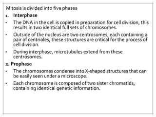
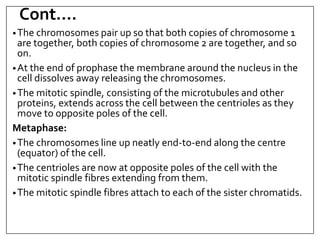
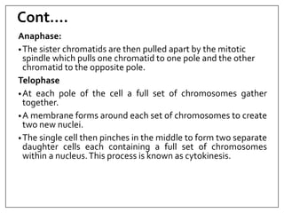
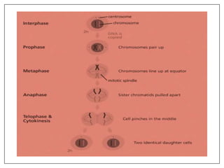
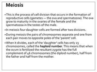
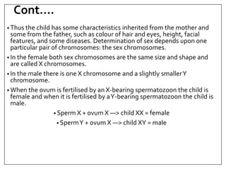
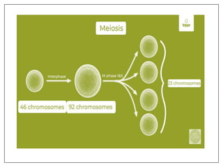
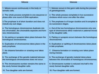
HUMAN ANATOMY & PHYSIOLOGY-I
CELL DIVISION
Presented By: Lovekesh Singh
Assistant Professor
ISF College of Pharmacy, MOGA
Introduction
• Most cells of the human body undergo cell division,
the process by which cells reproduce themselves.
• The two types of cell division
1. somatic cell division
2. reproductive cell division
A somatic cell is any cell of the body other than a
germ cell. A germ cell is a gamete (sperm or oocyte)
or any precursor cell destined to become a gamete.
Cell Division
In somatic cell division, a cell undergoes a nuclear division called
mitosis and a cytoplasmic division called cytokinesis to
produce two identical cells, each with the same number and
kind of chromosomes as the original cell.
•Somatic cell division replaces dead or injured cells and adds new
ones during tissue growth.
Reproductive cell division is the mechanism that produces
gametes, the cells needed to form the next generation of
sexually reproducing organisms.
•This process consists of a special two-step division called
meiosis, in which the number of chromosomes in the nucleus is
reduced by half.
Mitosis
•Beginning with the fertilised egg, or zygote, cell division is
an ongoing process. As the fetus develops in the mother's
uterus, its cells multiply and grow into all the specialties that
provide the sum total of the body's physiological functions.
•The life span of most individual cells is limited.
•Many become worn out and die, and are replaced by
identical cells by the process of mitosis.
•The organelles in the cytoplasm of the daughter cells
are incomplete at cell division but they develop as the
cell grows to maturity.
Mitosis is divided into five phases
1. Interphase
• The DNA in the cell is copied in preparation for cell division, this
results in two identical full sets of chromosomes.
• Outside of the nucleus are two centrosomes, each containing a
pair of centrioles, these structures are critical for the process of
cell division.
• During interphase, microtubules extend from these
centrosomes.
2. Prophase
• The chromosomes condense into X-shaped structures that can
be easily seen under a microscope.
• Each chromosome is composed of two sister chromatids,
containing identical genetic information.
Cont….
•The chromosomes pair up so that both copies of chromosome 1
are together, both copies of chromosome 2 are together, and so
on.
•At the end of prophase the membrane around the nucleus in the
cell dissolves away releasing the chromosomes.
•The mitotic spindle, consisting of the microtubules and other
proteins, extends across the cell between the centrioles as they
move to opposite poles of the cell.
Metaphase:
•The chromosomes line up neatly end-to-end along the centre
(equator) of the cell.
•The centrioles are now at opposite poles of the cell with the
mitotic spindle fibres extending from them.
•The mitotic spindle fibres attach to each of the sister chromatids.
Cont….
Anaphase:
•The sister chromatids are then pulled apart by the mitotic
spindle which pulls one chromatid to one pole and the other
chromatid to the opposite pole.
Telophase
•At each pole of the cell a full set of chromosomes gather
together.
•A membrane forms around each set of chromosomes to create
two new nuclei.
•The single cell then pinches in the middle to form two separate
daughter cells each containing a full set of chromosomes
within a nucleus.This process is known as cytokinesis.
Meiosis
•This is the process of cell division that occurs in the formation of
reproductive cells (gametes — the ova and spermatozoa).The ova
grow to maturity in the ovaries of the female and the
spermatozoa in the testes of the male.
•In meiosis four daughter cells are formed after two divisions.
•During meiosis the pairs of chromosomes separate and one from
each pair moves to opposite poles of the 'parent' cell.
•When it divides, each of the 'daughter' cells has only 23
chromosomes, called the haploid number.This means that when
the ovum is fertilized the resultant zygote has the full
complement of 46 chromosomes (the diploid number), half from
the father and half from the mother.
Cont….
• Thus the child has some characteristics inherited from the mother and
some from the father, such as colour of hair and eyes, height, facial
features, and some diseases. Determination of sex depends upon one
particular pair of chromosomes: the sex chromosomes.
• In the female both sex chromosomes are the same size and shape and
are called X chromosomes.
• In the male there is one X chromosome and a slightly smallerY
chromosome.
• When the ovum is fertilised by an X-bearing spermatozoon the child is
female and when it is fertilised by aY-bearing spermatozoon the child is
male.
• Sperm X + ovum X —> child XX = female
• SpermY + ovum X —> child XY = male
References
• Gerard J. Tortora (2003). Principles of Anatomy and Physiology.
Edition-10th.
• Anne Waugh and Alison Grant (2007) Anatomy and Physiology.
Edition-12th
THANKS

8. cell division.ppt

  • 1.
    HUMAN ANATOMY &PHYSIOLOGY-I CELL DIVISION Presented By: Lovekesh Singh Assistant Professor ISF College of Pharmacy, MOGA
  • 2.
    Introduction • Most cellsof the human body undergo cell division, the process by which cells reproduce themselves. • The two types of cell division 1. somatic cell division 2. reproductive cell division A somatic cell is any cell of the body other than a germ cell. A germ cell is a gamete (sperm or oocyte) or any precursor cell destined to become a gamete.
  • 3.
    Cell Division In somaticcell division, a cell undergoes a nuclear division called mitosis and a cytoplasmic division called cytokinesis to produce two identical cells, each with the same number and kind of chromosomes as the original cell. •Somatic cell division replaces dead or injured cells and adds new ones during tissue growth. Reproductive cell division is the mechanism that produces gametes, the cells needed to form the next generation of sexually reproducing organisms. •This process consists of a special two-step division called meiosis, in which the number of chromosomes in the nucleus is reduced by half.
  • 4.
    Mitosis •Beginning with thefertilised egg, or zygote, cell division is an ongoing process. As the fetus develops in the mother's uterus, its cells multiply and grow into all the specialties that provide the sum total of the body's physiological functions. •The life span of most individual cells is limited. •Many become worn out and die, and are replaced by identical cells by the process of mitosis. •The organelles in the cytoplasm of the daughter cells are incomplete at cell division but they develop as the cell grows to maturity.
  • 5.
    Mitosis is dividedinto five phases 1. Interphase • The DNA in the cell is copied in preparation for cell division, this results in two identical full sets of chromosomes. • Outside of the nucleus are two centrosomes, each containing a pair of centrioles, these structures are critical for the process of cell division. • During interphase, microtubules extend from these centrosomes. 2. Prophase • The chromosomes condense into X-shaped structures that can be easily seen under a microscope. • Each chromosome is composed of two sister chromatids, containing identical genetic information.
  • 6.
    Cont…. •The chromosomes pairup so that both copies of chromosome 1 are together, both copies of chromosome 2 are together, and so on. •At the end of prophase the membrane around the nucleus in the cell dissolves away releasing the chromosomes. •The mitotic spindle, consisting of the microtubules and other proteins, extends across the cell between the centrioles as they move to opposite poles of the cell. Metaphase: •The chromosomes line up neatly end-to-end along the centre (equator) of the cell. •The centrioles are now at opposite poles of the cell with the mitotic spindle fibres extending from them. •The mitotic spindle fibres attach to each of the sister chromatids.
  • 7.
    Cont…. Anaphase: •The sister chromatidsare then pulled apart by the mitotic spindle which pulls one chromatid to one pole and the other chromatid to the opposite pole. Telophase •At each pole of the cell a full set of chromosomes gather together. •A membrane forms around each set of chromosomes to create two new nuclei. •The single cell then pinches in the middle to form two separate daughter cells each containing a full set of chromosomes within a nucleus.This process is known as cytokinesis.
  • 9.
    Meiosis •This is theprocess of cell division that occurs in the formation of reproductive cells (gametes — the ova and spermatozoa).The ova grow to maturity in the ovaries of the female and the spermatozoa in the testes of the male. •In meiosis four daughter cells are formed after two divisions. •During meiosis the pairs of chromosomes separate and one from each pair moves to opposite poles of the 'parent' cell. •When it divides, each of the 'daughter' cells has only 23 chromosomes, called the haploid number.This means that when the ovum is fertilized the resultant zygote has the full complement of 46 chromosomes (the diploid number), half from the father and half from the mother.
  • 10.
    Cont…. • Thus thechild has some characteristics inherited from the mother and some from the father, such as colour of hair and eyes, height, facial features, and some diseases. Determination of sex depends upon one particular pair of chromosomes: the sex chromosomes. • In the female both sex chromosomes are the same size and shape and are called X chromosomes. • In the male there is one X chromosome and a slightly smallerY chromosome. • When the ovum is fertilised by an X-bearing spermatozoon the child is female and when it is fertilised by aY-bearing spermatozoon the child is male. • Sperm X + ovum X —> child XX = female • SpermY + ovum X —> child XY = male
  • 13.
    References • Gerard J.Tortora (2003). Principles of Anatomy and Physiology. Edition-10th. • Anne Waugh and Alison Grant (2007) Anatomy and Physiology. Edition-12th
  • 14.