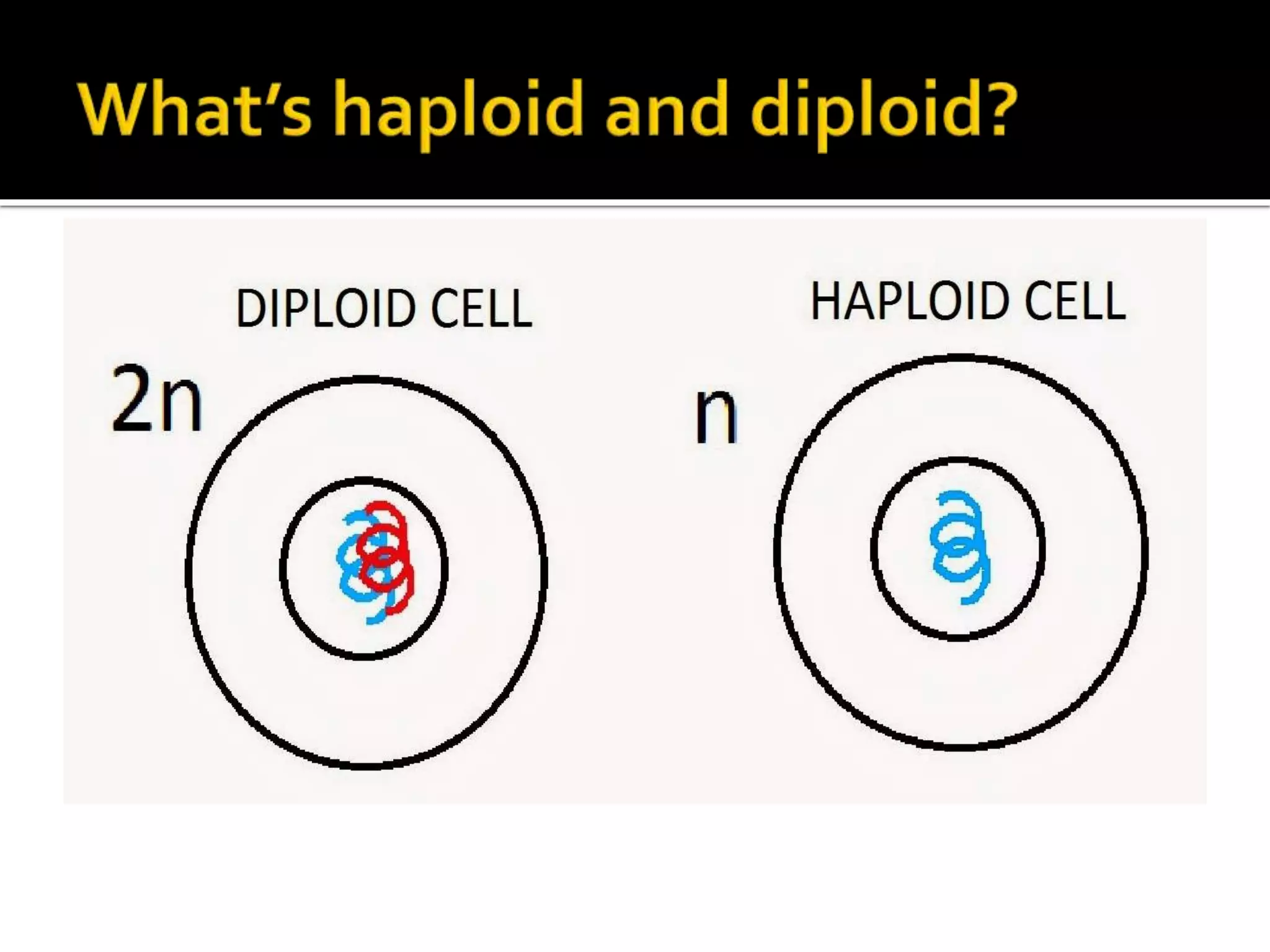
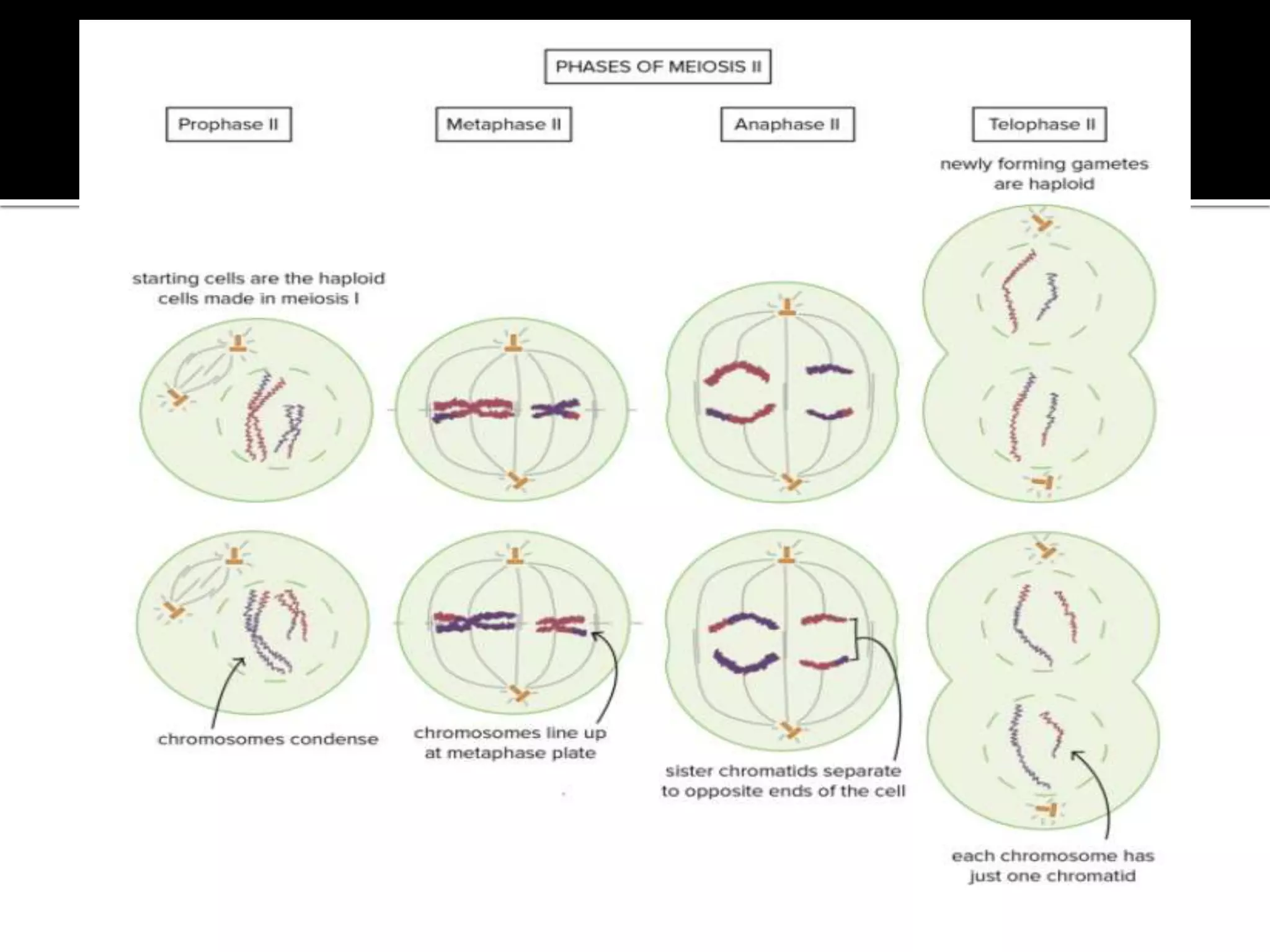
Meiosis is a double division which occurs in the diploid cells and give rise to four haploid cells ,each having half the number of chromosomes as compared to the parent cell.
Meiosis is the process in which a single cell divides twice to form four haploid daughter cells.
These cells are the gametes – sperms in males and egg in females.
For more details, visit @biOlOgy BINGE-insight learning
Mitosis is an equational division, dividing the mother cell into two daughter cells which are identical to one another and also to the mother cell in having the same number and kind of chromosome.
Structure and functions of endoplasmic reticulumICHHA PURAK
The presentation consists of 57 slides,describes following heads
• DISCOVERY
• INTRODUCTION
• BIOGENESIS OF ER
• ISOLATION OF MICROSOMES FROM E R
• STRUCTURE
• COMPONENTS OF ER
CISTERNAE
VESICLES
TUBULES
• MAIN FUNCTION OF ER
• TYPES OF ENDOPLASMIC RETICULUM
• SMOOTH ENDOPLASMIC RETICULUM (SER)
• FUNCTIONS OF SER
• ROUGH ENDOPLASMIC RETICULUM (RER)
• FUNCTIONS OF RER
• SUMMARY
• REFERENCES
• QUESTIONS
A cytological technique to detect the nature of adjacent chromosomal regions by using different staining technique assisted with some pre treatment of metaphase chromosomes prepared on the slides
Molecular diagnosis of genetic disease ppt for studentsthirupathiSathya
DEFINITION:
Dna analysis can be used for the identification of carriers of hereditary disorders.
For prenatal diagnosis of serious genetic conditions yearly diagnosis before the onset of symptoms is done MOLECULAR DIAGNOSIS OF GENETIC DISEASE
CYSTIC FIBROSIS:
Cystic fibrosis is a genetic disease that affect mostly lungs and also the pancreas.
Screening test:
It is a complex process
Large number of genetic alterations have to be done.
For eg : It is the one of the most common lethal autosomal recessive disorder in Europe.
It is caused by mutations to cystic fibrosis transmembrane conductance regulator(CFTR) gene .
Screening individuals who may be at risk for cystic fibrosis for 500 different mutations is a daunting task.
Diagnosis test that screen for a large number of mutations of a single gene in one assay being developed.
SICKLE CELL ANEMIA:
It is a disorder where red blood cells become rigid and sticky and are shaped like “sickle”.
This irregularly shaped cells stucks in small blood vessels which can slow and block the blood flow and oxygen to all the parts of the body.
There’s no cure for sickle cell anemia.
Screening for sickle cell anemia:
SCA is a genetic disease that is the result of a single nucleotide change in the codon for the sixth aminoacid of the β- chain of the hemoglobin molecule.
The anemia is caused by the inability of the mutated hemoglobin to carry sufficient oxygen.
Target – probe hybridasation is done.
Comparison between Replication of Prokaryotes and Eukaryotes mujahid hussain
Comparison between Replication of Prokaryotes and Eukaryotes
Mujahid Hussain
(M.Phil Botany)
Department of Botany
University of Sargodha, Sargodha ,Punjab, Pakistan
The slides contain all about meiosis. in this slides i collected all information about meiosis. which is useful for everyone.
so watch these slides and comment for any problems.
thanks
1. Describe how variation in meiosis happens and why it is beneficia.pdffashioncollection2
1. Describe how variation in meiosis happens and why it is beneficial? Steps and functions of
mitosis and meiosis? ( I am having a hard time with defining each step in mitosis and meiosis.
There is meiosis I and II and I get confused on the steps with functions. Any ideas how I can
remember?)
TIA :)
Solution
Explanation:-
Variation in meiosis:-
During meiosis in humans, 1 diploid cell (with 46 chromosomes or 23 pairs) undergoes 2 cycles
of cell division but only 1 round of DNA replication. The result is 4 haploidHaving one copy of
each chromosome, or having a single set of chromosomes. Gametes (egg and sperm cells) are
haploid. daughter cells known as gametes or egg and sperm cells (each with 23 chromosomes – 1
from each pair in the diploid cell).
At conception, an egg cell and a sperm cell combine to form a zygote (46 chromosomes or 23
pairs). This is the 1st cell of a new individual. The halving of the number of chromosomes in
gametes ensures that zygotes have the same number of chromosomes from one generation to the
next. This is critical for stable sexual reproduction through successive generations.
Benefits of Meiosis:-
1. New Cell Generation-
The chromosomes created during meiosis are composed of 50% copies of the parent cell and
50% new cells. ‘Â These new cells are produced during the cross-over stages of the cell division
process. ‘Â During this stage half of the genetic material from the parent cell is copied into the
new cells, with the other half having distinct properties and characteristics.
2. DNA Replication
The process of meiosis involves copying or replication of genetic material from the parent cell
into the new cells. ‘Â As much as half of the genetic properties of the parent cell are copied into
the newly-created cells. ‘Â When applied to humans for example, DNA from both parents will
partly be copied onto the cells of their offspring. ‘Â When DNA is copied or replicated, the
offspring will also have similar qualities with either or both of his/her parents.
3. Genetic Variation
With meiosis, only half of the genetic material is replicated into the new cells. ‘Â This simply
means that the remaining half will be composed of unique genetic properties making each cell
different from the other. ‘Â Through this process, humans for example will all have different
genetic material and structure.
With the process involved in meiosis, humans are able to reproduce similar yet distinct offspring.
‘Â The whole process basically explains the fact that babies may share some genetic traits from
their parents but they will also have unique sets of personalities because of their unique genetic
composition.
Meiosis Stages:-
Prophase I
Chromosomes condense, Crossing over occurs
Metaphase I
Homologous chromosomes pair up and align in middle of cell
Anaphase I
Homologous chromosomes pulled apart
Telophase I
Nuclear Envelope reforms
Cytokinesis I
Cell splits into two
Prophase II
Centrioles divide and move to opposite poles
Metaphase II
Chromoso.
Hershey and chase experiment-the blender experimentbiOlOgyBINGE
It was performed by Alfred Hershey and Martha Chase in 1952.
It led to confirmation of the genetic nature of DNA came from an experiment with E.coli phage T2.
for more details visit our youtube channel.
Oxidative phosphorylation or electron transport-linked phosphorylation)- the metabolic pathway in which cells use enzymes to oxidize nutrients, thereby releasing the chemical energy stored within in order to produce adenosine triphosphate (ATP).
every detail is available @biOlOgy BINGE-insight learning
Mitosis is the phase of the cell cycle where the nucleus of a cell is divided into two nuclei with an equal amount of genetic material in both the daughter nuclei.
every detail is available @biOlOgy BINGE-insight learning
The cell cycle, or cell-division cycle, is the series of events that take place in a cell that cause it to divide into two daughter cells. These events include the duplication of its DNA (DNA replication) and some of its organelles, and subsequently the partitioning of its cytoplasm and other components into two daughter cells in a process called cell division.
here u will find every detail of cell cycle.
for more details ,visit @biOlOgy BINGE-insight learning
The citric acid cycle (CAC) – also known as the TCA cycle (tricarboxylic acid cycle) or the Krebs cycle – is a series of chemical reactions used by all aerobic organisms to release stored energy through the oxidation of acetyl-CoA derived from carbohydrates, fats, and proteins.
here u will find every detail of KREBS cycle.
It is enzymatically –controlled catabolic pathway by which one molecule of Glucose(6C) breaks into two molecules of pyruvic acid(3C).
Glycolysis is a series of reactions that extract energy from glucose by splitting it into two three-carbon molecules called pyruvates.
for more details, visit @biOlOgy BINGE-insight learning(youtube channel)
Nucleus-the heart of the cell-cellular organellesbiOlOgyBINGE
In cell biology, the nucleus is a membrane-bound organelle found in eukaryotic cells.
The nucleus is found in all the eukaryotic cells of the plants and animals.
here u will find every detail of nucleus.
for more details ,visit @biOlOgy BINGE-insight learning (youtube channel)
The Golgi apparatus, also known as the Golgi complex, Golgi body, or simply the Golgi, is an organelle found in most eukaryotic cells.
It is of particular importance in processing proteins for secretion, containing a set of glycosylation enzymes that attach various sugar monomers to proteins as the proteins move through the apparatus.
here u will find every detail of golgi.
Mitochondria-the powerhouse of the cellbiOlOgyBINGE
Mitochondrion is a semi-autonomous, double-membrane-bound organelle found in most eukaryotic organisms.
The organelle is composed of compartments that carry out specialized functions.
here u will find every detail of mitochondria.
Phenomics assisted breeding in crop improvementIshaGoswami9
As the population is increasing and will reach about 9 billion upto 2050. Also due to climate change, it is difficult to meet the food requirement of such a large population. Facing the challenges presented by resource shortages, climate
change, and increasing global population, crop yield and quality need to be improved in a sustainable way over the coming decades. Genetic improvement by breeding is the best way to increase crop productivity. With the rapid progression of functional
genomics, an increasing number of crop genomes have been sequenced and dozens of genes influencing key agronomic traits have been identified. However, current genome sequence information has not been adequately exploited for understanding
the complex characteristics of multiple gene, owing to a lack of crop phenotypic data. Efficient, automatic, and accurate technologies and platforms that can capture phenotypic data that can
be linked to genomics information for crop improvement at all growth stages have become as important as genotyping. Thus,
high-throughput phenotyping has become the major bottleneck restricting crop breeding. Plant phenomics has been defined as the high-throughput, accurate acquisition and analysis of multi-dimensional phenotypes
during crop growing stages at the organism level, including the cell, tissue, organ, individual plant, plot, and field levels. With the rapid development of novel sensors, imaging technology,
and analysis methods, numerous infrastructure platforms have been developed for phenotyping.
Toxic effects of heavy metals : Lead and Arsenicsanjana502982
Heavy metals are naturally occuring metallic chemical elements that have relatively high density, and are toxic at even low concentrations. All toxic metals are termed as heavy metals irrespective of their atomic mass and density, eg. arsenic, lead, mercury, cadmium, thallium, chromium, etc.
Deep Behavioral Phenotyping in Systems Neuroscience for Functional Atlasing a...Ana Luísa Pinho
Functional Magnetic Resonance Imaging (fMRI) provides means to characterize brain activations in response to behavior. However, cognitive neuroscience has been limited to group-level effects referring to the performance of specific tasks. To obtain the functional profile of elementary cognitive mechanisms, the combination of brain responses to many tasks is required. Yet, to date, both structural atlases and parcellation-based activations do not fully account for cognitive function and still present several limitations. Further, they do not adapt overall to individual characteristics. In this talk, I will give an account of deep-behavioral phenotyping strategies, namely data-driven methods in large task-fMRI datasets, to optimize functional brain-data collection and improve inference of effects-of-interest related to mental processes. Key to this approach is the employment of fast multi-functional paradigms rich on features that can be well parametrized and, consequently, facilitate the creation of psycho-physiological constructs to be modelled with imaging data. Particular emphasis will be given to music stimuli when studying high-order cognitive mechanisms, due to their ecological nature and quality to enable complex behavior compounded by discrete entities. I will also discuss how deep-behavioral phenotyping and individualized models applied to neuroimaging data can better account for the subject-specific organization of domain-general cognitive systems in the human brain. Finally, the accumulation of functional brain signatures brings the possibility to clarify relationships among tasks and create a univocal link between brain systems and mental functions through: (1) the development of ontologies proposing an organization of cognitive processes; and (2) brain-network taxonomies describing functional specialization. To this end, tools to improve commensurability in cognitive science are necessary, such as public repositories, ontology-based platforms and automated meta-analysis tools. I will thus discuss some brain-atlasing resources currently under development, and their applicability in cognitive as well as clinical neuroscience.
Earliest Galaxies in the JADES Origins Field: Luminosity Function and Cosmic ...Sérgio Sacani
We characterize the earliest galaxy population in the JADES Origins Field (JOF), the deepest
imaging field observed with JWST. We make use of the ancillary Hubble optical images (5 filters
spanning 0.4−0.9µm) and novel JWST images with 14 filters spanning 0.8−5µm, including 7 mediumband filters, and reaching total exposure times of up to 46 hours per filter. We combine all our data
at > 2.3µm to construct an ultradeep image, reaching as deep as ≈ 31.4 AB mag in the stack and
30.3-31.0 AB mag (5σ, r = 0.1” circular aperture) in individual filters. We measure photometric
redshifts and use robust selection criteria to identify a sample of eight galaxy candidates at redshifts
z = 11.5 − 15. These objects show compact half-light radii of R1/2 ∼ 50 − 200pc, stellar masses of
M⋆ ∼ 107−108M⊙, and star-formation rates of SFR ∼ 0.1−1 M⊙ yr−1
. Our search finds no candidates
at 15 < z < 20, placing upper limits at these redshifts. We develop a forward modeling approach to
infer the properties of the evolving luminosity function without binning in redshift or luminosity that
marginalizes over the photometric redshift uncertainty of our candidate galaxies and incorporates the
impact of non-detections. We find a z = 12 luminosity function in good agreement with prior results,
and that the luminosity function normalization and UV luminosity density decline by a factor of ∼ 2.5
from z = 12 to z = 14. We discuss the possible implications of our results in the context of theoretical
models for evolution of the dark matter halo mass function.
Observation of Io’s Resurfacing via Plume Deposition Using Ground-based Adapt...Sérgio Sacani
Since volcanic activity was first discovered on Io from Voyager images in 1979, changes
on Io’s surface have been monitored from both spacecraft and ground-based telescopes.
Here, we present the highest spatial resolution images of Io ever obtained from a groundbased telescope. These images, acquired by the SHARK-VIS instrument on the Large
Binocular Telescope, show evidence of a major resurfacing event on Io’s trailing hemisphere. When compared to the most recent spacecraft images, the SHARK-VIS images
show that a plume deposit from a powerful eruption at Pillan Patera has covered part
of the long-lived Pele plume deposit. Although this type of resurfacing event may be common on Io, few have been detected due to the rarity of spacecraft visits and the previously low spatial resolution available from Earth-based telescopes. The SHARK-VIS instrument ushers in a new era of high resolution imaging of Io’s surface using adaptive
optics at visible wavelengths.
Professional air quality monitoring systems provide immediate, on-site data for analysis, compliance, and decision-making.
Monitor common gases, weather parameters, particulates.
The ability to recreate computational results with minimal effort and actionable metrics provides a solid foundation for scientific research and software development. When people can replicate an analysis at the touch of a button using open-source software, open data, and methods to assess and compare proposals, it significantly eases verification of results, engagement with a diverse range of contributors, and progress. However, we have yet to fully achieve this; there are still many sociotechnical frictions.
Inspired by David Donoho's vision, this talk aims to revisit the three crucial pillars of frictionless reproducibility (data sharing, code sharing, and competitive challenges) with the perspective of deep software variability.
Our observation is that multiple layers — hardware, operating systems, third-party libraries, software versions, input data, compile-time options, and parameters — are subject to variability that exacerbates frictions but is also essential for achieving robust, generalizable results and fostering innovation. I will first review the literature, providing evidence of how the complex variability interactions across these layers affect qualitative and quantitative software properties, thereby complicating the reproduction and replication of scientific studies in various fields.
I will then present some software engineering and AI techniques that can support the strategic exploration of variability spaces. These include the use of abstractions and models (e.g., feature models), sampling strategies (e.g., uniform, random), cost-effective measurements (e.g., incremental build of software configurations), and dimensionality reduction methods (e.g., transfer learning, feature selection, software debloating).
I will finally argue that deep variability is both the problem and solution of frictionless reproducibility, calling the software science community to develop new methods and tools to manage variability and foster reproducibility in software systems.
Exposé invité Journées Nationales du GDR GPL 2024
Nutraceutical market, scope and growth: Herbal drug technologyLokesh Patil
As consumer awareness of health and wellness rises, the nutraceutical market—which includes goods like functional meals, drinks, and dietary supplements that provide health advantages beyond basic nutrition—is growing significantly. As healthcare expenses rise, the population ages, and people want natural and preventative health solutions more and more, this industry is increasing quickly. Further driving market expansion are product formulation innovations and the use of cutting-edge technology for customized nutrition. With its worldwide reach, the nutraceutical industry is expected to keep growing and provide significant chances for research and investment in a number of categories, including vitamins, minerals, probiotics, and herbal supplements.
Seminar of U.V. Spectroscopy by SAMIR PANDASAMIR PANDA
Spectroscopy is a branch of science dealing the study of interaction of electromagnetic radiation with matter.
Ultraviolet-visible spectroscopy refers to absorption spectroscopy or reflect spectroscopy in the UV-VIS spectral region.
Ultraviolet-visible spectroscopy is an analytical method that can measure the amount of light received by the analyte.
2. Meiosis is a double division which occurs in the diploid cells and give rise to four haploid cells ,each having
half the number of chromosomes as compared to the parent cell.
Meiosis is the process in which a single cell divides twice to form four haploid daughter cells.
These cells are the gametes – sperms in males and egg in females.
Meiosis is a special type of cell division of germ cells in sexually-reproducing organisms used to produce
the gametes, such as sperm or egg cells.
It involves two rounds of division that ultimately result in four cells with only one copy of each paternal and
maternal chromosome (haploid).
Additionally, prior to the division, genetic material from the paternal and maternal copies of each
chromosome is crossed over, creating new combinations of code on each chromosome.
Later on, during fertilisation, the haploid cells produced by meiosis from a male and female will fuse to
create a cell with two copies of each chromosome again, the zygote.
3.
4. Mitosis is used for almost all of your body’s cell division needs. It adds new cells during development and
replaces old and worn-out cells throughout your life.
The goal of mitosis is to produce daughter cells that are genetically identical to their mothers, with not a
single chromosome more or less.
Meiosis, on the other hand, is used for just one purpose in the human body: the production of gametes—
sex cells, or sperm and eggs.
Its goal is to make daughter cells with exactly half as many chromosomes as the starting cell.
To put that another way, meiosis in humans is a division process that takes us from a diploid cell—one with
two sets of chromosomes—to haploid cells—ones with a single set of chromosomes.
In humans, the haploid cells made in meiosis are sperm and eggs. When a sperm and an egg join in
fertilization, the two haploid sets of chromosomes form a complete diploid set: a new genome.
5. “Meiosis is the type of cell division that
results in four daughter cells, each with half
the number of chromosomes of the parent
cell.”
6. THE PROCESS OF MEIOSIS IS DIVIDED INTO 2 STAGES. EACH STAGE
IS SUBDIVIDED INTO SEVERAL PHASES.
Meiosis I:
Prophase I
Metaphase I
Anaphase I
Telophase I
Cytokinesis I
Meiosis II:
Prophase II
Metaphase II
Anaphase II
Telophase II
Cytokinesis II
7. In many ways, meiosis is a lot like mitosis.
The cell goes through similar stages and uses similar strategies to organize and separate
chromosomes.
In meiosis, however, the cell has a more complex task. It still needs to separate sister
chromatids (the two halves of a duplicated chromosome), as in mitosis.
But it must also separate homologous chromosomes, the similar but nonidentical
chromosome pairs an organism receives from its two parents.
These goals are accomplished in meiosis using a two-step division process.
Homologue pairs separate during a first round of cell division, called meiosis I.
Sister chromatids separate during a second round, called meiosis II.
Since cell division occurs twice during meiosis, one starting cell can produce four gametes
(eggs or sperm).
8. A couple of homologous chromosomes,
or homologs, are a set of one maternal and
one paternal chromosome that pair up with
each other inside a cell during fertilization.
Homologs have the same genes in the
same loci where they provide points along
each chromosome which enable a pair of
chromosomes to align correctly with each
other before separating during meiosis.
9. It is called the heterotypic or reduction division.
It has 4 stages:
Prophase1
Metaphase1
Anaphase 1
Telophase1
10. Prophase I is by far the longest phase of meiosis . During prophase I, homologous
maternal and paternal chromosomes pair, synapse, and exchange genetic
information (by homologous recombination), forming at least one crossover per
chromosome.These crossovers become visible as chiasmata (plural;
singular chiasma).
Prophase is divided into 5 stages:
Leptotene
Zygotene
Pachytene
Diplotene
Diakinesis
11. The first stage of prophase I is
the leptotene stage, also known as leptonema.
In this stage of prophase I, individual
chromosomes—each consisting of two
replicated sister chromatids—become
"individualized" to form visible strands within
the nucleus.
12. Leptotene is followed by the zygotene stage, also
known as zygonema.
It is also called the bouquet stage because of the way
the telomeres cluster at one end of the nucleus.
The two homologous chromosomes get attached to
each other laterally.
The process of attachment is called synapsis.
Chromosomes form pairs called bivalents.
13.
14. The paired chromosomes or bivalents shorten.
Size of nucleolus reaches the maximum.
Each bivalent or chromosome pair is made up of 4 chromatids,two of each
chromosome.
The chromatids belonging to same chromosomes are called sister chromatids.
Chromatids belonging to two different chromosomes of a homologous pair are
called non-sister chromatids.
In pachytene stage,there occurs exchange of genetic material between non-
sister chromatids of two homologous chromosomes.
This is called crossing over.
After crossing over,the chromatids of same chromosomes become different.
15.
16. Diplotene is the stage when the synaptonemal
complex disassembles and homologous chromosomes separate
from one another a little.
However, the homologous chromosomes of each bivalent remain
tightly bound at chiasmata, the regions where crossing-over
occurred.
Therefore the homologous chromosomes undergo desynapsis
except in the region of crossing over.
The chromatids also separate and become distinguishable –tetrad
stage.
17.
18. Chromosomes condense further during the diakinesis stage.
This is the first point in meiosis where the four parts of the tetrads are actually
visible.
Sites of crossing over entangle together, effectively overlapping, making
chiasmata clearly visible.
Simultaneously,nuclear envelope disintegrates.
In animal cells,the two centrioles move to the opposite poles of the nucleus.
They develop a number of radiating microtubules from their surface called astral
rays.
Centrioles with astral rays is called aster.
19.
20. Homologous pairs move together along the
metaphase plate: As kinetochore
microtubules from both spindle poles attach
to their respective kinetochores, the paired
homologous chromosomes align along an
equatorial plane that bisects the spindle.
The centromeres slightly project towards
the pole.
Since there are 2 centromeres in each
bivalent ,the centromeres of all bivalents
produce a double metaphasic plate.
21. Kinetochore microtubules shorten, pulling homologous
chromosomes (which each consist of a pair of sister chromatids) to
opposite poles.
Nonkinetochore microtubules lengthen, pushing the centrosomes
farther apart.
The cell elongates in preparation for division down the center.
The homologous chromosomes break their connections and
separate out .This is called disjunction.
22.
23. The first meiotic division effectively ends when the chromosomes
arrive at the poles.
Each daughter cell now has half the number of chromosomes but
each chromosome consists of a pair of chromatids.
The microtubules that make up the spindle network disappear,
and a new nuclear membrane surrounds each haploid set.
The chromosomes uncoil back into chromatin. Sister chromatids
remain attached during telophase I.
24.
25. Cells may enter a period of rest known as interkinesis or
interphase II.
It is transitory stage of short duration between meiosis
1 and meiosis 2.
RNA , proteins and other biomolecules are synthesized
during this phase.
DNA replication is absent.
Centriole pair also replicates.
26. It is called homotypic or equational division.
It is divided into 4 phases:
Prophase
Metaphase
Anaphase
Telophase
29. Absence of long interphase and presence of interkinesis.
No chromosome replication in S-phase.
2 chromatids of a chromosome are genetically different.
Simpler and brief.
Daughter cells do not resemble each other .
Neither they resemble to their mother cell.