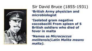
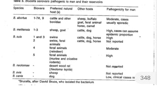
Sir David Bruce(1855-1931)
•British Army physician and
microbiologist
•Isolated gram negative
coccobacilli From spleen of 5
British soldiers who died of
fever in malta
•Names as Micrococcus
melitensis(Latin Malita means
malta).
6.
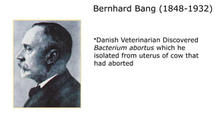
Bernhard Bang (1848-1932)
•DanishVeterinarian Discovered
Bacterium abortus which he
isolated from uterus of cow that
had aborted
Professor FEG Cox. The Wellcome Trust, Illustrated History of Tropical Diseases
7.
ALICE EVAN (1881 -1975 )
• American bacteriologist,She
discovered similar morphology and
pathology between bangs bacterium
abortus and Bruce’s micrococcuus
melitensis.
• As a memory of sir David Bruce called
it as Brucella genus
8.
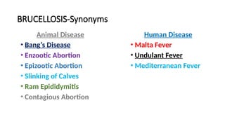
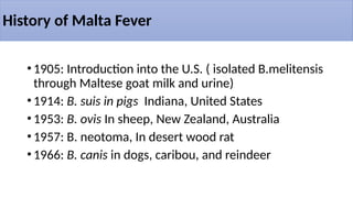
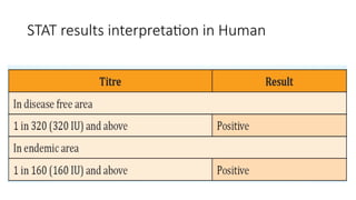
History of MaltaFever
•1905: Introduction into the U.S. ( isolated B.melitensis
through Maltese goat milk and urine)
•1914: B. suis in pigs Indiana, United States
•1953: B. ovis In sheep, New Zealand, Australia
•1957: B. neotoma, In desert wood rat
•1966: B. canis in dogs, caribou, and reindeer
9.
BIOLOGICAL WEAPON :
•Brucella Was One of the agents with which Japan
experimented In famous 731 manchu- ria unit before and
during second world war
• In US , B.suis was the first agent Weaponized in 1952And
extended the field testing With B.suis filled bombs
10.
•Human Brucellosis:
•Cost ofTreatment is more
•> 5.0 lakhs new human cases recorded annually.
•Potential biowar fare agent
•All Brucella species are classified as group 3
pathogens and handling requires biosafety level 3
precautions
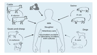
•Brucellae primarily affectcattle, buffaloes,
sheep, goats, pigs and dogs. Occasionally
horses, camels, water buffalo, yak and wild
animals are also affected.
•The risk of wildlife-to-human transmission is
low although a variety of wildlife may act as
natural reservoir of the infection.
14.
Brucellosis- ZOONOSIS
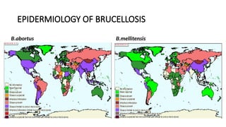
• Brucellosisis worldwide in distribution and is endemic in
certain areas such as Mediterranean countries(Arabian
peninsula , india, mexico ,central and north america)
• Human infection direct or indirect contact with infected
animal tissue.
• Person to person transmission - RARE in circumstances
implicating sexual contact, tissue transfer including blood
and bone marrow.
• Laboratory acquired Brucellosis accidental ingestion,
inhalation, injection, mucosal and skin Contamination.
15.
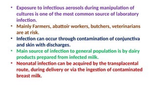
• Exposure toinfectious aerosols during manipulation of
cultures is one of the most common source of laboratory
infection.
• Mainly Farmers, abattoir workers, butchers, veterinarians
are at risk.
• Infection can occur through contamination of conjunctiva
and skin with discharges.
• Main source of infection to general population is by dairy
products prepared from infected milk.
• Neonatal infection can be acquired by the transplacental
route, during delivery or via the ingestion of contaminated
breast milk.
17.
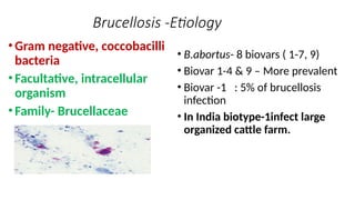
Brucellosis -Etiology
•Gram negative,coccobacilli
bacteria
•Facultative, intracellular
organism
•Family- Brucellaceae
• B.abortus- 8 biovars ( 1-7, 9)
• Biovar 1-4 & 9 – More prevalent
• Biovar -1 : 5% of brucellosis
infection
• In India biotype-1infect large
organized cattle farm.
20.
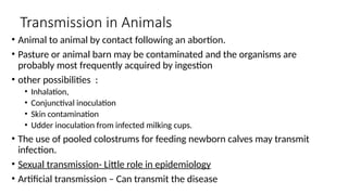
Transmission in Animals
•Animal to animal by contact following an abortion.
• Pasture or animal barn may be contaminated and the organisms are
probably most frequently acquired by ingestion
• other possibilities :
• Inhalation,
• Conjunctival inoculation
• Skin contamination
• Udder inoculation from infected milking cups.
• The use of pooled colostrums for feeding newborn calves may transmit
infection.
• Sexual transmission- Little role in epidemiology
• Artificial transmission – Can transmit the disease
21.
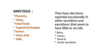
INFECTIOUS :
• Placenta,
•Fetus,
• Fetal Fluids
• Vaginal Discharges
• Semen
• Urine And
• Milk.
They have also been
reported occasionally in
other secretions and
excretions :that seem to
have little or no role
• Saliva,
• Faeces,
• Nasal or
• Ocular secretions
05/01/2025
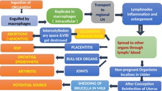
Engulfed by
macrophages
Lymphnodes
inflammation and
enlargement
Transport
to
regional
LN
Spreadto other
organs through
lymph/ blood
ULCERATIVE
ENDOMETRITIS
PLACENTITIS
SHEDDING OF
BRUCELLA IN MILK
ROP
ABORTIONS
(>6MONTHS)
PMN
Ingestion of
ORGANISM
Replicate in
macrophages
( intracellular )
ORCHITIS&
EPIDIDYMITIS
BULL-SEX ORGANS
JOINTS
ARTHRITIS
POTENTIAL SOURCE
Intercotylledon
ary space &Villi
get destroyed
After Conception
Reinfection of Uterus
Non-pregnant Organisms
localizes in Udder
24.
Clinical Signs: Horses
•B.abortus most common
• Susceptible to B. suis
•Fistulous Withers or Poll
Evil
• Inflammation of the
supraspinous bursa
• Abortions-Rare
25.
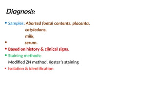
Diagnosis:
Samples: Abortedfoetal contents, placenta,
cotyledons,
milk,
serum.
Based on history & clinical signs.
Staining methods:
Modified ZN method, Koster’s staining
• Isolation & identification
26.
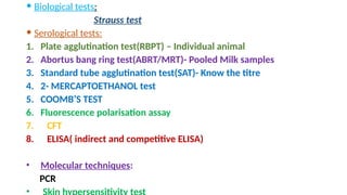
Biological tests:
Strausstest
Serological tests:
1. Plate agglutination test(RBPT) – Individual animal
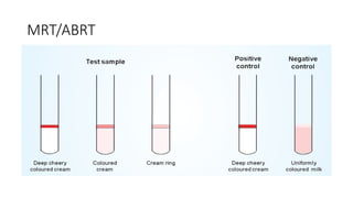
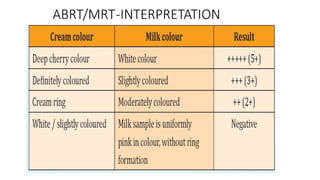
2. Abortus bang ring test(ABRT/MRT)- Pooled Milk samples
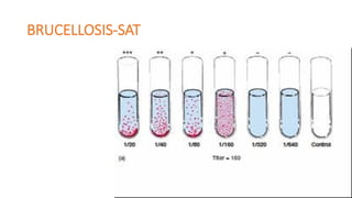
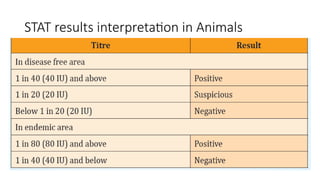
3. Standard tube agglutination test(SAT)- Know the titre
4. 2- MERCAPTOETHANOL test
5. COOMB’S TEST
6. Fluorescence polarisation assay
7. CFT
8. ELISA( indirect and competitive ELISA)
• Molecular techniques:
PCR
•
27.
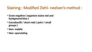
Staining : ModifiedZiehl- neelsen’s method :
• Gram negative ( organism stains red and
background blue )
• Coccobacilli / short rods ( pairs / small
groups )
• Non- mobile
• Non- sporulating
28.
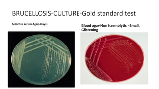
ISOLATION AND IDENTIFICATION:
• Catalase- positive
• Oxidase – positive
• Urease - positive
• Basal media :
• Potato dextrose agar
• serum dextrose agar
• Columbia agar ( 5% serum )
[ all supplemented with 5-10% CO2 and incubated at 37°C for about 24
– 48 hrs ]
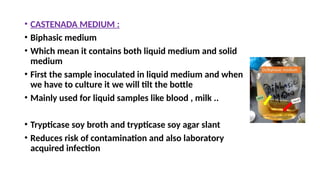
• CASTENADA MEDIUM:
• Biphasic medium
• Which mean it contains both liquid medium and solid
medium
• First the sample inoculated in liquid medium and when
we have to culture it we will tilt the bottle
• Mainly used for liquid samples like blood , milk ..
• Trypticase soy broth and trypticase soy agar slant
• Reduces risk of contamination and also laboratory
acquired infection
32.
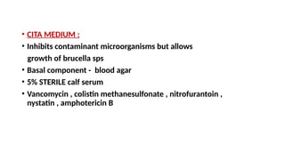
• CITA MEDIUM:
• Inhibits contaminant microorganisms but allows
growth of brucella sps
• Basal component - blood agar
• 5% STERILE calf serum
• Vancomycin , colistin methanesulfonate , nitrofurantoin ,
nystatin , amphotericin B
33.
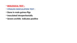
• BIOLOGICAL TEST:
• STRAUSS INOCULATION TEST :
• Done in male guinea Pigs
• Inoculated intraperitonially
• Severe orchitis indicates positive
34.
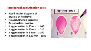
Rose bengal agglutinationtest :
• Rapid test for diagnosis of
brucella at field level
• No agglutination- negative
• Agglutination- positive
• If agglutination in 15sec - 1: 640
• If agglutination in 30sec - 1: 320
• If agglutination in 1 min - 1: 160
• If agglutination in 1.30 min – 1: 80
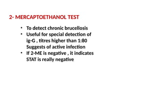
2- MERCAPTOETHANOL TEST
•To detect chronic brucellosis
• Useful for special detection of
ig-G , titres higher than 1:80
Suggests of active infection
• If 2-ME is negative , it indicates
STAT is really negative
42.
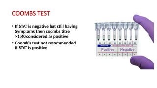
COOMBS TEST
• IfSTAT is negative but still having
Symptoms then coombs titre
>1:40 considered as positive
• Coomb‘s test not recommended
if STAT is positive
43.
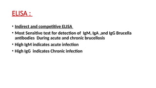
ELISA :
• Indirectand competitive ELISA
• Most Sensitive test for detection of IgM, IgA ,and IgG Brucella
antibodies During acute and chronic brucellosis
• High IgM indicates acute infection
• High IgG indicates Chronic infection
44.
MILK ELISA :
•Add 100 microliter of sample to Antigen coated plate
• Incubate at 25°C for 30 minutes
• Washed 4 times
• Add 100 microlitre of diluted antibody- peroxidase conjugate
• Incubate at 25°C for 30 minutes
• Wash and add 100 microliter of substrate solution
• Measure at 620nm
• Sp ratio - < 25 negative
• Sp ratio - > 25 positive
45.
Confirmation by PCR:
• Initial denaturation : 1 cycle × 95°C for 5 minutes
• Template denaturation : 30 cycles × 95°C for 30 seconds
• Primer annealing : 30 cycles × 54°C for 90 seconds
• Primer extension : 30 cycles × 72°C for 90 seconds
• Final extension : 1 cycle x 72°C for 6 minutes
46.
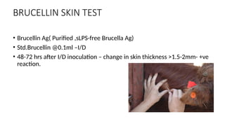
BRUCELLIN SKIN TEST
•Brucellin Ag( Purified ,sLPS-free Brucella Ag)
• Std.Brucellin @0.1ml –I/D
• 48-72 hrs after I/D inoculation – change in skin thickness >1.5-2mm- +ve
reaction.
47.
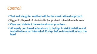
Control:
Test and slaughtermethod will be the most rational approach.
Hygienic disposal of uterine discharges,foetus,foetal membranes.
Clear and disinfect the contaminated premises .
All newly purchased animals are to be kept in strict isolation and
tested twice at an interval of 30 days before introduction into the
herd.
48.
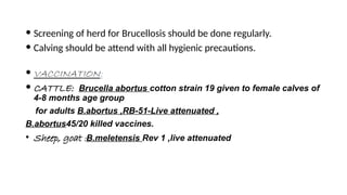
Screening ofherd for Brucellosis should be done regularly.
Calving should be attend with all hygienic precautions.
VACCINATION:
CATTLE: Brucella abortus cotton strain 19 given to female calves of
4-8 months age group
for adults B.abortus ,RB-51-Live attenuated ,
B.abortus45/20 killed vaccines.
• Sheep, goat :B.meletensis Rev 1 ,live attenuated
#2 Due to its illustrious history, brucellosis has many different names involving both humans and animals. Undulant Fever for humans and Bang’s Disease for animals are the two most widely recognized common names.
#4 J.A. Marston was an army surgeon (British) who, after contracting the Malta fever, wrote the first detailed account of the disease (his own illness). He was afflicted with an irregular fever for 30 to 90 days, gastrointestinal symptoms, and muscle and joint pains.
#5 The microorganism responsible for Malta fever was discovered by a British Army physician, Sir David Bruce, on July 9, 1887, which he called Micrococcus melitensis. It was isolated from the spleen of a British soldier who had died of the disease. He also identified that the organism grew best at higher temperatures and speculated that this accounted for the increased frequency of cases in hot summer months. He later established goats as the main reservoir for infection by identifying the organism in their blood, urine, and milk. This discovery helped explain the epidemiology of the disease. For example, officers were three times more likely to become ill because they drank more milk than private soldiers, and large numbers of cases were found in hospitals where milk was widely distributed.
#6 A Danish physician and veterinarian, Bernhard Bang discovered Brucella abortus 1897 while investigating contagious abortion that had been affecting cattle in Denmark for over a century. He also discovered the organism affected horses, sheep, and goats. Thus the disease become known as “Bang’s disease”.
#8 In his book Epidemics, Hippocrates first described a condition of reccurring fever and death with a duration of 4 months in 450 B.C. Undulant fever did not enter into the United States until 1905 through the shipping of 65 Maltese goats on the S.S. Joshua Nicholson. B. suis was isolated in 1914 by Traum in the U.S. from aborting swine in Indiana. B. ovis was isolated in 1953 from sheep with ram epididymitis in New Zealand and Australia. B. canis was discovered in 1966 from dogs, caribou, and reindeer.
#17 This organism is an aerobic, small, Gram-negative coccobacillus or short rod that can persist in the environment invariably depending on temperature, pH, and humidity. Brucella spp. can persist indefinitely if frozen or protected in aborted fetuses or placentas. It is a facultative, intracellular pathogen and thus requires prolonged treatment with clinically effective antibiotics.
#24 Horses are susceptible to B. abortus or B. suis from infectious or traumatic origin. Clinically, these animals have an inflammation in the supraspinous bursa or supra-atlantal bursa; this is referred to as Fistulous Withers or Poll Evil, respectively. The bursal sac becomes distended by a clear, viscous, straw-colored exudate and develops a thickened wall. It can rupture, leading to secondary inflammation. In chronic cases, nearby ligaments and the dorsal vertebral spines may become necrotic. Brucella-associated abortions are rare in horses.


![ISOLATION AND IDENTIFICATION :
• Catalase- positive
• Oxidase – positive
• Urease - positive
• Basal media :
• Potato dextrose agar
• serum dextrose agar
• Columbia agar ( 5% serum )
[ all supplemented with 5-10% CO2 and incubated at 37°C for about 24
– 48 hrs ]](https://image.slidesharecdn.com/brucellosis-250501134320-ba7f340c/85/brucellosis-veterinary-microbiology-pptx-28-320.jpg)


![Brucellosis916[1]](https://cdn.slidesharecdn.com/ss_thumbnails/brucellosis9161-200524052427-thumbnail.jpg?width=640&height=640&fit=bounds)


