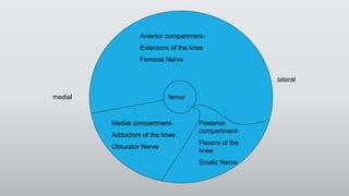
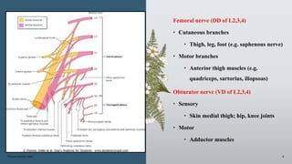
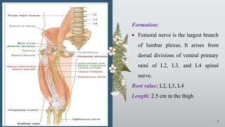
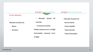
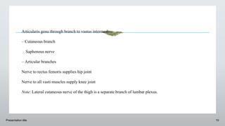
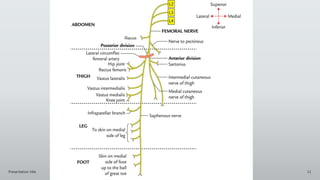
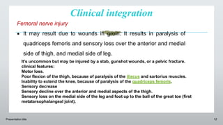
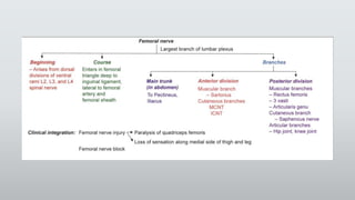
Femoral Nerve
Dr Sushma Daripelli
femur
medial
lateral
Anterior compartment-
Extensors of the knee
Femoral Nerve
Medial compartment-
Adductors of the knee
Obturator Nerve
Posterior
compartment-
Flexors of the
knee
Sciatic Nerve
Introduction
Presentation title 3
• Femoral Nerve is the main nerve of anterior compartment of thigh.
• It originates from the dorsal sections of the anterior primary rami of L2, L3, L4
nerves and is the largest branch of lumbar plexus.
• It goes into the thigh behind the inguinal ligament and lateral to femoral
sheath while descending via psoas major and appearing on its lateral order
between psoas and illiacus.
Presentation title 4
Femoral nerve (DD of L2,3,4)
• Cutaneous branches
• Thigh, leg, foot (e.g. saphenous nerve)
• Motor branches
• Anterior thigh muscles (e.g.
quadriceps, sartorius, iliopsoas)
Obturator nerve (VD of L2,3,4)
• Sensory
• Skin medial thigh; hip, knee joints
• Motor
• Adductor muscles
Presentation title 5
Introduction: Femoral nerve is the
chief nerve of the anterior
compartment of thigh
Presentation title 6
Formation:
 Femoral nerve is the largest branch
of lumbar plexus. It arises from
dorsal divisions of ventral primary
rami of L2, L3, and L4 spinal
nerve.
Root value: L2, L3, L4
Length: 2.5 cm in the thigh
Course and relations
Presentation title 7
In the abdomen
 Femoral nerve begins on the posterior abdominal wall. It emerges out
along the lateral border of psoas major muscle and runs further downward
on the iliacus under the cover of fascia iliaca.
Note: Lateral circumflex femoral artery passes in between anterior and
posterior divisions of femoral nerve.
Presentation title 8
In the thigh
 It enters the femoral triangle by passing behind the inguinal ligament, just
lateral to the femoral artery.
 Femoral nerve lies outside the femoral sheath because femoral nerve lies
posterior to fascia iliaca which forms the posterior wall of femoral sheath.
 At about 2.5 cm below the inguinal ligament, the femoral nerve divides into
anterior and posterior divisions.
In the abdomen
Muscular branches for
 Iliacus
 Pectineus
In thigh Anterior division
– Muscular branch for
sartorius
– Cutaneous branches
Medial cutaneous nerve of thigh
Intermediate cutaneous nerve
of thigh
Presentation title 9
In thigh Posterior division
– Muscular branches for
Rectus femoris
Vastus medialis
Vastus lateralis
Vastus intermedius
Presentation title 10
Articularis genu through branch to vastus intermedius
– Cutaneous branch
Saphenous nerve
– Articular branches
Nerve to rectus femoris supplies hip joint
Nerve to all vasti muscles supply knee joint
Note: Lateral cutaneous nerve of the thigh is a separate branch of lumbar plexus.
Presentation title 11
Clinical integration
Presentation title 12
Femoral nerve injury
 It may result due to wounds in groin. It results in paralysis of
quadriceps femoris and sensory loss over the anterior and medial
side of thigh, and medial side of leg.
It’s uncommon but may be injured by a stab, gunshot wounds, or a pelvic fracture.
clinical features:
Motor loss.
Poor flexion of the thigh, because of paralysis of the iliacus and sartorius muscles.
Inability to extend the knee, because of paralysis of the quadriceps femoris.
Sensory decrease
Sensory decline over the anterior and medial aspects of the thigh.
Sensory loss on the medial side of the leg and foot up to the ball of the great toe (first
metatarsophalangeal joint).
Presentation title 13
Femoral nerve block
 Local anaesthetic agent is injected around the femoral nerve to achieve
the anaesthetic agent that bocks nerve condition by inactivation of Na+
channels. It is available as gels for local applications or injection for
nerve blocks.
Presentation title 14
FEMORAL NERVE NEUROPATHY
The primary trunk of the femoral nerve isn’t subject to an entrapment neuropathy .
However, it might be compressed by the retroperitoneal tumors. A localized
neuropathy of the femoral nerve may happen in diabetes mellitus. Listed here are
the characteristic clinical features:
Wasting and weakness of quadriceps resulting in significant trouble in walking.
Pain and paraesthesia on the anterior and medial aspects of the thigh going down
along the medial aspect of the leg and foot along the distribution of the saphenous
nerve.
Thank
you…
Presentation title 17
ANTERIOR COMPARTMENT OF THIGH
 The anterior compartment of thigh lies between medial and
lateral intermuscular septa.
 It is also called extensor compartment as it contains extensor
muscles of thigh.
Contents
Anterior compartment of thigh contains:
1. Muscles
- Rectus femoris
- 3 vasti (vastus medialis, intemedius, and lateralis)
- Sartorius
- Articularis genu
- Tensor fascia latae
Nerve: Femoral nerve
Artery: Femoral artery
Fascial space: Femoral triangle and adductor canal.
Presentation title 18
Presentation title 19
Presentation title 20
Presentation title 21
Presentation title 22
Presentation title 23
Presentation title 24

Femoral Nerve gmc.pptx

  • 1.
  • 2.
    femur medial lateral Anterior compartment- Extensors ofthe knee Femoral Nerve Medial compartment- Adductors of the knee Obturator Nerve Posterior compartment- Flexors of the knee Sciatic Nerve
  • 3.
    Introduction Presentation title 3 •Femoral Nerve is the main nerve of anterior compartment of thigh. • It originates from the dorsal sections of the anterior primary rami of L2, L3, L4 nerves and is the largest branch of lumbar plexus. • It goes into the thigh behind the inguinal ligament and lateral to femoral sheath while descending via psoas major and appearing on its lateral order between psoas and illiacus.
  • 4.
    Presentation title 4 Femoralnerve (DD of L2,3,4) • Cutaneous branches • Thigh, leg, foot (e.g. saphenous nerve) • Motor branches • Anterior thigh muscles (e.g. quadriceps, sartorius, iliopsoas) Obturator nerve (VD of L2,3,4) • Sensory • Skin medial thigh; hip, knee joints • Motor • Adductor muscles
  • 5.
    Presentation title 5 Introduction:Femoral nerve is the chief nerve of the anterior compartment of thigh
  • 6.
    Presentation title 6 Formation: Femoral nerve is the largest branch of lumbar plexus. It arises from dorsal divisions of ventral primary rami of L2, L3, and L4 spinal nerve. Root value: L2, L3, L4 Length: 2.5 cm in the thigh
  • 7.
    Course and relations Presentationtitle 7 In the abdomen  Femoral nerve begins on the posterior abdominal wall. It emerges out along the lateral border of psoas major muscle and runs further downward on the iliacus under the cover of fascia iliaca. Note: Lateral circumflex femoral artery passes in between anterior and posterior divisions of femoral nerve.
  • 8.
    Presentation title 8 Inthe thigh  It enters the femoral triangle by passing behind the inguinal ligament, just lateral to the femoral artery.  Femoral nerve lies outside the femoral sheath because femoral nerve lies posterior to fascia iliaca which forms the posterior wall of femoral sheath.  At about 2.5 cm below the inguinal ligament, the femoral nerve divides into anterior and posterior divisions.
  • 9.
    In the abdomen Muscularbranches for  Iliacus  Pectineus In thigh Anterior division – Muscular branch for sartorius – Cutaneous branches Medial cutaneous nerve of thigh Intermediate cutaneous nerve of thigh Presentation title 9 In thigh Posterior division – Muscular branches for Rectus femoris Vastus medialis Vastus lateralis Vastus intermedius
  • 10.
    Presentation title 10 Articularisgenu through branch to vastus intermedius – Cutaneous branch Saphenous nerve – Articular branches Nerve to rectus femoris supplies hip joint Nerve to all vasti muscles supply knee joint Note: Lateral cutaneous nerve of the thigh is a separate branch of lumbar plexus.
  • 11.
  • 12.
    Clinical integration Presentation title12 Femoral nerve injury  It may result due to wounds in groin. It results in paralysis of quadriceps femoris and sensory loss over the anterior and medial side of thigh, and medial side of leg. It’s uncommon but may be injured by a stab, gunshot wounds, or a pelvic fracture. clinical features: Motor loss. Poor flexion of the thigh, because of paralysis of the iliacus and sartorius muscles. Inability to extend the knee, because of paralysis of the quadriceps femoris. Sensory decrease Sensory decline over the anterior and medial aspects of the thigh. Sensory loss on the medial side of the leg and foot up to the ball of the great toe (first metatarsophalangeal joint).
  • 13.
    Presentation title 13 Femoralnerve block  Local anaesthetic agent is injected around the femoral nerve to achieve the anaesthetic agent that bocks nerve condition by inactivation of Na+ channels. It is available as gels for local applications or injection for nerve blocks.
  • 14.
    Presentation title 14 FEMORALNERVE NEUROPATHY The primary trunk of the femoral nerve isn’t subject to an entrapment neuropathy . However, it might be compressed by the retroperitoneal tumors. A localized neuropathy of the femoral nerve may happen in diabetes mellitus. Listed here are the characteristic clinical features: Wasting and weakness of quadriceps resulting in significant trouble in walking. Pain and paraesthesia on the anterior and medial aspects of the thigh going down along the medial aspect of the leg and foot along the distribution of the saphenous nerve.
  • 16.
  • 17.
    Presentation title 17 ANTERIORCOMPARTMENT OF THIGH  The anterior compartment of thigh lies between medial and lateral intermuscular septa.  It is also called extensor compartment as it contains extensor muscles of thigh. Contents Anterior compartment of thigh contains: 1. Muscles - Rectus femoris - 3 vasti (vastus medialis, intemedius, and lateralis) - Sartorius - Articularis genu - Tensor fascia latae Nerve: Femoral nerve Artery: Femoral artery Fascial space: Femoral triangle and adductor canal.
  • 18.
  • 19.
  • 20.
  • 21.
  • 22.
  • 23.
  • 24.