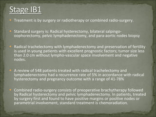
Dr. Adnan Hassan, MD, FACOG Dept. Of Obstetrics and Gynecology Jordan hospital  Consultant in Obstetrics,  Gynecology, and Gynecologic Oncology American Board
Nearly 500,000 cervical cancer new cases are occurring world wide each year. Cervical cancer represents the third most common cause of female mortality from cancer with 274,000 deaths annually. The mortality is higher in developing countries, where 80% of new cases occur. Screening and treatment programs are frequently inaccessible to women in developing countries. The crude incidence of cervical cancer in the European Union is 13.2/100,000 and the crude mortality is 5.9/100,000women year.
Infection with sexually transmitted oncogenic human papilloma virus is responsible for virtually all cases of cervical cancer. HPV 16 and 18 are the most prevalent of the oncogenic types. Early age at first coitus and early pregnancy. Multiple partners Smoking Decreased immunity
Screening includes Pap smear and HPV DNA testing. Primary prevention via vaccination is now available. The high efficacy of the vaccine may dramatically decrease cervical cancer, preventing up to 70% of newly diagnosed cases. The cost of the vaccine however, precludes its widespread implementation.
Diagnosis of invasive carcinoma of the cervix is based on surgical biopsy and histopathological examination .
FIGO classification is the most widely used classification. FIGO classification is based on clinical assessment of tumor extension, tumor size, vaginal and parametrial involvement, and extension to the bladder and rectum . FIGO classification has been reviewed and has integrated sub-division in stage IIA tumors, based on clinical tumor size assessment. FIGO classification requires basic radiological examinations including chest X-ray and IVU. MRI is considered the reference complementary examination. It is superior to CT scan for tumor extension assessment and equal to CT scam for node involvement. MRI and CT have low sensitivity for nodal involvement. Ultra small particles of iron oxide (USPIO) used as MRI contrast agent improves sensitivity. Chest CT scan may be included for metastases assessment. Surgical staging which includes pelvic and para-aortic lymphadenectomy is used in certain cases. Sentinel lymph node procedure  for lymph nodes assessment is currently under study. It seems sensitive and feasible..
0  Carcinoma  in situ  (preinvasive carcinoma)  I  Cervical carcinoma confined to the uterus; invasive carcinoma diagnosed by microscopy. IA All macroscopically visible lesions—even with superficial invasion—are stage IB IA1 Stromal invasion ≤3.0 mm in depth and ≤7.0 mm in horizontal spread IA2 Stromal invasion >3.0 mm and ≤5.0 mm with a horizontal spread ≤7.0 mm      IB Clinically visible lesion confined to the cervix or microscopic lesion greater than IA2     IB1 Clinically visible lesion ≤4.0 cm in greatest dimension IB2 Clinically visible lesion >4.0 cm in greatest dimension II  Tumour beyond the uterus but not to pelvic wall or to lower third of the vagina IIA  without parametrial extension      IIA1 clinically visible lesion ≤4.0 cm in greatest dimension      IIA2 clinically visible lesion >4.0 cm in greatest dimension      IIB with parametrial extension III Tumour extends to pelvic wall and/or involves lower third of the vagina and/or with hydronephrosis or non-functioning kidney    IIIA Tumour involvement of the lower third of the vagina without extension to the pelvic wall      IIIB Tumour extension to the pelvic wall and/or causes hydronephrosis or non-functioning kidney IVA Tumour involvement of rectal and/or bladder mucosa  IVB Distant metastasis  
Includes tumor size, stage, nodal involvement, lympho-vascular space involvement, and histological subtype. Squamous cell carcinoma is the most frequent histological type accounting for 80-85% of the cases. Adenocarcinoma represents 15-20% of cases. Adenocarcinoma has lower survival rate compared with squamous cell carcinoma, stage to stage with higher distant failure rate.
Multidisciplinary treatment planning is mandatory, based on tumor size and extension.
Cold knife conization with free margins or simple hysterectomy, according to the patient’s age and parity. In case of lympho-vascular space involvement, pelvic lymphadenectomy is recommended.
Surgery is the standard treatment and consists of conization or trachelectomy in young patients and simple or radical hysterectomy in older patients. Pelvic lymphadenectomy is required. In patients with positive margins, parametrial involvement, or pelvic nodes metastases, chemoradiation is required.
Treatment is by surgery or radiotherapy or combined radio-surgery. Standard surgery is: Radical hysterectomy, bilateral salpingo-oophorectomy, pelvic lymphadenectomy, and para-aortic nodes biopsy Radical trachelectomy with lymphadenectomy and preservation of fertility is used in young patients with excellent prognostic factors; tumor size less than 2.0 cm without lympho-vascular space involvement and negative nodes. A review of 548 patients treated with radical trachelectomy and lymphadenectomy had a recurrence rate of 5% in accordance with radical hysterectomy and pregnancy outcome with a range of 41-78% Combined radio-surgery consists of preoperative brachytherapy followed by Radical hysterectomy and pelvic lymphadenectomy. In patients, treated by surgery first and found to have positive margins or positive nodes or parametrial involvement, standard treatment is chemoradiation.
Concomitant chemoradiation is the standard treatment. This modality is superior to radiotherapy alone. A meta-analysis of 18 trials with 3452 patients, showed that chemoradiation with cisplatin-based chemotherapy demonstrated 6% improvement in absolute 5-year survival from 60-66% and 8% improvement in 5 years disease free survival. The most common regimen is cisplatin monotherapy  40 mg/sq. m on weekly schedule. External irradiation is combined with brachytherapy and the total treatment remains < 55 days. Complementary extrafascial hysterectomy was evaluated  with the frame of a randomized trial by RTOG group. There was no survival difference with potential benefit for hysterectomy for patients with persistent disease.
Platinum-based combination has a potential benefit. Combination of cisplatin and toptecan seems effective, but no regimen was superior to cisplatin and paclitaxel. Differences in chemotherapy should take into account preexisting morbidity and potential toxicity for the patient
Neoadjuvant chemotherapy remains controversial and is currently under investigation by the EORTIC (55994). The results of the GOG 141 study, showing no advantage of neoadjuvant chemotherapy with vincristine and cisplatin before Radical hysterectomy and pelvic and  para-aortic lymphadenectomy in bulky stage IB.
Clinical and pelvic examination, including Pap smear are usually performed every 3 months for the first 2 years, every 6 months for the next 3 years and yearly thereafter PET/CT scan is the most sensitive in detecting early local recurrence and metastasis
Radiotherapy should be considered for patients with pelvic recurrence and without prior irradiation Pelvic surgery (exenteration) is an option for selected patients with central pelvic recurrence and prior irradiation. For most patients, palliative chemotherapy is the only option

A. Hassan - Cervical cancer - Guidelines and clinical case presentation (2-3 cases)

  • 1.
    Dr. Adnan Hassan,MD, FACOG Dept. Of Obstetrics and Gynecology Jordan hospital Consultant in Obstetrics, Gynecology, and Gynecologic Oncology American Board
  • 2.
    Nearly 500,000 cervicalcancer new cases are occurring world wide each year. Cervical cancer represents the third most common cause of female mortality from cancer with 274,000 deaths annually. The mortality is higher in developing countries, where 80% of new cases occur. Screening and treatment programs are frequently inaccessible to women in developing countries. The crude incidence of cervical cancer in the European Union is 13.2/100,000 and the crude mortality is 5.9/100,000women year.
  • 3.
    Infection with sexuallytransmitted oncogenic human papilloma virus is responsible for virtually all cases of cervical cancer. HPV 16 and 18 are the most prevalent of the oncogenic types. Early age at first coitus and early pregnancy. Multiple partners Smoking Decreased immunity
  • 4.
    Screening includes Papsmear and HPV DNA testing. Primary prevention via vaccination is now available. The high efficacy of the vaccine may dramatically decrease cervical cancer, preventing up to 70% of newly diagnosed cases. The cost of the vaccine however, precludes its widespread implementation.
  • 5.
    Diagnosis of invasivecarcinoma of the cervix is based on surgical biopsy and histopathological examination .
  • 6.
    FIGO classification isthe most widely used classification. FIGO classification is based on clinical assessment of tumor extension, tumor size, vaginal and parametrial involvement, and extension to the bladder and rectum . FIGO classification has been reviewed and has integrated sub-division in stage IIA tumors, based on clinical tumor size assessment. FIGO classification requires basic radiological examinations including chest X-ray and IVU. MRI is considered the reference complementary examination. It is superior to CT scan for tumor extension assessment and equal to CT scam for node involvement. MRI and CT have low sensitivity for nodal involvement. Ultra small particles of iron oxide (USPIO) used as MRI contrast agent improves sensitivity. Chest CT scan may be included for metastases assessment. Surgical staging which includes pelvic and para-aortic lymphadenectomy is used in certain cases. Sentinel lymph node procedure for lymph nodes assessment is currently under study. It seems sensitive and feasible..
  • 7.
    0 Carcinoma in situ (preinvasive carcinoma) I Cervical carcinoma confined to the uterus; invasive carcinoma diagnosed by microscopy. IA All macroscopically visible lesions—even with superficial invasion—are stage IB IA1 Stromal invasion ≤3.0 mm in depth and ≤7.0 mm in horizontal spread IA2 Stromal invasion >3.0 mm and ≤5.0 mm with a horizontal spread ≤7.0 mm      IB Clinically visible lesion confined to the cervix or microscopic lesion greater than IA2     IB1 Clinically visible lesion ≤4.0 cm in greatest dimension IB2 Clinically visible lesion >4.0 cm in greatest dimension II Tumour beyond the uterus but not to pelvic wall or to lower third of the vagina IIA without parametrial extension      IIA1 clinically visible lesion ≤4.0 cm in greatest dimension      IIA2 clinically visible lesion >4.0 cm in greatest dimension      IIB with parametrial extension III Tumour extends to pelvic wall and/or involves lower third of the vagina and/or with hydronephrosis or non-functioning kidney   IIIA Tumour involvement of the lower third of the vagina without extension to the pelvic wall      IIIB Tumour extension to the pelvic wall and/or causes hydronephrosis or non-functioning kidney IVA Tumour involvement of rectal and/or bladder mucosa IVB Distant metastasis  
  • 8.
    Includes tumor size,stage, nodal involvement, lympho-vascular space involvement, and histological subtype. Squamous cell carcinoma is the most frequent histological type accounting for 80-85% of the cases. Adenocarcinoma represents 15-20% of cases. Adenocarcinoma has lower survival rate compared with squamous cell carcinoma, stage to stage with higher distant failure rate.
  • 9.
    Multidisciplinary treatment planningis mandatory, based on tumor size and extension.
  • 10.
    Cold knife conizationwith free margins or simple hysterectomy, according to the patient’s age and parity. In case of lympho-vascular space involvement, pelvic lymphadenectomy is recommended.
  • 11.
    Surgery is thestandard treatment and consists of conization or trachelectomy in young patients and simple or radical hysterectomy in older patients. Pelvic lymphadenectomy is required. In patients with positive margins, parametrial involvement, or pelvic nodes metastases, chemoradiation is required.
  • 12.
    Treatment is bysurgery or radiotherapy or combined radio-surgery. Standard surgery is: Radical hysterectomy, bilateral salpingo-oophorectomy, pelvic lymphadenectomy, and para-aortic nodes biopsy Radical trachelectomy with lymphadenectomy and preservation of fertility is used in young patients with excellent prognostic factors; tumor size less than 2.0 cm without lympho-vascular space involvement and negative nodes. A review of 548 patients treated with radical trachelectomy and lymphadenectomy had a recurrence rate of 5% in accordance with radical hysterectomy and pregnancy outcome with a range of 41-78% Combined radio-surgery consists of preoperative brachytherapy followed by Radical hysterectomy and pelvic lymphadenectomy. In patients, treated by surgery first and found to have positive margins or positive nodes or parametrial involvement, standard treatment is chemoradiation.
  • 13.
    Concomitant chemoradiation isthe standard treatment. This modality is superior to radiotherapy alone. A meta-analysis of 18 trials with 3452 patients, showed that chemoradiation with cisplatin-based chemotherapy demonstrated 6% improvement in absolute 5-year survival from 60-66% and 8% improvement in 5 years disease free survival. The most common regimen is cisplatin monotherapy 40 mg/sq. m on weekly schedule. External irradiation is combined with brachytherapy and the total treatment remains < 55 days. Complementary extrafascial hysterectomy was evaluated with the frame of a randomized trial by RTOG group. There was no survival difference with potential benefit for hysterectomy for patients with persistent disease.
  • 14.
    Platinum-based combination hasa potential benefit. Combination of cisplatin and toptecan seems effective, but no regimen was superior to cisplatin and paclitaxel. Differences in chemotherapy should take into account preexisting morbidity and potential toxicity for the patient
  • 15.
    Neoadjuvant chemotherapy remainscontroversial and is currently under investigation by the EORTIC (55994). The results of the GOG 141 study, showing no advantage of neoadjuvant chemotherapy with vincristine and cisplatin before Radical hysterectomy and pelvic and para-aortic lymphadenectomy in bulky stage IB.
  • 16.
    Clinical and pelvicexamination, including Pap smear are usually performed every 3 months for the first 2 years, every 6 months for the next 3 years and yearly thereafter PET/CT scan is the most sensitive in detecting early local recurrence and metastasis
  • 17.
    Radiotherapy should beconsidered for patients with pelvic recurrence and without prior irradiation Pelvic surgery (exenteration) is an option for selected patients with central pelvic recurrence and prior irradiation. For most patients, palliative chemotherapy is the only option