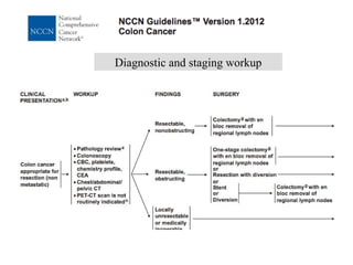
Stefano Cascinu  Department of Medical Oncology Università Politecnica delle Marche  Ancona, Italy Colorectal Cancer: ESMO or NCCN Guidelines?
Screening Diagnosis Staging Adjuvant Advanced disease Follow-up
Screening: general population FOBT:  reduces mortality by 25% it should be offered to men and women aged 50 years until 74 years. The screening interval shoud be 1-2 years.
Screening: high risk population Lynch syndrome Colonoscopy starting at age 20-25 every 1-2 years.  Endometrial and ovarian cancer screening may be performed yearly starting at 30-35 years. Surveillance for other Lynch-associated cancers is recommended based on the family history (upper endoscopy and abdominal US). FAP Classic FAP:  Sigmoidoscopy every 2 years starting at age 12-14. Once adenomas are detected colonoscopy should be performed annually Attenuated form: Colonoscopy every two years starting at 18-20, lifelong. Once adenomas are detected it should be performed annually.
Diagnostic and staging workup
Diagnosis Endoscopy plus biopsy Virtual colonoscopy or CT colonography:  not standard procedures but useful to precisely locate the tumor for the surgical approach in patients candidates for laparoscopy.
Physical examination Laboratory data Blood count; CEA, liver chemistry Instrumental work-up Preoperative: CT scan (chest and abdomen) Surgical staging: assessment of the liver intraoperative US, nodal spread Staging
A  F D  O J  L U  L V  O A  W N  U T  P and T  H E R A P Y
Adjuvant therapy Stage III, fit patients: 6 months FOLFOX or XELOX Stage III, unfit patients 6 months Capecitabine or 5-fluorouracil Stage III elderly patients (>70Years) 6 months 5fluorouracil (?)
Adjuvant therapy Stage II patients low risk No adjuvant therapy Stage II patients high risk  (lymph-node sampling<12; poorly differentiated tumors; vascular or lymphatic or perineural invasion; tumor presentation with obstruction or perforation; pT4)  Adjuvant therapy with 5FU or FOLFOX Stage II patients MSI No adjuvant therapy or, if at high risk, FOLFOX
Follow-up CT scan of chest and abdomen every 6-12 months CEUS could substitute CT scan Colonoscopy performed at 1 year and then every 3-5 years PET/scan is  not recommended
Advanced disease Aggressive therapy Symptomatic disease Borderline resectable liver metastases No aggressive therapy Indolent disease Elderly patients
Synchronous metastases: Resectable unresectable
Metachronous metastases Resectable Unresectable
Rectal cancer
 
 
Metastatic at diagnosis
 
staging Endoscopic ultrasound MRI for all rectal tumours
Preoperative therapy Postoperative therapy cT1-2 and some cT3 (a) above elevators, surgery alone cT3 (b); cT4, N+ preoperative chemo- radiotherapy Tis, T1 N0  local excision T2 N0 wide surgical resection T3/T4 N0 surgery and adjuvant therapy T any N1/2 surgery and adjuvant therapy

S. Cascinu - Colorectal cancer - Guidelines and clinical case presentation (2-3 cases)

  • 1.
    Stefano Cascinu Department of Medical Oncology Università Politecnica delle Marche Ancona, Italy Colorectal Cancer: ESMO or NCCN Guidelines?
  • 2.
    Screening Diagnosis StagingAdjuvant Advanced disease Follow-up
  • 3.
    Screening: general populationFOBT: reduces mortality by 25% it should be offered to men and women aged 50 years until 74 years. The screening interval shoud be 1-2 years.
  • 4.
    Screening: high riskpopulation Lynch syndrome Colonoscopy starting at age 20-25 every 1-2 years. Endometrial and ovarian cancer screening may be performed yearly starting at 30-35 years. Surveillance for other Lynch-associated cancers is recommended based on the family history (upper endoscopy and abdominal US). FAP Classic FAP: Sigmoidoscopy every 2 years starting at age 12-14. Once adenomas are detected colonoscopy should be performed annually Attenuated form: Colonoscopy every two years starting at 18-20, lifelong. Once adenomas are detected it should be performed annually.
  • 5.
  • 6.
    Diagnosis Endoscopy plusbiopsy Virtual colonoscopy or CT colonography: not standard procedures but useful to precisely locate the tumor for the surgical approach in patients candidates for laparoscopy.
  • 7.
    Physical examination Laboratorydata Blood count; CEA, liver chemistry Instrumental work-up Preoperative: CT scan (chest and abdomen) Surgical staging: assessment of the liver intraoperative US, nodal spread Staging
  • 8.
    A FD O J L U L V O A W N U T P and T H E R A P Y
  • 9.
    Adjuvant therapy StageIII, fit patients: 6 months FOLFOX or XELOX Stage III, unfit patients 6 months Capecitabine or 5-fluorouracil Stage III elderly patients (>70Years) 6 months 5fluorouracil (?)
  • 10.
    Adjuvant therapy StageII patients low risk No adjuvant therapy Stage II patients high risk (lymph-node sampling<12; poorly differentiated tumors; vascular or lymphatic or perineural invasion; tumor presentation with obstruction or perforation; pT4) Adjuvant therapy with 5FU or FOLFOX Stage II patients MSI No adjuvant therapy or, if at high risk, FOLFOX
  • 11.
    Follow-up CT scanof chest and abdomen every 6-12 months CEUS could substitute CT scan Colonoscopy performed at 1 year and then every 3-5 years PET/scan is not recommended
  • 12.
    Advanced disease Aggressivetherapy Symptomatic disease Borderline resectable liver metastases No aggressive therapy Indolent disease Elderly patients
  • 13.
  • 14.
  • 15.
  • 16.
  • 17.
  • 18.
  • 19.
  • 20.
    staging Endoscopic ultrasoundMRI for all rectal tumours
  • 21.
    Preoperative therapy Postoperativetherapy cT1-2 and some cT3 (a) above elevators, surgery alone cT3 (b); cT4, N+ preoperative chemo- radiotherapy Tis, T1 N0 local excision T2 N0 wide surgical resection T3/T4 N0 surgery and adjuvant therapy T any N1/2 surgery and adjuvant therapy