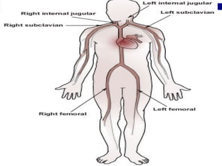
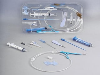
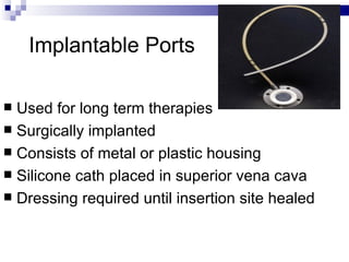
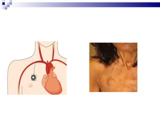
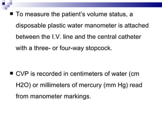
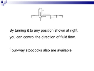
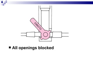
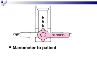
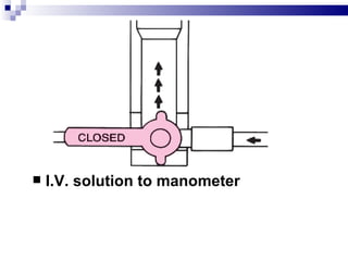
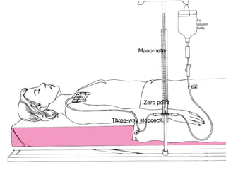
CENTRAL VENOUS
PRESSURE …

  BY LT KALAIVANI R
CVP

Central venous pressure (CVP) (also
known as: right atrial pressure; RAP)
describes the pressure of blood in the
thoracic vena cava, near the right atrium of
the heart
Normal CVP can be measured from
two points of reference:

 •Sternum: 0–14     cm H2O

 •Mid axillary   line: 8–15 cm H2O
Factors that decrease CVP include

  Hypovolemia

  Deep inhalation

   shock
Factors that increase CVP include
•Hypervolemia

•Tension pneumothorax

•Heart failure

•Pleural effusion
Central Venous
Catheters
Indications
 Long term intravenous antibiotics
 Chemotherapy
 Total paranteral nutrition
 Dialysis
 Monitoring CVP
 Limited vascular access
 Administering large amount of blood &
  blood products.
Types Of Central Venous Catheters

 Nontunneled central catheters
 Tunneled central catheters
 Peripherally inserted central catheters
  (PICC)
 Implantable ports
Nontunneled Central Venous
            Catheters
 Used for short-term therapy
 Inserted percutaneously
     Subclavian  vein
     Internal jugular vein
     Femoral vein
 Has from 1 to 4 lumens or ports
 Usually from 6 to 8 inches in length
Tunneled Central Venous
               Catheters
   Used for long term therapy

   Inserted surgically

   Small Dacron cuff sits in subcutaneous tunnel

   Initially sutured but removed in 7 to 10 days

   External portion of the cath can be repaired
Tunneled CVC
Peripherally Inserted Central
           Catheters (PICC)
 Used for intermediate to long term therapy
 May be single or double lumen
 Inserted percutaneously
     Basalicvein
     Cephalic vein
 Threaded into the superior vena cava
 May be inserted by specially trained RN
Implantable Ports

 Used for long term therapies
 Surgically implanted
 Consists of metal or plastic housing
 Silicone cath placed in superior vena cava
 Dressing required until insertion site healed
Equipment
For intermittent CVP
        monitoring

       Disposable CVP manometer set
       leveling device (such as a rod from a reusable
       CVP pole holder or a carpenter’s level or rule) ,
       additional stopcock (to attach the CVP
    manometer to the catheter)
 extension tubing (if needed)
 I.V. Pole,
 I.V. solution ,
 I.V. drip chamber and tubing
 dressing materials
 and tape.
Implementation

   Gather the necessary equipment.

   Explain the procedure to the patient to reduce his
    anxiety.

   Assist the physician as he inserts the CV catheter.
Complications
   Infection
     Phlebitis


     Septicemia   or pyrogenic reaction

 Air   embolism
 Thrombosis/occlusion

 Extravasations

 pneumothorax
CENTRAL VENOUS PRESSURE
     MONITORING
   CVP monitoring helps
    to assess cardiac function,
    to evaluate venous return to the heart,
     to indirectly know how well the heart is
    pumping
   To measure the patient’s volume status, a
    disposable plastic water manometer is attached
    between the I.V. line and the central catheter
    with a three- or four-way stopcock.


   CVP is recorded in centimeters of water (cm
    H2O) or millimeters of mercury (mm Hg) read
    from manometer markings.
MEASURING CVP WITH
A WATER MANOMETER


To ensure accurate (CVP) readings, make sure
the manometer base is aligned with the patient’s
right atrium (the zero reference point).
The manometer set usually contains a
leveling rod to allow you to determine this
quickly.


After adjusting the manometer’s position,
examine the typical three-way stopcock.
By turning it to any position shown at right,
you can control the direction of fluid flow.


Four-way stopcocks also are available
   All openings blocked
   Manometer to patient
   I.V. solution to manometer
   I.V. solution to patient
I.V.
                        solution
                        bottle


         Manometer


           Zero point


Three-way stopcock
 Turn the stopcock off to the patient, and
slowly fill the manometer with I.V. solution until
the fluid level is 10 to 20 cm H2O higher than
the patient’s expected CVP value.


Don’t overfill the tube because fluid that spills
over the top can become a source of
contamination.
    Turn the stopcock off to the I.V. solution and
    open to the patient.

   The fluid level in the manometer will drop.

   When the fluid level comes to rest, it will
    fluctuate slightly with respirations.

   Expect it to drop during inspiration and to rise
    during expiration.

    Record CVP at the end of expiration, when
    intrathoracic pressure has a negligible effect.
Depending on the type of water manometer
used, note the value either at the bottom of the
meniscus ‫ أسفل السطح المحدب‬or at the midline
of the small floating ball.

 After you’ve obtained the CVP value, turn the
stopcock to resume the I.V. infusion.

Adjust the I.V. drip rate as required.

Place the patient in a comfortable position.
General Nursing Care Of Patient
              With CVC

    Before insertion, lines are initially
    flushed with saline
    During percutaneous insertion of CVC in
    the subclavian or jugular, place patient in
    Trendlenberg or have him perform
    Valsalva maneuver
   After insertion, an occlusive gauze or
    transparent dressing is applied
   Blood is aspirated through all lumens to
    verify patency
   Chest xray must be performed before use
   Flushing of lines
     Each   lumen is treated as a separate cath
     Injection   caps are vigorously cleaned with
      alcohol
     Use   10cc or larger syringe for administration
      of meds or flush
     Turbulent    flush technique is recommended
   Site assessment and determination of
    external cath length is performed and
    documented with each dressing change


   Tubings are changed per protocol – 72hrs


   Caps and connections are changed per
    protocol – 3-7 days
   When to call the physician

     Temp      of 100.5F or greater
     Chills,   dyspnea, dizziness
     Pain,   redness, swelling, or drainage at site
     Unresolved     resistance, pain or fluid leaking while
      flushing
     Hole    or tear in cath
     Excessive     bleeding at site
     Change      in length of external cath
     Swelling    in neck, face, chest, or arm
   General safety measures
     No   sharp objects near cath
     Clamp   cath when not in use
     No   pulling or tension on the cath
     Activity   limitations
Discontinuing A CVC
   Follow the institution’s policy and procedure
   For percutaneous internal jugular or subclavian
    insertion sites, place patient in trendlenburg
    position and have him perform the Valsalva
    maneuver
•Remove cath and apply pressure with an
occlusive dressing over a petroleum gauze
•Check cath to ensure tip is intact
•Document how patient tolerated procedure,
placement of dressing and cath tip intact.
Documentation
   Document all dressing, tubing, and solution
    changes.

   Document the patient’s tolerance of the
    procedure,
   the date and time of catheter removal, and the
    type of dressing applied.

   Note the condition of the catheter insertion site
    and whether a culture specimen was collected.
    Note any complications and actions taken.
CONCLUSION


   C   Central Venous you.flv
Central venous catheterization

Central venous catheterization

  • 1.
  • 2.
    CVP Central venous pressure(CVP) (also known as: right atrial pressure; RAP) describes the pressure of blood in the thoracic vena cava, near the right atrium of the heart
  • 3.
    Normal CVP canbe measured from two points of reference: •Sternum: 0–14 cm H2O •Mid axillary line: 8–15 cm H2O
  • 4.
    Factors that decreaseCVP include Hypovolemia Deep inhalation  shock
  • 5.
    Factors that increaseCVP include •Hypervolemia •Tension pneumothorax •Heart failure •Pleural effusion
  • 7.
  • 9.
    Indications  Long termintravenous antibiotics  Chemotherapy  Total paranteral nutrition  Dialysis  Monitoring CVP  Limited vascular access  Administering large amount of blood & blood products.
  • 12.
    Types Of CentralVenous Catheters  Nontunneled central catheters  Tunneled central catheters  Peripherally inserted central catheters (PICC)  Implantable ports
  • 13.
    Nontunneled Central Venous Catheters  Used for short-term therapy  Inserted percutaneously  Subclavian vein  Internal jugular vein  Femoral vein  Has from 1 to 4 lumens or ports  Usually from 6 to 8 inches in length
  • 15.
    Tunneled Central Venous Catheters  Used for long term therapy  Inserted surgically  Small Dacron cuff sits in subcutaneous tunnel  Initially sutured but removed in 7 to 10 days  External portion of the cath can be repaired
  • 17.
  • 18.
    Peripherally Inserted Central Catheters (PICC)  Used for intermediate to long term therapy  May be single or double lumen  Inserted percutaneously  Basalicvein  Cephalic vein  Threaded into the superior vena cava  May be inserted by specially trained RN
  • 20.
    Implantable Ports  Usedfor long term therapies  Surgically implanted  Consists of metal or plastic housing  Silicone cath placed in superior vena cava  Dressing required until insertion site healed
  • 22.
  • 23.
    For intermittent CVP monitoring  Disposable CVP manometer set  leveling device (such as a rod from a reusable CVP pole holder or a carpenter’s level or rule) ,  additional stopcock (to attach the CVP manometer to the catheter)
  • 24.
     extension tubing(if needed)  I.V. Pole,  I.V. solution ,  I.V. drip chamber and tubing  dressing materials  and tape.
  • 25.
    Implementation  Gather the necessary equipment.  Explain the procedure to the patient to reduce his anxiety.  Assist the physician as he inserts the CV catheter.
  • 26.
    Complications  Infection  Phlebitis  Septicemia or pyrogenic reaction  Air embolism  Thrombosis/occlusion  Extravasations  pneumothorax
  • 27.
  • 28.
    CVP monitoring helps to assess cardiac function, to evaluate venous return to the heart,  to indirectly know how well the heart is pumping
  • 29.
    To measure the patient’s volume status, a disposable plastic water manometer is attached between the I.V. line and the central catheter with a three- or four-way stopcock.  CVP is recorded in centimeters of water (cm H2O) or millimeters of mercury (mm Hg) read from manometer markings.
  • 30.
    MEASURING CVP WITH AWATER MANOMETER To ensure accurate (CVP) readings, make sure the manometer base is aligned with the patient’s right atrium (the zero reference point).
  • 31.
    The manometer setusually contains a leveling rod to allow you to determine this quickly. After adjusting the manometer’s position, examine the typical three-way stopcock.
  • 32.
    By turning itto any position shown at right, you can control the direction of fluid flow. Four-way stopcocks also are available
  • 33.
    All openings blocked
  • 34.
    Manometer to patient
  • 35.
    I.V. solution to manometer
  • 36.
    I.V. solution to patient
  • 37.
    I.V. solution bottle Manometer Zero point Three-way stopcock
  • 38.
     Turn thestopcock off to the patient, and slowly fill the manometer with I.V. solution until the fluid level is 10 to 20 cm H2O higher than the patient’s expected CVP value. Don’t overfill the tube because fluid that spills over the top can become a source of contamination.
  • 39.
    Turn the stopcock off to the I.V. solution and open to the patient.  The fluid level in the manometer will drop.  When the fluid level comes to rest, it will fluctuate slightly with respirations.  Expect it to drop during inspiration and to rise during expiration.  Record CVP at the end of expiration, when intrathoracic pressure has a negligible effect.
  • 40.
    Depending on thetype of water manometer used, note the value either at the bottom of the meniscus ‫ أسفل السطح المحدب‬or at the midline of the small floating ball.  After you’ve obtained the CVP value, turn the stopcock to resume the I.V. infusion. Adjust the I.V. drip rate as required. Place the patient in a comfortable position.
  • 41.
    General Nursing CareOf Patient With CVC  Before insertion, lines are initially flushed with saline  During percutaneous insertion of CVC in the subclavian or jugular, place patient in Trendlenberg or have him perform Valsalva maneuver
  • 42.
    After insertion, an occlusive gauze or transparent dressing is applied  Blood is aspirated through all lumens to verify patency  Chest xray must be performed before use
  • 44.
    Flushing of lines  Each lumen is treated as a separate cath  Injection caps are vigorously cleaned with alcohol  Use 10cc or larger syringe for administration of meds or flush  Turbulent flush technique is recommended
  • 45.
    Site assessment and determination of external cath length is performed and documented with each dressing change  Tubings are changed per protocol – 72hrs  Caps and connections are changed per protocol – 3-7 days
  • 46.
    When to call the physician  Temp of 100.5F or greater  Chills, dyspnea, dizziness  Pain, redness, swelling, or drainage at site  Unresolved resistance, pain or fluid leaking while flushing  Hole or tear in cath  Excessive bleeding at site  Change in length of external cath  Swelling in neck, face, chest, or arm
  • 47.
    General safety measures  No sharp objects near cath  Clamp cath when not in use  No pulling or tension on the cath  Activity limitations
  • 48.
    Discontinuing A CVC  Follow the institution’s policy and procedure  For percutaneous internal jugular or subclavian insertion sites, place patient in trendlenburg position and have him perform the Valsalva maneuver
  • 49.
    •Remove cath andapply pressure with an occlusive dressing over a petroleum gauze •Check cath to ensure tip is intact •Document how patient tolerated procedure, placement of dressing and cath tip intact.
  • 50.
    Documentation  Document all dressing, tubing, and solution changes.  Document the patient’s tolerance of the procedure,  the date and time of catheter removal, and the type of dressing applied.  Note the condition of the catheter insertion site and whether a culture specimen was collected.  Note any complications and actions taken.
  • 51.
    CONCLUSION  C Central Venous you.flv