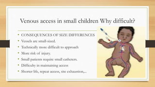
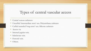
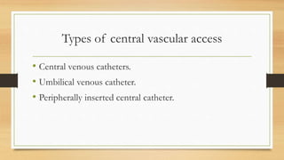
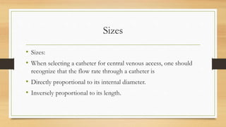
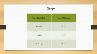
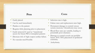
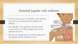
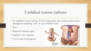
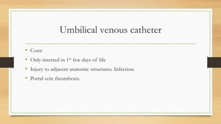
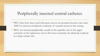
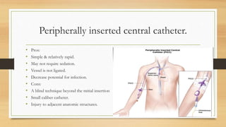
Vascular Access site
consideration for
Neonatal and
Pediatrics
Mr. Prem Kumar
Karthikeyan
Lecturer
Introduction
• Generally Peritoneal dialysis is considered the preferred method of dialysis
for Neonatal and Pediatrics.
• The ideal vascular access delivers an adequate flow rate for the dialysis
prescription, has a long use-life, and has a low rate of complications (e.g.
Infection, stenosis, thrombosis, aneurysm, and limb ischemia)”.
• The arteriovenous fistula (AVF) best approximates this definition.
• The ideal vascular access delivers an adequate flow rate for the dialysis
prescription, has a long use-life, and has a low rate of complications (e.g.
Infection, stenosis, thrombosis, aneurysm, and limb ischemia)”.
• The arteriovenous fistula (AVF) best approximates this definition, however
• - Not useful in acute cases
• - Not feasible in neonates and small children
• Despite some efforts at microsurgical approaches for smaller children.
Introduction
Venous access in small children Why difficult?
• CONSEQUENCES OF SIZE DIFFERENCES
• Vessels are small-sized.
• Technically more difficult to approach
• More risk of injury.
• Small patients require small catheters.
• Difficulty in maintaining access
• Shorter life, repeat access, site exhaustion,...
Types of central vascular access
• Central venous catheters:
• Uncuffed ‘intermediate term’ usu. Polyurethane catheters
• Cuffed tunneled ‘long-term’ usu. Silicone catheters
• Access via
• Internal jugular vein.
• Subclavian vein.
• Femoral vein.
• Others.
Types of central vascular access
• Central venous catheters.
• Umbilical venous catheter.
• Peripherally inserted central catheter.
Sizes
• Sizes:
• When selecting a catheter for central venous access, one should
recognize that the flow rate through a catheter is
• Directly proportional to its internal diameter.
• Inversely proportional to its length.
Sizes
Size of the Child Size of Catheter
Neonate 5 Fr
3-6 Kg 7 Fr
6-12 Kg 8 Fr
Pros
• Easily placed.
• Can be used immediately
• Painless to the patient
• Requires little planning prior to placement
• Easily removed if used as “transitional
access for future PD or transplant patients
• Decrease risk of high-output cardiac failure.
• No vascular steal Possible
Cons
• Infection rate is high.
• Failure rates and replacement rates high.
• Permanent damage to central venous
system (stenosis/ thrombosis) may occur.
• Blood flow rates are variable, leading to
potentially poor clearance.
• Damage to central vessels can prohibit
future AVF/AVG placement in ipsilateral
extremity.
• Arrhythmia.
Internal jugular vein catheter
• Central venous access via the IJV is safe, relatively
easy and very commonly used in infants and
children.
• The right IJV is the preferred vein for central
venous access as it offers straight access to the
superior vena cava.
• The rate of complications are lower compared to
the femoral and the subclavian access.
Technique
• A shoulder roll provides an appropriate degree of neck extension with a slight
contralateral head rotation.
• The carotid artery is palpated with one hand, then the skin is usually punctured at
the level of the cricoid ring just lateral to the carotid artery. The needle is advanced
at a 30-40° angle to the skin towards the ipsilateral nipple.
• Ultrasound can be used to visualize the diameter and position of the IJV in relation
to the carotid artery. In particular in patients with expected difficulties or with
previous central lines in that position an ultrasound improves the likelihood of a
successful puncture. In addition obstruction of the vein can be excluded.
Difficulties and complications during insertion
• Injury to the carotid artery.
• Very rarely, a haematoma causing tracheal compression can occur.
• The risk of pneumothorax or hemothorax (lower than that in subclavian
access).
• Malpositioning occurs more often when the left IJV is used. The guidewire
may form a bow and end up in the right IJV.
Subclavian vein catheter
• The subclavian approach remains the most
commonly used blind approach for
subclavian vein cannulation.
• The physician’s experience and comfort level
with the procedure, however, are the main
determinants as to the success of the line
placement in cases with no other patient-
related factors that may increase the
incidence of complications.
Technique
• The child is placed in the Trendelenburg position with a rolled towel underneath the
shoulders between the scapulae to slightly hyperextend the back. The neck should
not be overextended.
• The puncture site should be at the junction of the medial and middle thirds of the
clavicle near the depression created by the deltoid and the pectoralis major muscles.
• The introducer needle is inserted through the puncture site. It is held parallel to the
frontal plane and directed toward the posterior aspect of the sternal notch.
• The needle should be advanced along the inferior surface of the clavicle (parallel to
the anterior chest wall as possible) to decrease the risk of puncturing the pleura.
Contraindications to subclavian venous catheter
insertion
Absolute
• Trauma to the ipsilateral clavicle,
anterior proximal rib, or subclavian
vessels.
• Coagulopathy (direct pressure to stop
bleeding cannot be applied to the
subclavian vein or artery, because of
their location beneath the clavicle).
Relative
• Chest wall deformity.
• Future AV access planned in ipsilateral
arm
Femoral vein catheter
• The femoral vein is not often used as the primary site for central venous access.
• The rare occasions for using the femoral site include the following:
• Placement of a temporary hemodialysis or pheresis catheter.
• Inaccessibility of other primary central veins as a consequence of thrombosis or
stenosis.
• During cardiopulmonary resuscitation (CPR), in that this approach does not interfere
with chest compressions or defibrillation.
Technique
• The leg is restrained with the hip abducted and in slight external rotation.
• The pelvis is elevated slightly by placing a towel under the hips to improve exposure
of the vein.
• The femoral artery is palpated with one hand, then the skin is usually punctured 0.3-
1 cm medially.
• The needle is advanced at a 30-40° angle to the skin towards the umbilicus.
• Ultrasound guidance should be used for this procedure when equipment and
operator expertise is available.
Complications
• Injury to the femoral artery.
• Hematoma formation.
• Thrombosis.
• Infection.
Umbilical venous catheter
• An umbilical venous catheter (UVC) is placed into the cardiovascular system
through the remaining “tail” of a cut umbilical cord.
• Pros :
• Simple & relatively rapid.
• Sedation is not required.
• Can be used in emergency.
Umbilical venous catheter
• Cons:
• Only inserted in 1st few days of life
• Injury to adjacent anatomic structures. Infection.
• Portal vein thrumbosis.
Peripherally inserted central catheter.
• PICC lines have been used with great success in neonatal intensive care units
(NICUs) and are considered a mainstay of vascular access in this setting.
• PICC are inserted peripherally, usually in the cephalic vein in the upper
extremity or the saphenous vein in the lower extremity, the distal tip is placed
in a large central vein.
Peripherally inserted central catheter.
• Pros:
• Simple & relatively rapid.
• May not require sedation.
• Vessel is not ligated.
• Decrease potential for infection.
• Cons:
• A blind technique beyond the mitial insertion
• Small caliber catheter.
• Injury to adjacent anatomic structures.

Pediatrics vascular access and their types.pptx

  • 1.
    Vascular Access site considerationfor Neonatal and Pediatrics Mr. Prem Kumar Karthikeyan Lecturer
  • 2.
    Introduction • Generally Peritonealdialysis is considered the preferred method of dialysis for Neonatal and Pediatrics. • The ideal vascular access delivers an adequate flow rate for the dialysis prescription, has a long use-life, and has a low rate of complications (e.g. Infection, stenosis, thrombosis, aneurysm, and limb ischemia)”. • The arteriovenous fistula (AVF) best approximates this definition.
  • 3.
    • The idealvascular access delivers an adequate flow rate for the dialysis prescription, has a long use-life, and has a low rate of complications (e.g. Infection, stenosis, thrombosis, aneurysm, and limb ischemia)”. • The arteriovenous fistula (AVF) best approximates this definition, however • - Not useful in acute cases • - Not feasible in neonates and small children • Despite some efforts at microsurgical approaches for smaller children. Introduction
  • 4.
    Venous access insmall children Why difficult? • CONSEQUENCES OF SIZE DIFFERENCES • Vessels are small-sized. • Technically more difficult to approach • More risk of injury. • Small patients require small catheters. • Difficulty in maintaining access • Shorter life, repeat access, site exhaustion,...
  • 5.
    Types of centralvascular access • Central venous catheters: • Uncuffed ‘intermediate term’ usu. Polyurethane catheters • Cuffed tunneled ‘long-term’ usu. Silicone catheters • Access via • Internal jugular vein. • Subclavian vein. • Femoral vein. • Others.
  • 6.
    Types of centralvascular access • Central venous catheters. • Umbilical venous catheter. • Peripherally inserted central catheter.
  • 7.
    Sizes • Sizes: • Whenselecting a catheter for central venous access, one should recognize that the flow rate through a catheter is • Directly proportional to its internal diameter. • Inversely proportional to its length.
  • 8.
    Sizes Size of theChild Size of Catheter Neonate 5 Fr 3-6 Kg 7 Fr 6-12 Kg 8 Fr
  • 9.
    Pros • Easily placed. •Can be used immediately • Painless to the patient • Requires little planning prior to placement • Easily removed if used as “transitional access for future PD or transplant patients • Decrease risk of high-output cardiac failure. • No vascular steal Possible Cons • Infection rate is high. • Failure rates and replacement rates high. • Permanent damage to central venous system (stenosis/ thrombosis) may occur. • Blood flow rates are variable, leading to potentially poor clearance. • Damage to central vessels can prohibit future AVF/AVG placement in ipsilateral extremity. • Arrhythmia.
  • 10.
    Internal jugular veincatheter • Central venous access via the IJV is safe, relatively easy and very commonly used in infants and children. • The right IJV is the preferred vein for central venous access as it offers straight access to the superior vena cava. • The rate of complications are lower compared to the femoral and the subclavian access.
  • 11.
    Technique • A shoulderroll provides an appropriate degree of neck extension with a slight contralateral head rotation. • The carotid artery is palpated with one hand, then the skin is usually punctured at the level of the cricoid ring just lateral to the carotid artery. The needle is advanced at a 30-40° angle to the skin towards the ipsilateral nipple. • Ultrasound can be used to visualize the diameter and position of the IJV in relation to the carotid artery. In particular in patients with expected difficulties or with previous central lines in that position an ultrasound improves the likelihood of a successful puncture. In addition obstruction of the vein can be excluded.
  • 12.
    Difficulties and complicationsduring insertion • Injury to the carotid artery. • Very rarely, a haematoma causing tracheal compression can occur. • The risk of pneumothorax or hemothorax (lower than that in subclavian access). • Malpositioning occurs more often when the left IJV is used. The guidewire may form a bow and end up in the right IJV.
  • 13.
    Subclavian vein catheter •The subclavian approach remains the most commonly used blind approach for subclavian vein cannulation. • The physician’s experience and comfort level with the procedure, however, are the main determinants as to the success of the line placement in cases with no other patient- related factors that may increase the incidence of complications.
  • 14.
    Technique • The childis placed in the Trendelenburg position with a rolled towel underneath the shoulders between the scapulae to slightly hyperextend the back. The neck should not be overextended. • The puncture site should be at the junction of the medial and middle thirds of the clavicle near the depression created by the deltoid and the pectoralis major muscles. • The introducer needle is inserted through the puncture site. It is held parallel to the frontal plane and directed toward the posterior aspect of the sternal notch. • The needle should be advanced along the inferior surface of the clavicle (parallel to the anterior chest wall as possible) to decrease the risk of puncturing the pleura.
  • 15.
    Contraindications to subclavianvenous catheter insertion Absolute • Trauma to the ipsilateral clavicle, anterior proximal rib, or subclavian vessels. • Coagulopathy (direct pressure to stop bleeding cannot be applied to the subclavian vein or artery, because of their location beneath the clavicle). Relative • Chest wall deformity. • Future AV access planned in ipsilateral arm
  • 16.
    Femoral vein catheter •The femoral vein is not often used as the primary site for central venous access. • The rare occasions for using the femoral site include the following: • Placement of a temporary hemodialysis or pheresis catheter. • Inaccessibility of other primary central veins as a consequence of thrombosis or stenosis. • During cardiopulmonary resuscitation (CPR), in that this approach does not interfere with chest compressions or defibrillation.
  • 17.
    Technique • The legis restrained with the hip abducted and in slight external rotation. • The pelvis is elevated slightly by placing a towel under the hips to improve exposure of the vein. • The femoral artery is palpated with one hand, then the skin is usually punctured 0.3- 1 cm medially. • The needle is advanced at a 30-40° angle to the skin towards the umbilicus. • Ultrasound guidance should be used for this procedure when equipment and operator expertise is available.
  • 18.
    Complications • Injury tothe femoral artery. • Hematoma formation. • Thrombosis. • Infection.
  • 19.
    Umbilical venous catheter •An umbilical venous catheter (UVC) is placed into the cardiovascular system through the remaining “tail” of a cut umbilical cord. • Pros : • Simple & relatively rapid. • Sedation is not required. • Can be used in emergency.
  • 20.
    Umbilical venous catheter •Cons: • Only inserted in 1st few days of life • Injury to adjacent anatomic structures. Infection. • Portal vein thrumbosis.
  • 21.
    Peripherally inserted centralcatheter. • PICC lines have been used with great success in neonatal intensive care units (NICUs) and are considered a mainstay of vascular access in this setting. • PICC are inserted peripherally, usually in the cephalic vein in the upper extremity or the saphenous vein in the lower extremity, the distal tip is placed in a large central vein.
  • 22.
    Peripherally inserted centralcatheter. • Pros: • Simple & relatively rapid. • May not require sedation. • Vessel is not ligated. • Decrease potential for infection. • Cons: • A blind technique beyond the mitial insertion • Small caliber catheter. • Injury to adjacent anatomic structures.