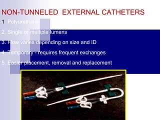
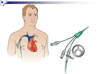
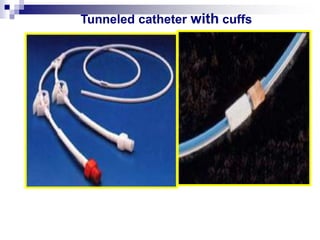
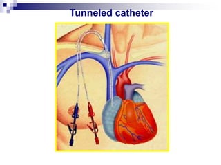
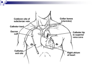
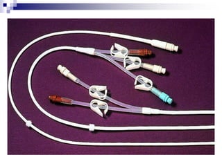
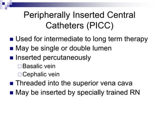
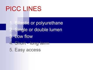
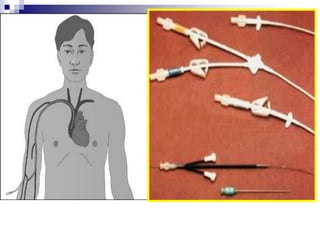
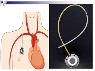
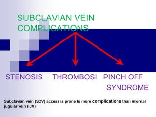
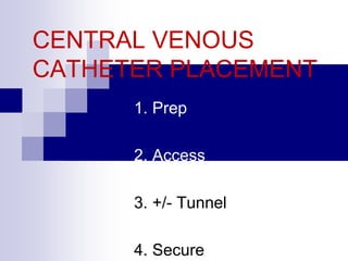
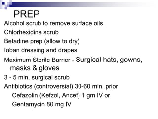
Precautions for Central Venous Catheters in NeonatesKing_maged
Includes: different methods of venous access, CDC guidelines for prevention of catheter-related infections as well as precautions for umbilical catheters use .. Prepared by Dr. Maged Zakaria, NICU Resident, Ain-Shams University Maternity Hospital
It is the process of removing waste from the blood. Ppt would help to learn especially for Nursing students.
Hemodialysis, Peritoneal dialysis, Renal transplantation
Central Venous Access and Catheters. Their indications and contraindications, Different types of central catheters and their advantages and disadvantages, Technique of insertion, and Complications related to central venous lines.
Central Venous Access Devices Made Incredibly Easy!Cathy Lewis
Target audience: RNs during New Hire Orientation and nurses needing additional training on identifying, assessing, and maintaining central lines.
Developed in conjunction with subject matter experts (SMEs) from IV Team. Principles based on practice at this particular institution.
Global launch of the Healthy Ageing and Prevention Index 2nd wave – alongside...ILC- UK
The Healthy Ageing and Prevention Index is an online tool created by ILC that ranks countries on six metrics including, life span, health span, work span, income, environmental performance, and happiness. The Index helps us understand how well countries have adapted to longevity and inform decision makers on what must be done to maximise the economic benefits that comes with living well for longer.
Alongside the 77th World Health Assembly in Geneva on 28 May 2024, we launched the second version of our Index, allowing us to track progress and give new insights into what needs to be done to keep populations healthier for longer.
The speakers included:
Professor Orazio Schillaci, Minister of Health, Italy
Dr Hans Groth, Chairman of the Board, World Demographic & Ageing Forum
Professor Ilona Kickbusch, Founder and Chair, Global Health Centre, Geneva Graduate Institute and co-chair, World Health Summit Council
Dr Natasha Azzopardi Muscat, Director, Country Health Policies and Systems Division, World Health Organisation EURO
Dr Marta Lomazzi, Executive Manager, World Federation of Public Health Associations
Dr Shyam Bishen, Head, Centre for Health and Healthcare and Member of the Executive Committee, World Economic Forum
Dr Karin Tegmark Wisell, Director General, Public Health Agency of Sweden
How many patients does case series should have In comparison to case reports.pdfpubrica101
Pubrica’s team of researchers and writers create scientific and medical research articles, which may be important resources for authors and practitioners. Pubrica medical writers assist you in creating and revising the introduction by alerting the reader to gaps in the chosen study subject. Our professionals understand the order in which the hypothesis topic is followed by the broad subject, the issue, and the backdrop.
https://pubrica.com/academy/case-study-or-series/how-many-patients-does-case-series-should-have-in-comparison-to-case-reports/
Defecation
Normal defecation begins with movement in the left colon, moving stool toward the anus. When stool reaches the rectum, the distention causes relaxation of the internal sphincter and an awareness of the need to defecate. At the time of defecation, the external sphincter relaxes, and abdominal muscles contract, increasing intrarectal pressure and forcing the stool out
The Valsalva maneuver exerts pressure to expel faeces through a voluntary contraction of the abdominal muscles while maintaining forced expiration against a closed airway. Patients with cardiovascular disease, glaucoma, increased intracranial pressure, or a new surgical wound are at greater risk for cardiac dysrhythmias and elevated blood pressure with the Valsalva maneuver and need to avoid straining to pass the stool.
Normal defecation is painless, resulting in passage of soft, formed stool
CONSTIPATION
Constipation is a symptom, not a disease. Improper diet, reduced fluid intake, lack of exercise, and certain medications can cause constipation. For example, patients receiving opiates for pain after surgery often require a stool softener or laxative to prevent constipation. The signs of constipation include infrequent bowel movements (less than every 3 days), difficulty passing stools, excessive straining, inability to defecate at will, and hard feaces
IMPACTION
Fecal impaction results from unrelieved constipation. It is a collection of hardened feces wedged in the rectum that a person cannot expel. In cases of severe impaction the mass extends up into the sigmoid colon.
DIARRHEA
Diarrhea is an increase in the number of stools and the passage of liquid, unformed feces. It is associated with disorders affecting digestion, absorption, and secretion in the GI tract. Intestinal contents pass through the small and large intestine too quickly to allow for the usual absorption of fluid and nutrients. Irritation within the colon results in increased mucus secretion. As a result, feces become watery, and the patient is unable to control the urge to defecate. Normally an anal bag is safe and effective in long-term treatment of patients with fecal incontinence at home, in hospice, or in the hospital. Fecal incontinence is expensive and a potentially dangerous condition in terms of contamination and risk of skin ulceration
HEMORRHOIDS
Hemorrhoids are dilated, engorged veins in the lining of the rectum. They are either external or internal.
FLATULENCE
As gas accumulates in the lumen of the intestines, the bowel wall stretches and distends (flatulence). It is a common cause of abdominal fullness, pain, and cramping. Normally intestinal gas escapes through the mouth (belching) or the anus (passing of flatus)
FECAL INCONTINENCE
Fecal incontinence is the inability to control passage of feces and gas from the anus. Incontinence harms a patient’s body image
PREPARATION AND GIVING OF LAXATIVESACCORDING TO POTTER AND PERRY,
An enema is the instillation of a solution into the rectum and sig
CHAPTER 1 SEMESTER V - ROLE OF PEADIATRIC NURSE.pdfSachin Sharma
Pediatric nurses play a vital role in the health and well-being of children. Their responsibilities are wide-ranging, and their objectives can be categorized into several key areas:
1. Direct Patient Care:
Objective: Provide comprehensive and compassionate care to infants, children, and adolescents in various healthcare settings (hospitals, clinics, etc.).
This includes tasks like:
Monitoring vital signs and physical condition.
Administering medications and treatments.
Performing procedures as directed by doctors.
Assisting with daily living activities (bathing, feeding).
Providing emotional support and pain management.
2. Health Promotion and Education:
Objective: Promote healthy behaviors and educate children, families, and communities about preventive healthcare.
This includes tasks like:
Administering vaccinations.
Providing education on nutrition, hygiene, and development.
Offering breastfeeding and childbirth support.
Counseling families on safety and injury prevention.
3. Collaboration and Advocacy:
Objective: Collaborate effectively with doctors, social workers, therapists, and other healthcare professionals to ensure coordinated care for children.
Objective: Advocate for the rights and best interests of their patients, especially when children cannot speak for themselves.
This includes tasks like:
Communicating effectively with healthcare teams.
Identifying and addressing potential risks to child welfare.
Educating families about their child's condition and treatment options.
4. Professional Development and Research:
Objective: Stay up-to-date on the latest advancements in pediatric healthcare through continuing education and research.
Objective: Contribute to improving the quality of care for children by participating in research initiatives.
This includes tasks like:
Attending workshops and conferences on pediatric nursing.
Participating in clinical trials related to child health.
Implementing evidence-based practices into their daily routines.
By fulfilling these objectives, pediatric nurses play a crucial role in ensuring the optimal health and well-being of children throughout all stages of their development.
We understand the unique challenges pickleball players face and are committed to helping you stay healthy and active. In this presentation, we’ll explore the three most common pickleball injuries and provide strategies for prevention and treatment.
CHAPTER 1 SEMESTER V PREVENTIVE-PEDIATRICS.pdfSachin Sharma
This content provides an overview of preventive pediatrics. It defines preventive pediatrics as preventing disease and promoting children's physical, mental, and social well-being to achieve positive health. It discusses antenatal, postnatal, and social preventive pediatrics. It also covers various child health programs like immunization, breastfeeding, ICDS, and the roles of organizations like WHO, UNICEF, and nurses in preventive pediatrics.
2. INSERTION OF PERIPHERAL IV LINE
Aims
1. To gain peripheral venous access in order to:
• administer fluids
• administer blood products, medications and nutritional
components
2. To minimise the risk of complications when initiating IV
therapy through:
• judicious choice of equipment
• careful choice of IV site
• good insertion technique
• aseptic preparation of infusions
3. Key points
1. Only nurses who have been certified as competent in the
insertion of IV cannula will perform this procedure.
2. Where the patient is less than 14 years of age, the IV
cannula will be inserted by a medical practitioner. The
exception will be in the case of neonates where neonatal
trained nurses may insert an IV cannula if directed by a
medical officer
3. In the case of two unsuccessful attempts at insertion, the
operator will seek the assistance of another experienced nurse
for one additional attempt. After a total three unsuccessful
attempts the assistance of a medical practitioner will be
sought.
5. Phlebitis
Contributing factors:
• Catheter material • Catheter size
• Site of insertion • Skill of operator
• Duration of cannula • Type of infusion
• Dilution of solution • Host factors
• Insertion in ED • Type of skin prep
• Frequency of dressing change
• Presence of infection
8. Selection of Equipment
Cannula selection
1. Select cannula based on purpose and duration
of use, and age of patient.
2. Consider risk of infection and extravasation.
3. Cannulae made from polyurethanes are
associated with decreased risk of phlebitis.
4. Steel needles have higher risk of extravasation
and should be avoided where tissue necrosis is
likely if extravasation occurs.
9. Skin prep
Antiseptic solution - 70% isopropyl alcohol,
0.5 - 1% Chlorhexidine.
Use an aqueous based alternative if there is a
known allergy to alcohol
10. Selection of Catheter Site
Choose a suitable vein. In adults, use long straight veins in an
upper extremity away from the joints for catheter insertion - in
preference to sites on the lower extremities. If possible avoid
veins in the dominant hand and use distal veins first.
Do not insert cannula on the side of mastectomy or AV
shunts/Gortex. Transfer catheter inserted in a lower
extremity site to an upper extremity site as soon as the latter is
available.
In paediatric patients, it is recommended that the cannula be
inserted into the scalp, hand, or foot site in preference to a leg,
arm, or ante cubital fossa site (Category II)
11. Reasons For Inserting Central
Venous Catheters
Limited vascular access
Administration of highly osmotic or caustic fluids
or medications
Frequent administration of blood and blood
products
Frequent blood sampling
Measurement of CVP
Hemodialysis
12. Type of CVC Inserted Depends On
Patient’s condition
Anticipated length of therapy
13. Types Of Central Venous
Catheters
Nontunneled central catheters
Tunneled central catheters
Peripherally inserted central catheters
(PICC)
Implantable ports
14. NON-TUNNELED EXTERNAL CATHETERS
1. Polyurethane
2. Single or multiple lumens
3. Flow varies depending on size and ID
4. Temporary - requires frequent exchanges
5. Easier placement, removal and replacement
15.
16. Nontunneled Central Venous
Catheters
Used for short-term therapy
Inserted percutaneously
Subclavian vein
Internal jugular vein
Femoral vein
Has from 1 to 4 lumens or ports
Usually from 6 to 8 inches in length
17. Can be quickly inserted
Not flexible and may break
Dislodged more easily
Has the highest infection rate
Dressing changes required using aseptic
technique
Unused ports must be routinely flushed with
heparin solution and clamped
18. TUNNELED CATHETERS
1. Single or multiple lumens
2. Flow - variable
3. Long term
4. Easy access (no skin puncture)
5. Cuff - Dacron, vita
21. Tunneled Central Venous
Catheters
Used for long term therapy
Inserted surgically
Small Dacron cuff sits in subcutaneous tunnel
No dressing is required after cuff heals unless
the patient is immunocompromised
Initially sutured but removed in 7 to 10 days
External portion of the cath can be repaired
22.
23.
24. Peripherally Inserted Central
Catheters (PICC)
Used for intermediate to long term therapy
May be single or double lumen
Inserted percutaneously
Basalic vein
Cephalic vein
Threaded into the superior vena cava
May be inserted by specially trained RN
25. PICC LINES
1. Silastic or polyurethane
2. Single or double lumen
3. Low flow
4. Short - long term
5. Easy access
26.
27. Infusing or drawing blood from smaller gauged
PICC may be more difficult
Small gauged PICC infuse fluids slower and
occlude faster
Measure and document external length of PICC
with each dressing change
Dressing acts as a bacterial shield and helps
anchor cath
Unused ports must be flushed with Heparin
solution and clamped
28. SUBCUTANEOUS PORTS
1. Single or double lumen
2. Flow - most commonly slow
3. Long term
4. Access requires needle puncture
29. 5. Less maintenance
6. Activity is unlimited after site heals
7. Cosmetically more appealing
8. Concealed pocket retards infection (?)
SUBCUTANEOUS PORTS
30. Minimizes infection
Huber needle must be used to access port
Must always confirm needle placement before
med administration
Transparent dressing covers Huber needle and
port
Unused port is flushed every 28 days with
Heparin solution
34. ADVANTAGES OF THE
RIGHT IJ
1. Larger
2. More superficial
3. Further from the lung
4. More direct route to the heart
5. Acute and chronic complications are reduced
36. Alcohol scrub to remove surface oils
Chlorhexidine scrub
Betadine prep (allow to dry)
Ioban dressing and drapes
Maximum Sterile Barrier - Surgical hats, gowns,
masks & gloves
3 - 5 min. surgical scrub
Antibiotics (controversial) 30-60 min. prior
Cefazolin (Kefzol, Ancef) 1 gm IV or
Gentamycin 80 mg IV
PREP
37. General Nursing Care Of Patient
With CVC
Always follow the institution’s policy and
procedure
Before insertion, lines are initially flushed
with saline
During percutaneous insertion of CVC in
the subclavian or jugular, place patient in
Trendlenberg or have him perform
Valsalva maneuver
38. After insertion, an occlusive gauze or
transparent dressing is applied
Blood is aspirated through all lumens to
verify patency
Chest xray must be performed before use
Each lumen of the cath is secured with a
Leur-lok cap or CLC 2000 device
39. Use only needless system to access ports
Infusing devices are used for all infusions
TPN is administered exclusively through a
dedicated line and port.
Catheters must be clamped when
removing the cap and when not in use
40. Flushing of lines
Each lumen is treated as a separate cath
Injection caps are vigorously cleaned with
alcohol
Use 10cc or larger syringe for administration
of meds or flush
Turbulent flush technique is recommended
41. For med administration, use SAS method
If port is not to be maintained with a
continuous infusion, end with Heparin flush
solution
Peds 10kg> and adults – 100 units
Heparin/ml with preservatives
Neonates and peds <10kg – 10 units
Heparin/ml without preservatives
For specific amounts see procedure
Clamp cath while infusing last ½ cc of flush
If CLC 2000 used, do not clamp cath until
syringe disconnected
42. Site assessment and determination of
external cath length is performed and
documented with each dressing change
Tubings are changed per protocol – 72hrs
Caps and connections are changed per
protocol – 3-7 days
43. Dressing changes per protocol
Use sterile technique
Change when damp, soiled or loosened
Change every 7 days if transparent
Change every other day if gauze is used
Clean skin around insertion site with
alcohol in a circular motion. Also clean
cath with alcohol
44. Use antmicrobial disk if indicated
Form a loop of the tubing or cath outside
the dressing and anchor securely with
tape
Label site with date, time and initials
Document dressing change, condition of
site and length of external cath when
appropriate
45. For drawing blood specimen
Discard initial sample of blood
Collect specimen
Flush with 10cc saline
Flush with Heparin solution if
indicated
46. Monitor for complications
Infection
Phlebitis
Septicemia or pyrogenic reaction
Air embolism
Thrombosis/occlusion
Extravasation
Damaged cath
49. AIR EMBOLUS: SYMPTOMS
1. Respiratory distress
2. Increased heart rate
3. pulse
5. Cyanosis
4. Poore in the level of consciousness
50. AIR EMBOLUS:
TREATMENT
1. Left lateral decubitus (Durant’s) Position
2 100% O2
3. Vasopressin if necessary
4. Chest compression
5. Aspiration through catheter +/-
Mortality decreases from 90% 30%
with conventional treatment
52. Risk Factors
Four major risk factors are
associated with increased catheter-
related infection rates:
Cutaneous colonization of the
insertion site
Moisture under the dressing
Prolonged catheter time
Technique of care and placement of
the central line
53. Evidence-Based Strategies
Selected to Reduce CLA-BSIs
1. Central line-associated bloodstream infections
bundle
2. Hand hygiene
3. Maximal sterile barriers
4. Chlorhexidine for skin asepsis
5. Avoid femoral lines
6. Avoid/remove unnecessary lines
54. Hand Hygiene
Cornerstone of any infection
prevention program
Many studies have shown
that improvement in hand
hygiene significantly
decreases a variety of
infectious complications
Insufficient or ineffective hand
hygiene contributes
significantly to a greater
bacterial burden and
subsequent spread of
microorganisms within the
environment
55. Hand Hygiene
Use of waterless alcohol-
base hand rub
Most effective and efficient
method for hand antisepsis
against bacterial pathogens
When hands are visibly
soiled, they should be
washed with soap and
water
56. Efficacy of Hand Hygiene
Preparations in Killing
Bacteria
Good Better Best
Plain Soap Antimicrobial
soap
Alcohol-based
handrub
57. Maximal Sterile Barriers
One study found a 6-fold
higher rate of catheter-
related septicemia when
minimal sterile barriers
(sterile gloves and small
drape) were used instead
of maximal sterile barriers
Raad II, Hohn H, Gilbreath J, et al. Prevention of central venous
catheter-related infections by using maximal sterile barrier precautions
during insertion. Infect Control Hosp Epidemiol. 1994;15:231–238.
58. Chlorhexidine for Skin Asepsis
Studies have compared chlorhexidine gluconate
(CHG) versus povidone iodine as a skin
antiseptic for catheter insertion and routine
insertion site care
Recent meta-analysis, the use of CHG rather than
povidone iodine was found to reduce the risk of CLA-
BSIs by approximately 50% in hospitalized patients
who required short term catheterization
Chaiyakunapruk N, Veenstra, DL, Lipsky BA, Saint S. Chlorhexidine
compared with povidone-iodine solution for vascular catheter-site care: a
meta-analysis. Ann Intern Med. 2002;136:792–801.
59. Benefits of CHG
2% CHG in tincture of isopropyl alcohol
has rapid bactericidal activity and is
effective within 30 seconds after
application versus 2-minute period for
povidone iodine
CHG provides persistent bactericidal
activity on the skin and maintains its
activity in the presence of other organic
material
Minimal systemic absorption
60. Site Selection: Avoid Femoral Lines
Insertion of CVCs can lead to serious and
sometimes life-threatening complications,
whether of mechanical, infectious, or thrombotic
origin
Higher rate of infectious complications in study
comparing femoral lines versus subclavian lines
19.8% vs 4.5%
61. Avoid and Remove Unnecessary
Lines
Once placed, there should be periodic, if
not daily assessment, of its continued
need, with emphasis on prompt removal
62. Empowerment of Nursing
One of the most important steps in
preventing CLA-BSIs is to empower the
nursing staff to stop the central line
insertion procedure if the guidelines were
not followed
63. TYPES OF INFECTION
EXIT SITE, TUNNEL/POCKET or CATHETER
1. Cutaneous - pain, erythema, swelling,
+/- exudate
2. Bacteremia - fever, leukocytosis and
positive blood cultures
3. Septic thrombophlebitis - bacteremia,
thrombosis and purulent discharge
65. INFECTION
1. Septic thrombophlebitis - remove catheter
2. Cutaneous - local treatment
3. Bacteremia -
1. IV antibiotics 48 -72 hours
if improved - keep catheter
if no change, worse or recurs
remove catheter
or
2. Exchange catheter over wire,
85% cure with treatment
66. Continue to treat infection for 10 - 14
days
If ineffective - try locking with
thrombolytics between antibiotic doses
and administer antibiotics through
catheters
INFECTION
67. Discharge Teaching For The
Patient With A CVC
Proper handwashing and principles of
sterile technique
Dressing change procedure and frequency
Flushing and cap change procedure and
frequency
Observation of cath and insertion site
68. When to call the physician
Temp of 100.5F or greater
Chills, dyspnea, dizziness
Pain, redness, swelling, or drainage at
site
Unresolved resistance, pain or fluid
leaking while flushing
Hole or tear in cath
Excessive bleeding at site
Change in length of external cath
Swelling in neck, face, chest, or arm
69. General safety measures
No sharp objects near cath
Clamp cath when not in use
No pulling or tension on the cath
Discard syringes and needles in sharps
container
Activity limitations
Use a stress loop
Home health referral
70. Discontinuing A CVC
Follow the institution’s policy and procedure
For percutaneous internal jugular or subclavian
insertion sites, place patient in trendlenburg
position and have him perform the Valsalva
maneuver
Remove cath and apply pressure with an
occlusive dressing over a petroleum gauze
Check cath to ensure tip is intact
Document how patient tolerated procedure,
placement of dressing and cath tip intact