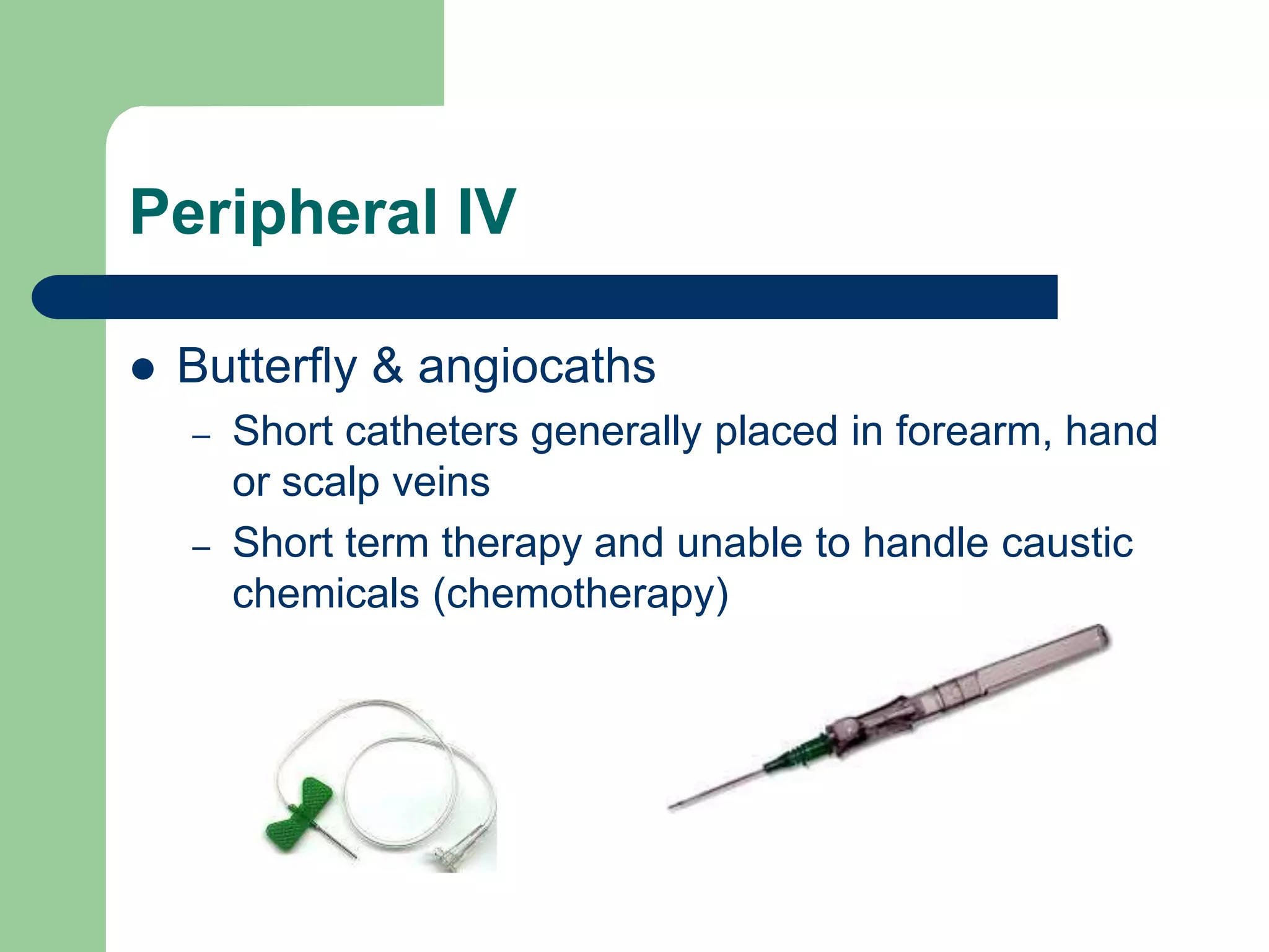
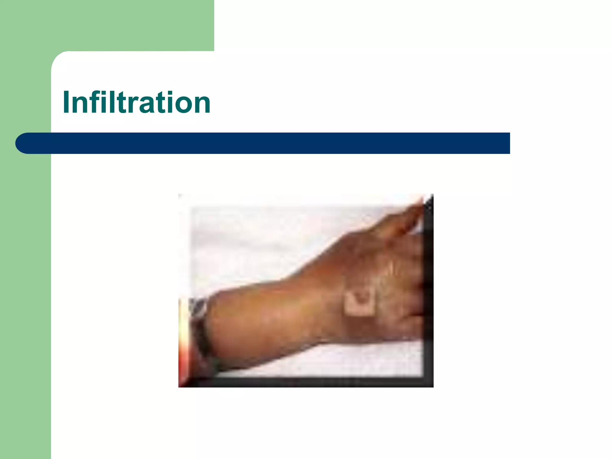
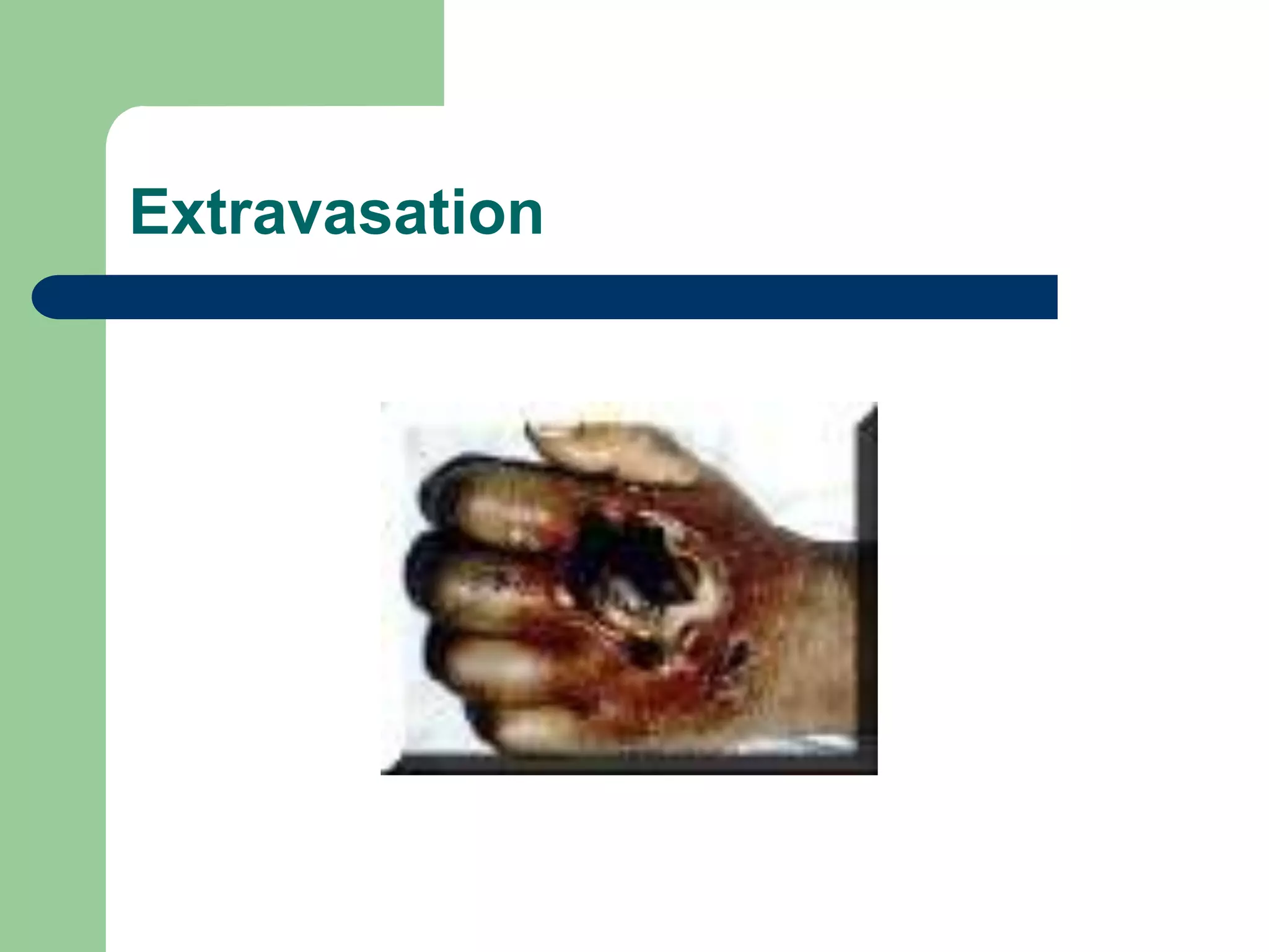
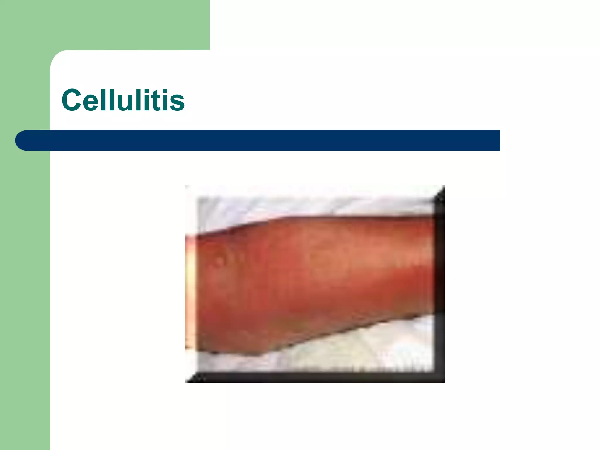
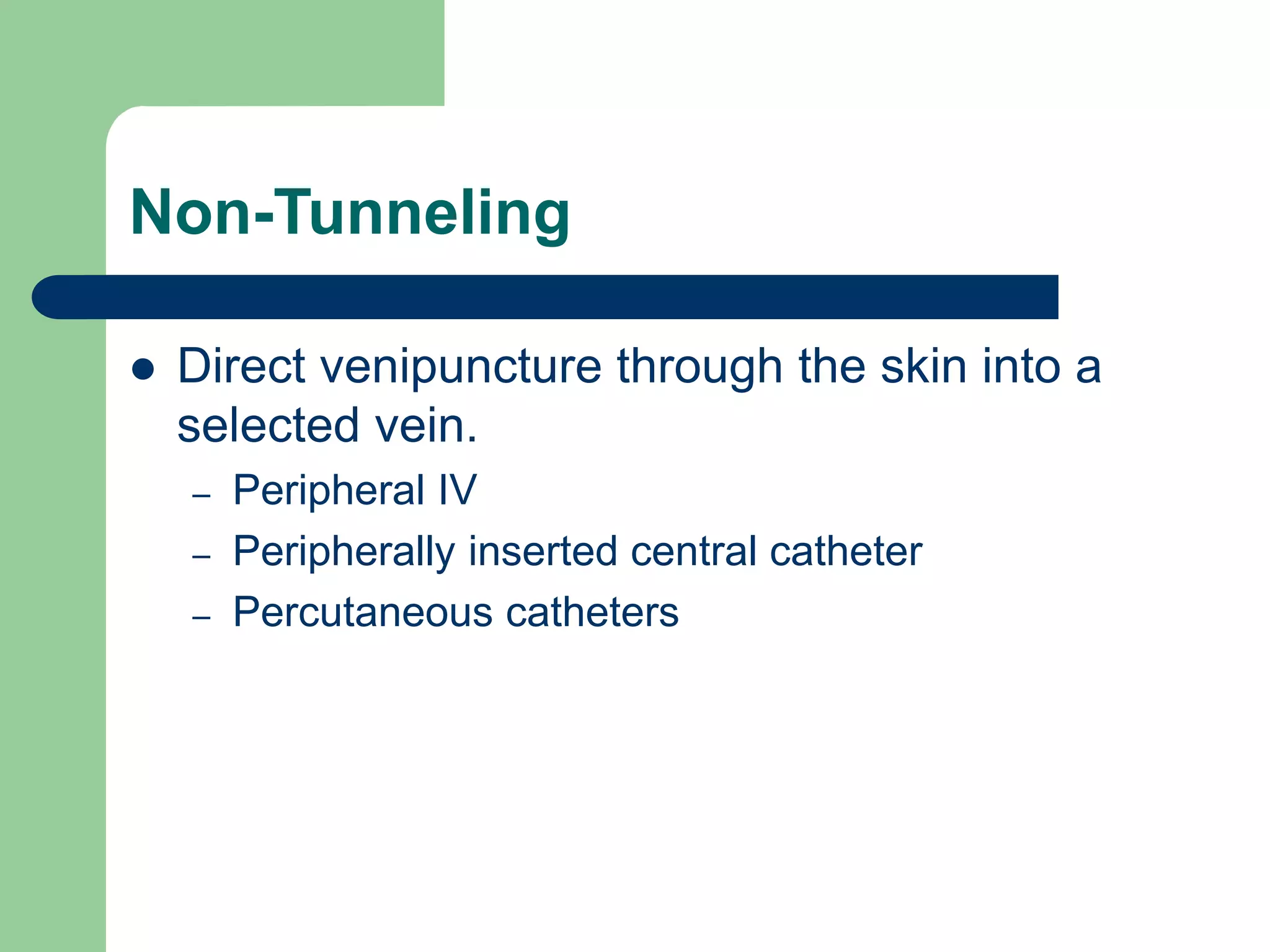
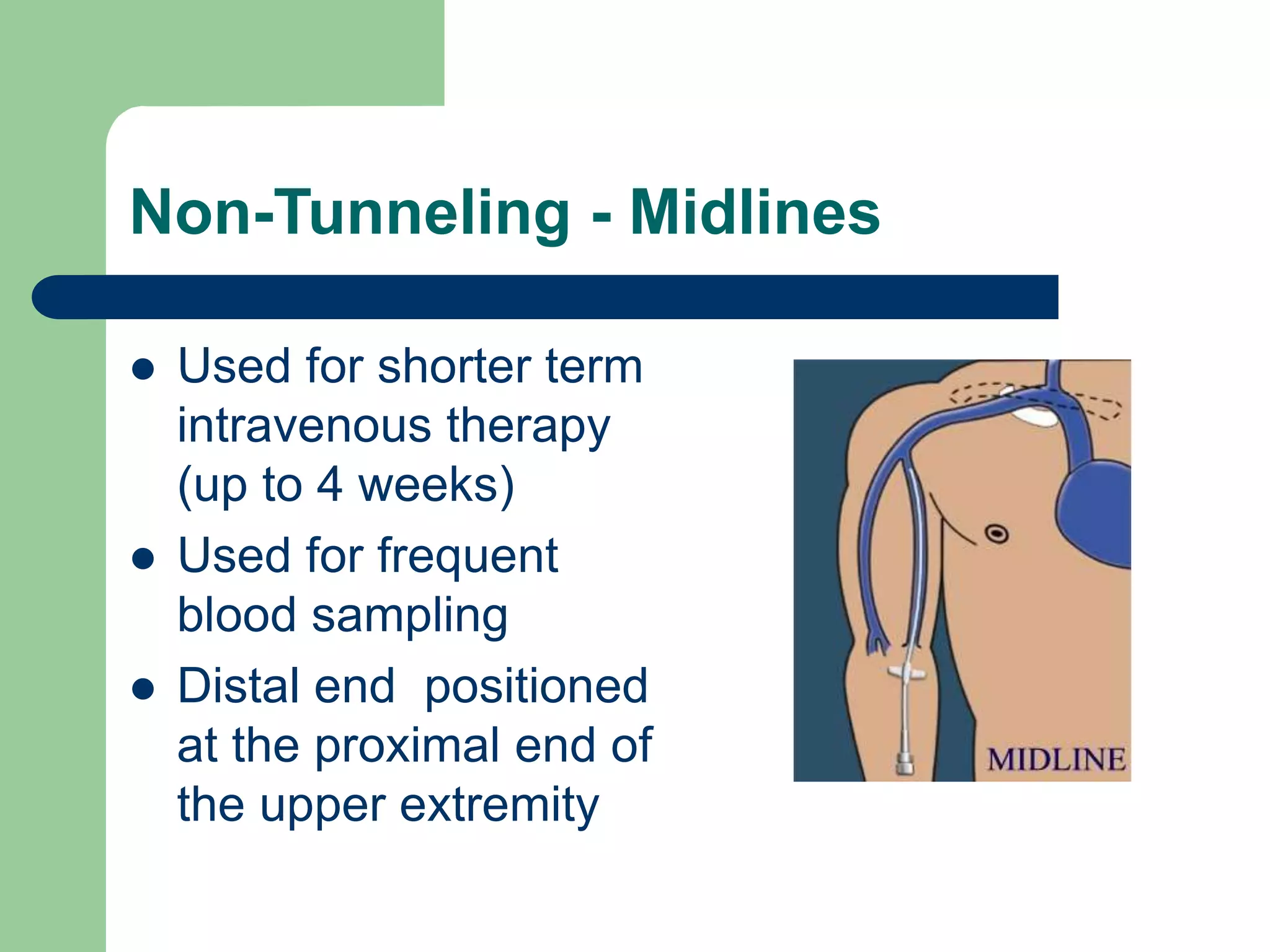
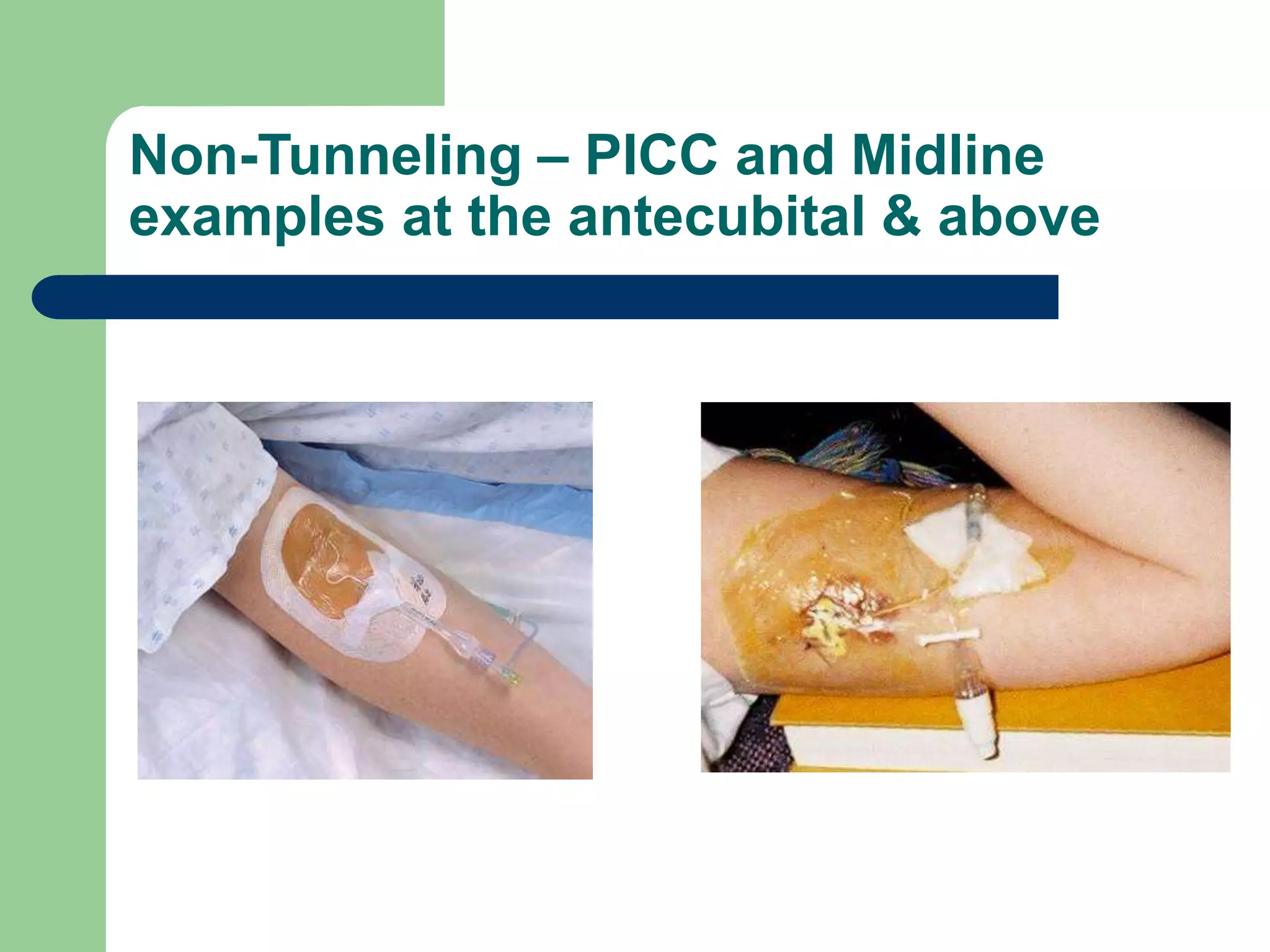
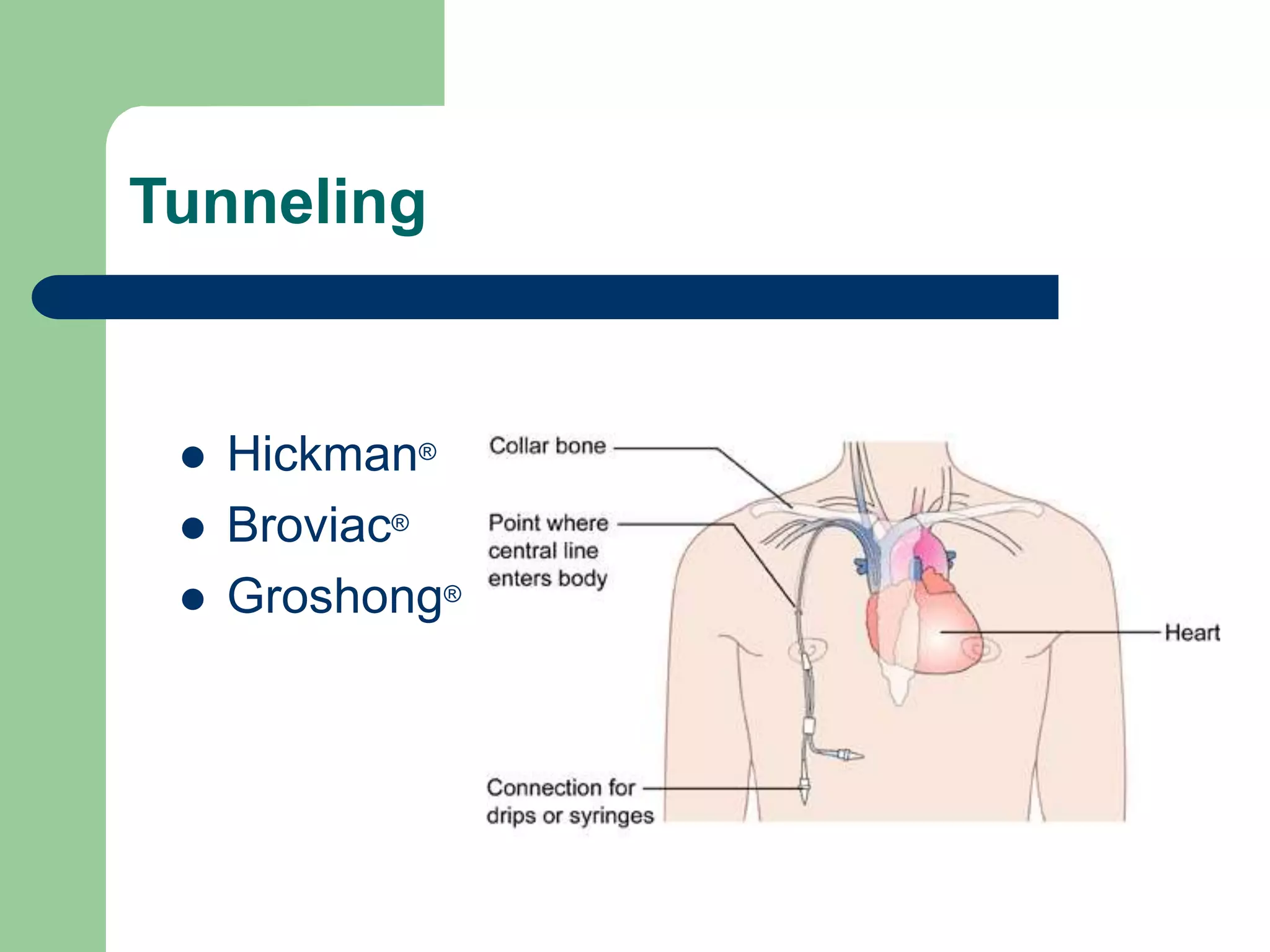
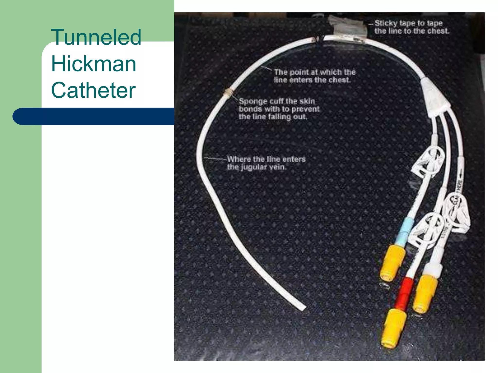
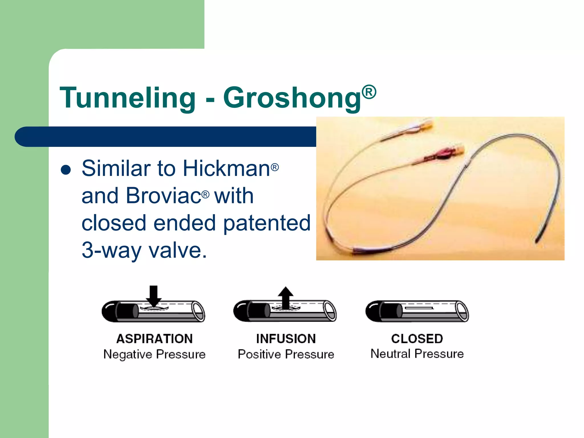
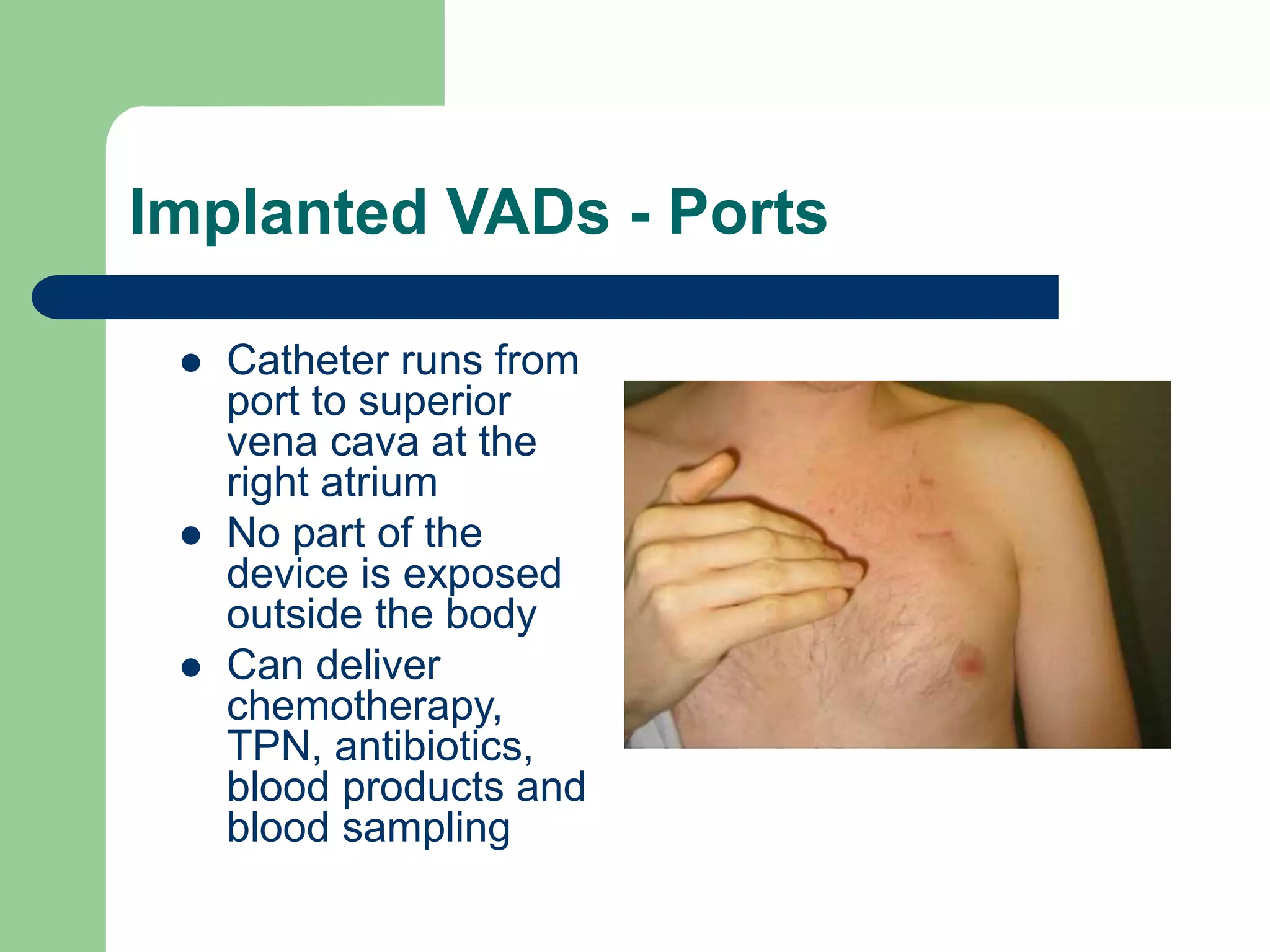
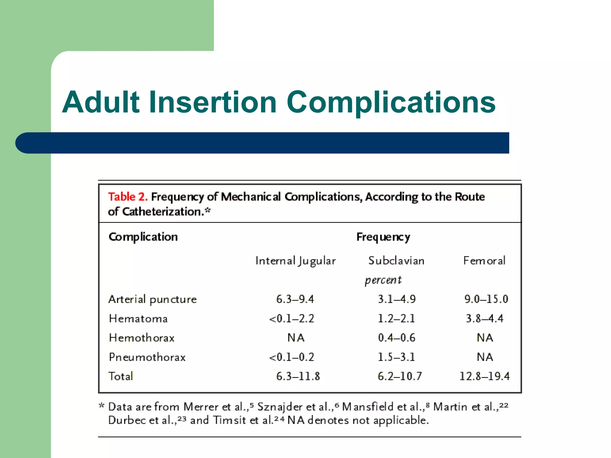
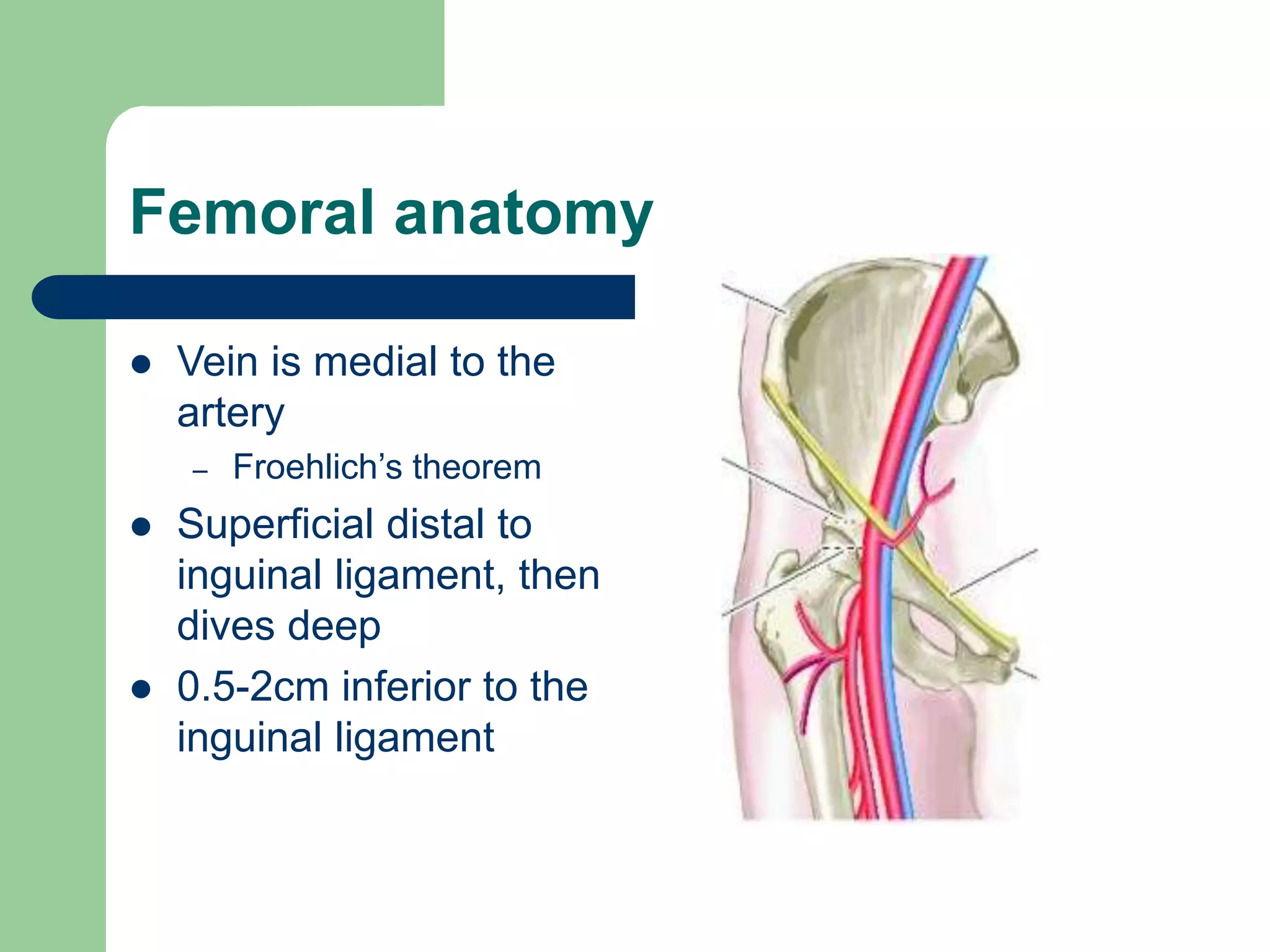
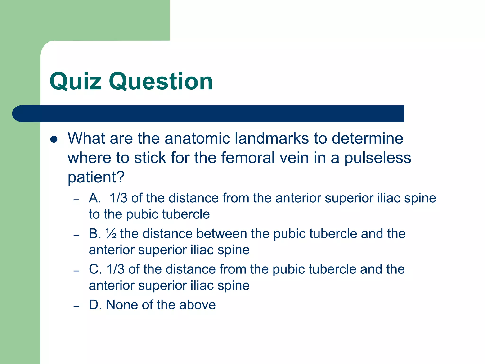
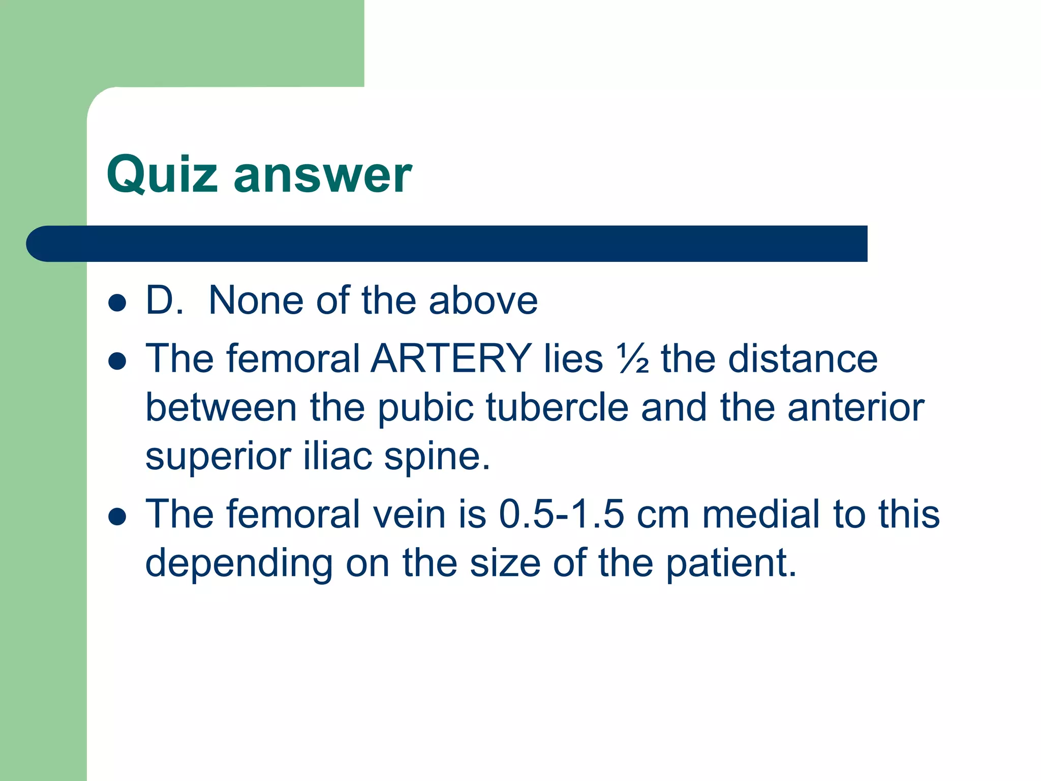
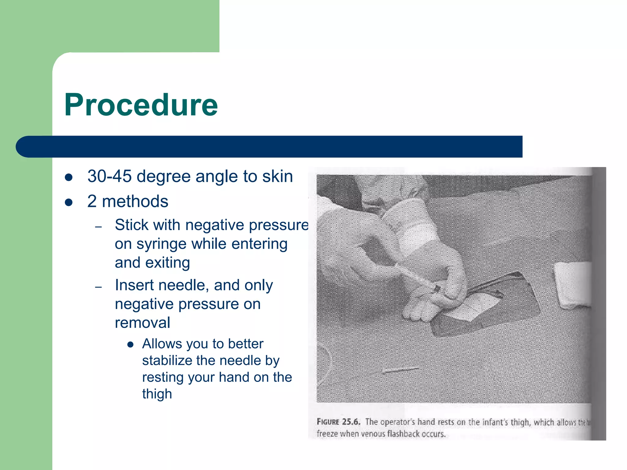
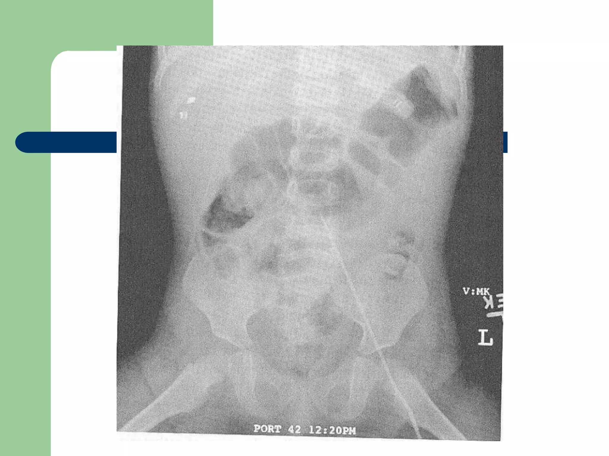
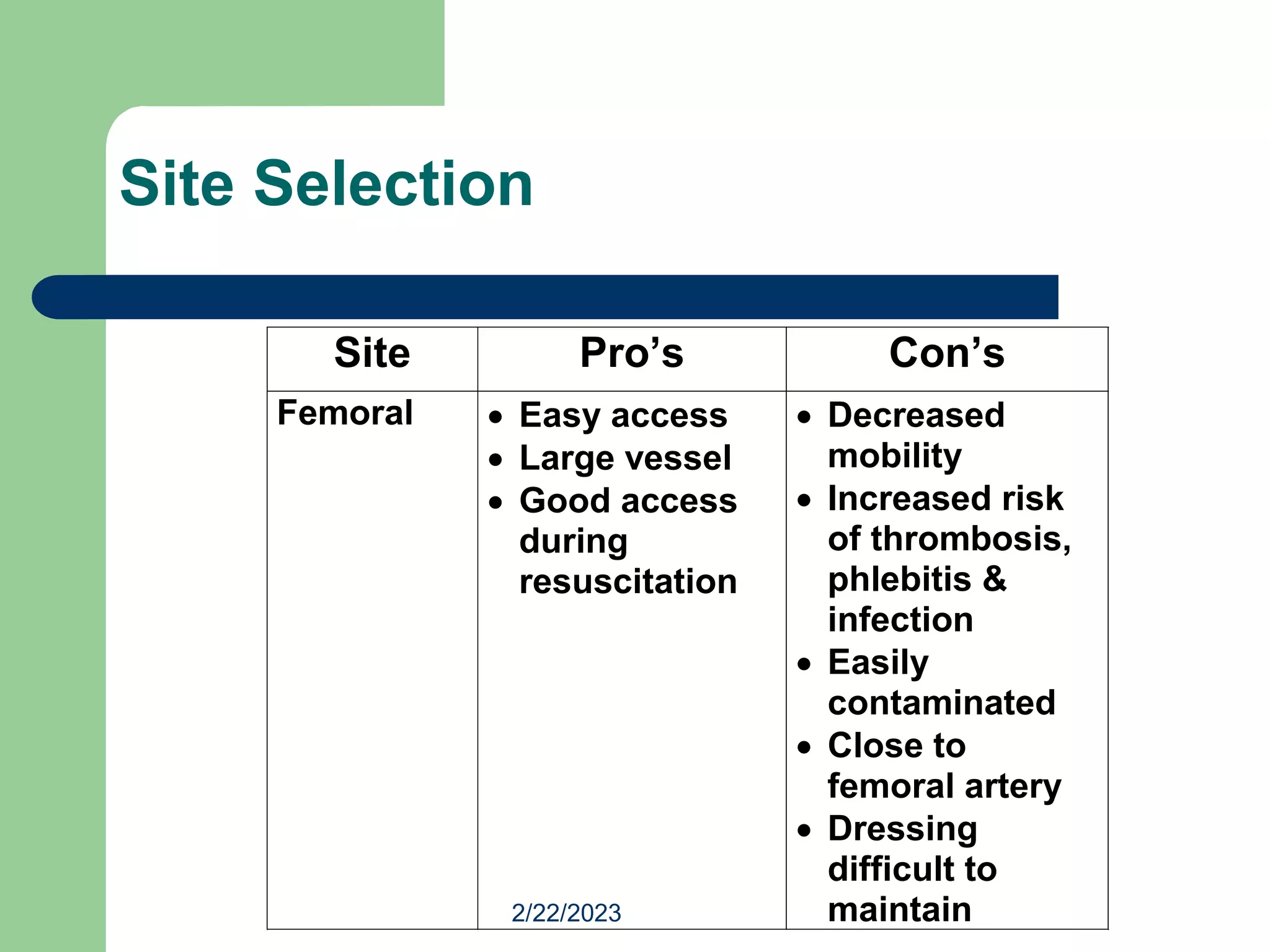
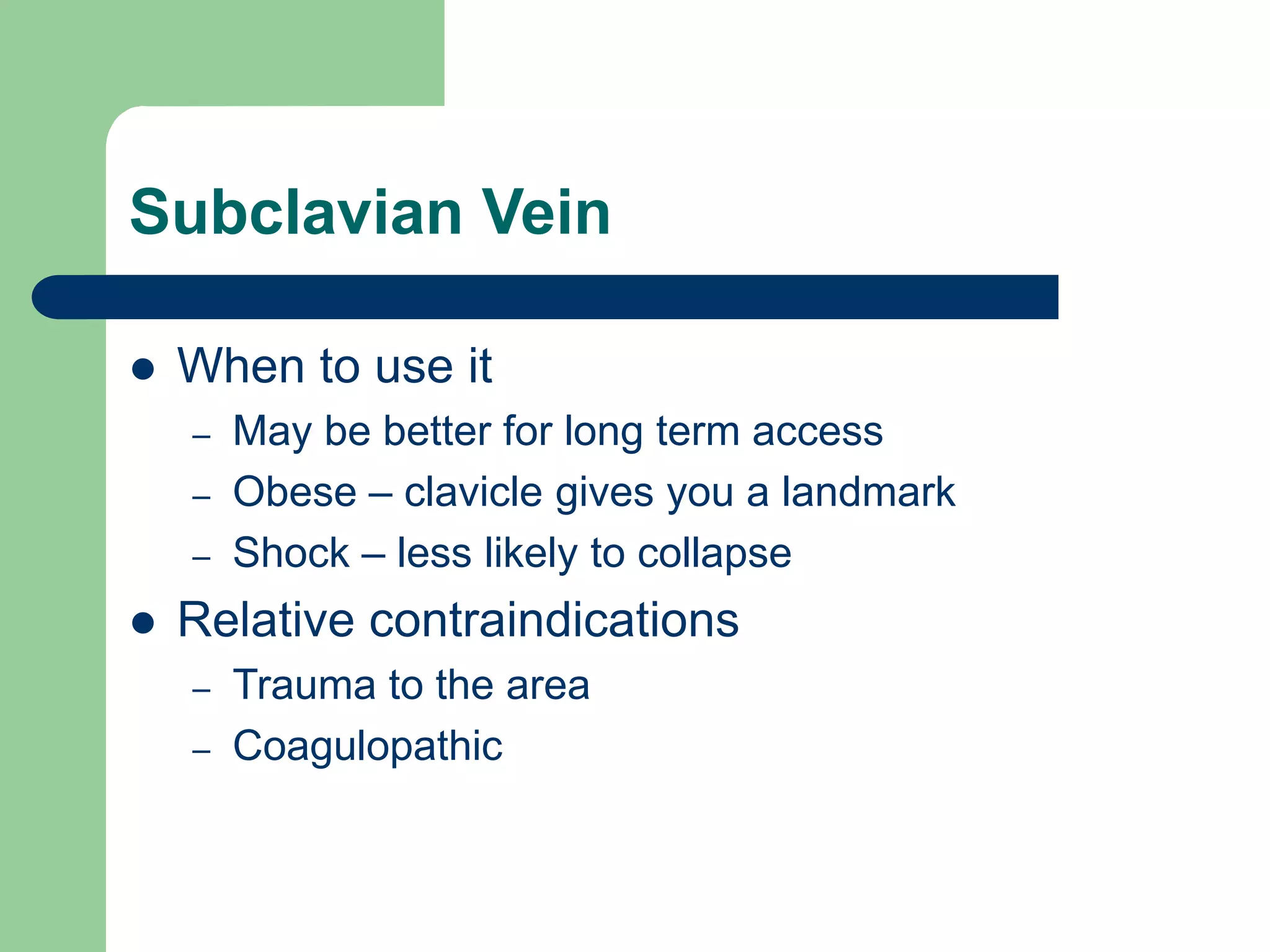
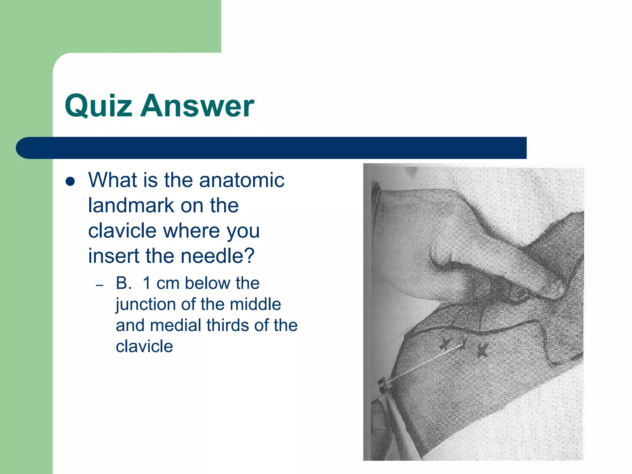
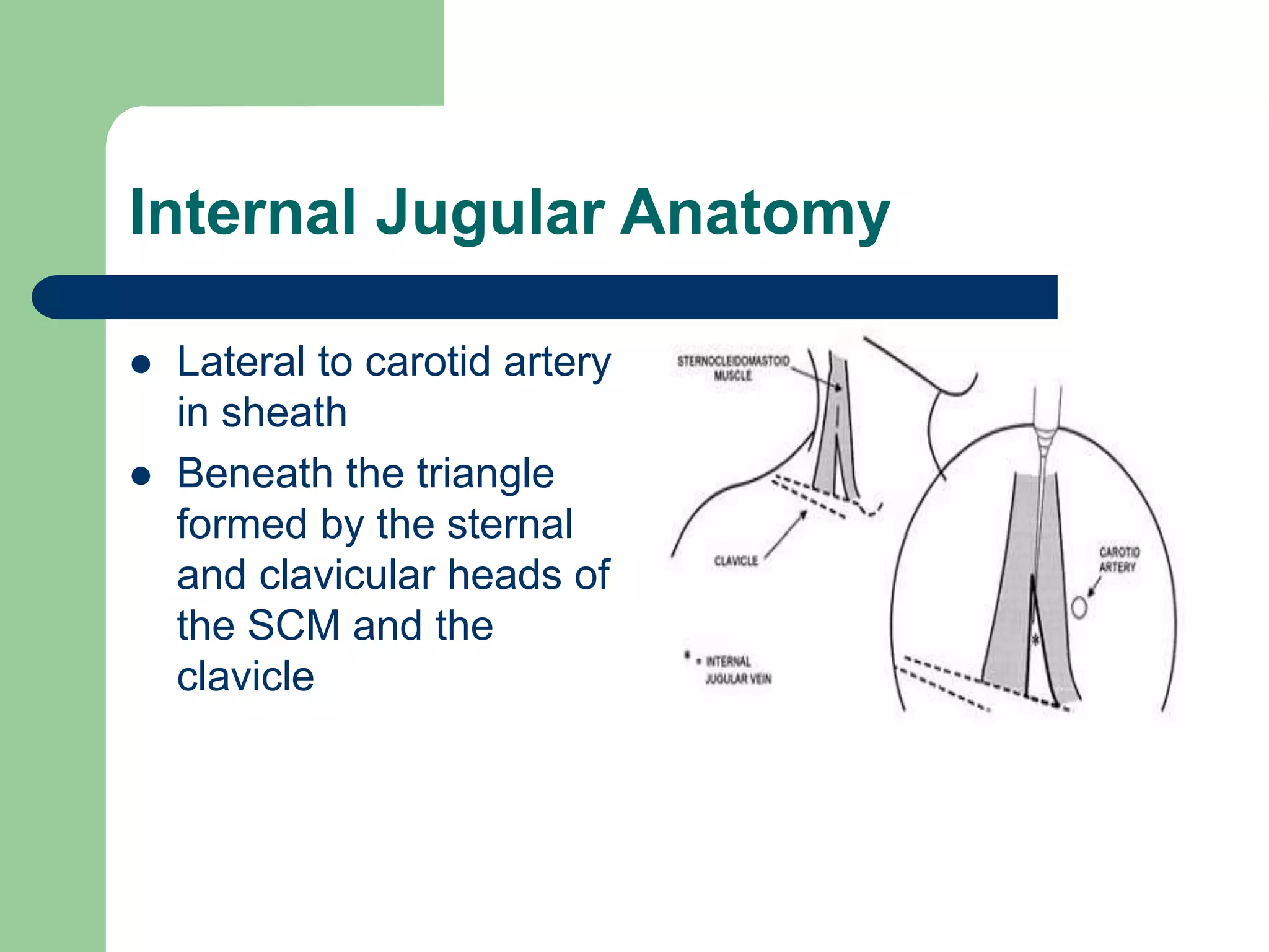
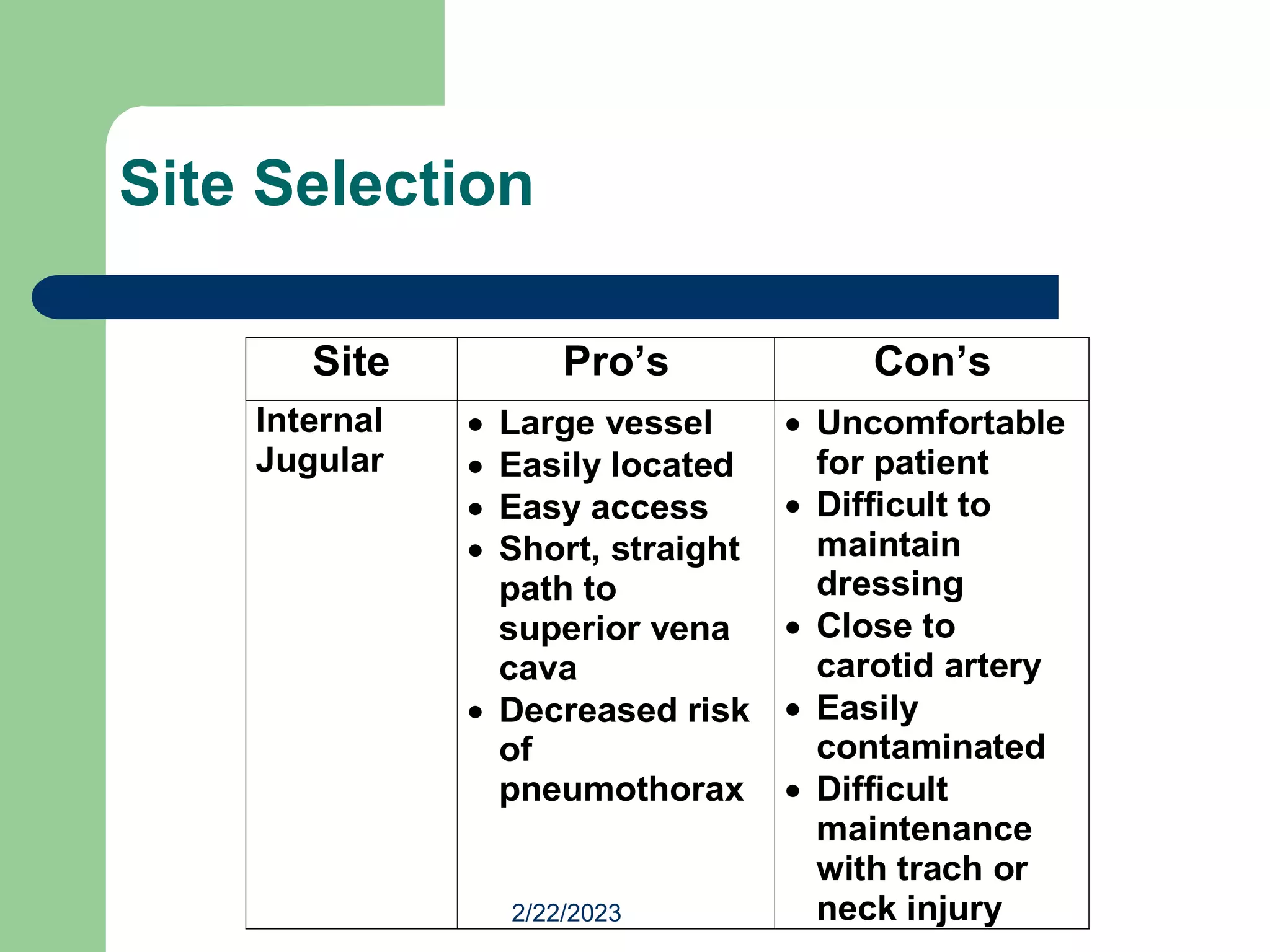
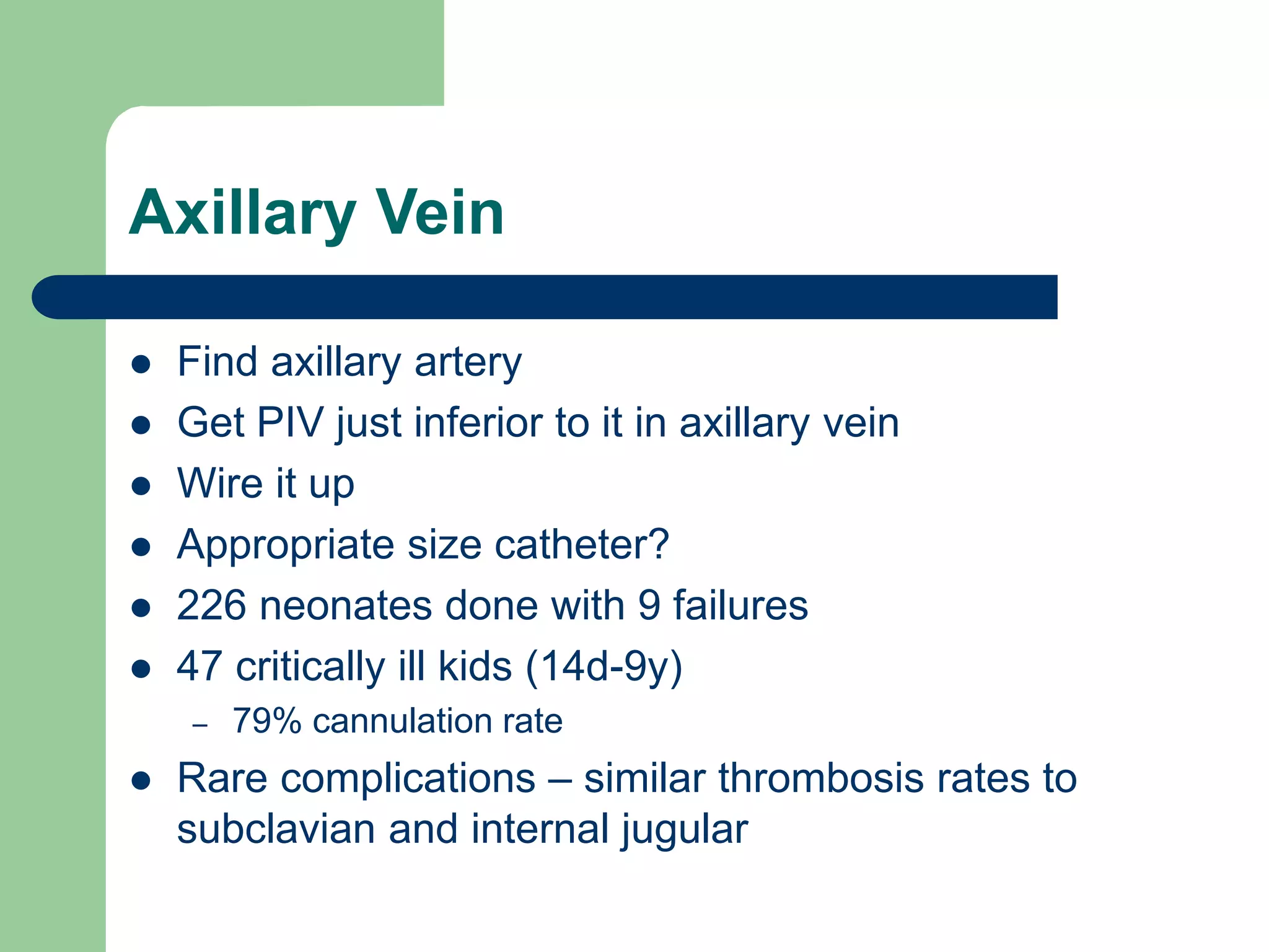
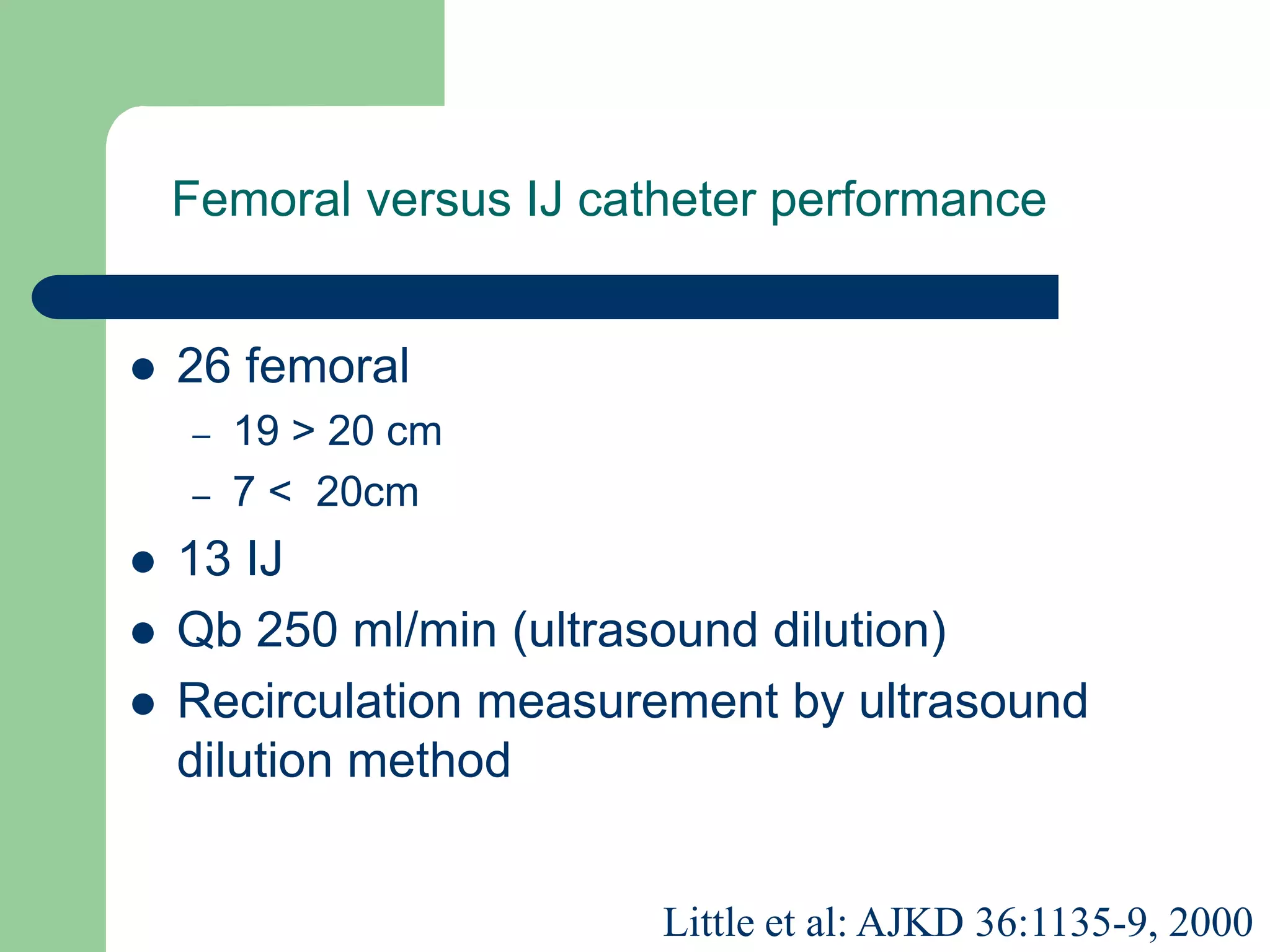
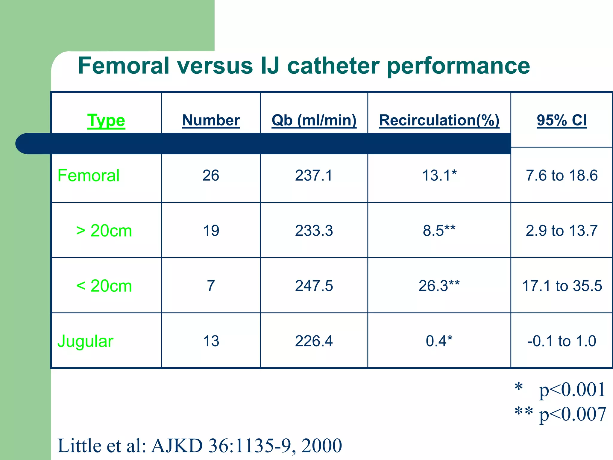
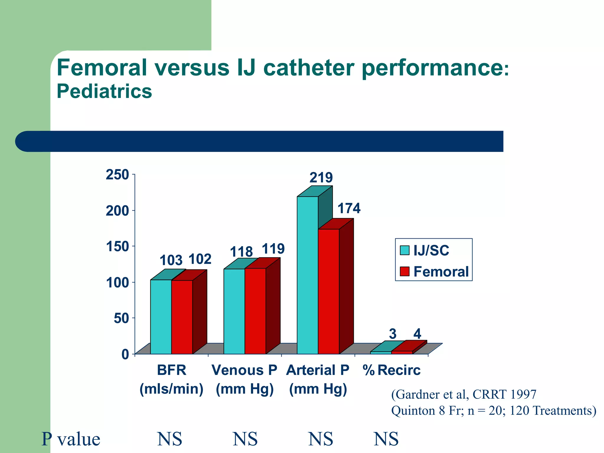
Venous Access
Matthew L. Paden, MD
Emory University
Children’s Healthcare of Atlanta at Egleston
Peripheral IV
 Butterfly & angiocaths
– Short catheters generally placed in forearm, hand
or scalp veins
– Short term therapy and unable to handle caustic
chemicals (chemotherapy)
Peripheral Sites
 Veins of the Forearm
 1. Cephalic vein
2. Median Cubital vein
3. Accessory Cephalic
vein
4. Basilic vein
5. Cephalic vein
6. Median antebrachial
vein
Peripheral Sites
 Veins of the Hand
 1. Digital Dorsal veins
2. Dorsal Metacarpal veins
3. Dorsal venous network
4. Cephalic vein
5. Basilic vein
Peripheral IVs
 Try to cannulate the most distal veins first
– Drugs or fluids put through the cannula may
extravasate at the upstream failed cannula site
 Transillumination
 Topical nitropaste
Infiltration
Extravasation
Extravasation
Phlebitis
Cellulitis
Types of Central Vascular Access
Devices
 Non-tunneling
 Tunneling
 Implanted
Non-Tunneling
 Direct venipuncture through the skin into a
selected vein.
– Peripheral IV
– Peripherally inserted central catheter
– Percutaneous catheters
Non-Tunneling - PICC
 Peripherally inserted central catheters
(PICC)
– Midline
 Central venous catheter inserted at or
above the antecubital space and then
advanced until the distal tip of the
catheter is positioned at the superior vena
cava or superior vena cava and right atrial
junction.
Non-tunneling - PICC
 Useful for patient receiving
long term medication
therapy, chemotherapy or
TPN
 Used for frequent blood
sampling
 Distal end positioned at the
superior vena cava or
junction of superior vena
cava and right atrium
Non-Tunneling - PICC
 Peripherally inserted central catheters
(PICC)
Non-Tunneling - Midlines
 Used for shorter term
intravenous therapy
(up to 4 weeks)
 Used for frequent
blood sampling
 Distal end positioned
at the proximal end of
the upper extremity
PICC versus Midline
Non-Tunneling – PICC and Midline
examples at the antecubital & above
Non-Tunneling – CVC
 Percutaneous catheters
 Also known as: Central Venous Catheters
(CVC)
– Subclavian, femoral or internal jugular
– Single, double or triple lumen
Non-tunneling - CVC
 Tip advanced to superior
vena cava or SVC and
right atrium junction
 As with PICC, appropriate
for patients requiring long
term chemotherapy or
TPN
Non-tunneling CVC subclavian site
Tunneling
 Hickman®
 Broviac®
 Groshong®
Tunneling
 Inserted into a central
vein via percutaneous
venipuncture or cut down
 Catheter then tunneled
under the skin in the
subcutaneous tissue and
exited in a convenient
location
 Dacron cuff hold the
catheter in place
Tunneled
Hickman
Catheter
Tunneling - Broviac®
 Similar to the Hickman
catheter, but is of smaller
size.
 This catheter is mostly
used for pediatric
patients.
Tunneling - Groshong®
 Similar to Hickman®
and Broviac® with
closed ended patented
3-way valve.
Implanted VADs - Ports
 Catheter attached to a
self-sealing silicone
septum surrounded by a
titanium, stainless steal
or plastic port
 Port sutured under the
skin
 Some brand names:
– Port-a-cath®
– Infus-a-port®
– Power Port ®
Implanted VADs - Ports
 Catheter runs from
port to superior
vena cava at the
right atrium
 No part of the
device is exposed
outside the body
 Can deliver
chemotherapy,
TPN, antibiotics,
blood products and
blood sampling
Implanted VADs - Ports
 Can only be accessed
with special needle
called a HUBER needle
 Contains a deflecting,
non-coring point
Insertion Complications
 Inadvertent Arterial Puncture
 Hematoma Formation
 Extravasation
 Infection
 Phlebitis
 Pneumothorax
Adult Insertion Complications
Systemic Complications
 Infection
 Deep Vein Thrombosis
 Pulmonary Embolism
 Superior Vena Cava Syndrome
Mechanical Complication
 Catheter tip migration
 Broken or damaged catheter
 Catheter occlusion
Femoral Vein
 Adults –
– DVT
– Excess infection risk
– “Potentially inaccurate CVP”
Femoral Vein
 Kids –
– Better risk profile
– Ease of insertion, compressible
– No difference in DVT – ref 1-2
– Same infection risk (maybe lower) – ref 3-5
– Accurately reflects RAP if no increase in
abdominal pressures – ref 6-8
1. Beck C, et al. J Ped 1998;133:237-41.
2. Jacobs B, et al. Crit Care Med 1999;27:A29
3. Casado-Flores J, et al. Ped Crit Care Med 2001;2:57-
62.
4. Richards M, et al. NNIS Pediatrics 1999;103:103-9
5. Stenzel JP, et al. J Ped 1989;114:411-5.
6. Fernendez E, et al. Ped Crit Care Med 2004;5:14-18
7. Lloyd R, et al. Pediatrics 1992;89:506-8.
8. Ho K, et al. Crit Care Med 1998;26:461-4.
Femoral anatomy
 Vein is medial to the
artery
– Froehlich’s theorem
 Superficial distal to
inguinal ligament, then
dives deep
 0.5-2cm inferior to the
inguinal ligament
Quiz Question
 What are the anatomic landmarks to determine
where to stick for the femoral vein in a pulseless
patient?
– A. 1/3 of the distance from the anterior superior iliac spine
to the pubic tubercle
– B. ½ the distance between the pubic tubercle and the
anterior superior iliac spine
– C. 1/3 of the distance from the pubic tubercle and the
anterior superior iliac spine
– D. None of the above
Quiz answer
 D. None of the above
 The femoral ARTERY lies ½ the distance
between the pubic tubercle and the anterior
superior iliac spine.
 The femoral vein is 0.5-1.5 cm medial to this
depending on the size of the patient.
Straight vs. Frog leg
 “The optimal positioning
of the leg can vary
according to the
preference of the
operator.”
– Discuss
Procedure
 30-45 degree angle to skin
 2 methods
– Stick with negative pressure
on syringe while entering
and exiting
– Insert needle, and only
negative pressure on
removal
 Allows you to better
stabilize the needle by
resting your hand on the
thigh
Procedure
 Blood flash - Insert wire
– Wire not going smoothly
 Needle no longer in vessel
 False tracking in subcutaneous tissue
 Thrombus
 Advancing into lumbar veins
 Small incision
– Blade directed away from wire
Procedure
 Twisting motion of dilation
 Remove dilator
 Advance catheter
 Remove wire
 Aspirate and flush all ports
 Secure line with sutures
 Sterile dressing
Seldinger Technique
Procedure
 Wheeler – “Confirmation of proper CVC
position is required after placement of all
CVC’s”
Warnings
 If you hit the artery – pressure until hemostatic
 Wire should float – should never have resistance
 If can’t pull the wire through the needle – remove
both wire and needle together so you don’t sheer off
the wire
 Never let go of the wire
 Catheter tip “pointing too cephalad” – in lumbar veins
Complications
 74 of 89 (83%) – no complications
 Other 15 – minor bleeding/hematoma
 94.4% success rate
 Median duration 5 days
– 21% <3 days 26% 7-14 days
– 43% 4-7 days 10% >14 days
 Long term – 8 leg swelling, 11 BSI
Venkataraman, et al. Clin Ped 1997;36:311-9.
Complications
 45 months – 395 CVL – 162 femoral
 No insertion complications
 Mean duration 8.9 days
 9 noninfectious complications
– 4 thrombosis, 1 perforation, 1 embolism, 2 bleeding
 “The low incidence of complications in this study
suggests that the femoral vein is the preferred site in
most critically ill children when CVC is indicated.”
Stenzel JP, et al. J Ped 1989;114:411-5
2/22/2023
Site Selection
Site Pro’s Con’s
Femoral  Easy access
 Large vessel
 Good access
during
resuscitation
 Decreased
mobility
 Increased risk
of thrombosis,
phlebitis &
infection
 Easily
contaminated
 Close to
femoral artery
 Dressing
difficult to
maintain
Subclavian Vein
 When to use it
– May be better for long term access
– Obese – clavicle gives you a landmark
– Shock – less likely to collapse
 Relative contraindications
– Trauma to the area
– Coagulopathic
Subclavian Anatomy
 Begins as axillary vein,
eventually joins the IJ
to become the
inominate or
brachiocephalic
 Anterior scalene
separates the SCA from
SCV
 Most common is
infraclavicular approach
Positioning
 Head down 15-30
degrees
 Rolled towel placed
longitudinally between
scapulae
 Tilt head toward side of
catheterization
– Reduced catheter
malposition in infants
Quiz Question
 What is the anatomic landmark on the
clavicle where you insert the needle?
– A. 1 cm below the junction of the middle and
lateral thirds of the clavicle
– B. 1 cm below the junction of the middle and
medial thirds of the clavicle
– C. 1 cm below the middle third of the clavicle
– D. 1 cm below the lateral third of the clavicle
Quiz Answer
 What is the anatomic
landmark on the
clavicle where you
insert the needle?
– B. 1 cm below the
junction of the middle
and medial thirds of the
clavicle
Procedure
 Needle inserted 1 cm below
junction of middle and
medial thirds of the clavicle
 Marched down clavicle and
parallel to frontal plane
 Bevel directed caudal
 Blood flash during insertion
or withdrawal
Procedure
 Regular Seldinger
technique
 Watch for dysrhythmias
with wire insertion
Confirmation
 Position should be in
the distal SVC
 FDA – “the catheter tip
should not be placed in
or allowed to migrate
into the heart”
 34% mortality rate with
CVC related pericardial
effusions in pediatrics
Complications
 Inability to cannulate
 SCA puncture/cannulation
 Catheter misplacement
 Pneumothorax
 Hemothorax
 Nerve injury
Complications
 100 patients - 1/3 of patients <1 year
 92% overall success rate
– 89% in emergencies
 Major complications
– 4 pneumothorax, 2 BSI
Venkataraman, et al. J peds 1998;113:480-5.
2/22/2023
Site Selection
Site Pro’s Con’s
Subclavian  Large vessel
 Can tolerate
high flow
 Dressing easy
to maintain
 Less
restrictive for
patient
 Lowest sepsis
rate
 Close to lung
apex, risk of
pneumothorax
 Close to
subclavian
artery
 Hard to control
bleeding
Internal Jugular Vein
 When to use it
– High rate of success
– Compressible if coagulopathic
– Lung hyperinflation (less likely to get
pneumothorax than subclavian)
– Transvenous pacing via RIJ
 Relative contraindications
– Ongoing CPR – difficult to access
– Cervical trauma/increased ICP
Internal Jugular Anatomy
 Lateral to carotid artery
in sheath
 Beneath the triangle
formed by the sternal
and clavicular heads of
the SCM and the
clavicle
Quiz Question
 All of the following are correct about a left internal
jugular cannulation EXCEPT:
– A. LIJ has a more acute angle at connection with
subclavian
– B. Lower pneumothorax risk compared to right because
right pleural dome is higher
– C. Lymphatic duct adjacent to junction of LIJ and
innominate vein
– D. Reduced risk of carotid puncture because of its caudo-
cephalad structure
Quiz answer
 B is the correct answer to the question
 Reasons to go right –
 The left has :
– More acute angle at connection with subclavian
– Left pleural dome is higher (more pneumothorax
risk)
– Lymphatic duct adjacent to junction of LIJ and
innominate
Internal Jugular Positioning
 Trendelenberg 15-30
degrees
 Shoulder roll
 Head turned away from
side of insertion
Procedure – Median approach
 Needle insertion –
approximately one half
the distance between
the mastoid and the
sternal notch
 20-30 degree needle
angle
 Seldinger technique –
watch for dysrhythmias
Posterior Approach
Anterior Approach
Procedure
 Finder needle
techniques
– Consider when:
 Poor landmarks (obese)
 Coagulopathic
 Carotid artery disease
in adults
 Ultrasound
Ultrasound Image of Right Neck
Correct IJ placement
CXR provided by Jeremy P. Feldman, MD
E-Bay Fellow in Pulmonary Vascular Disease
Complications
 Arterial puncture more common than
subclavian
 Pneumo/hemo thorax very rare
 Catheter malpositioning similar to subclavian
2/22/2023
Site Selection
Site Pro’s Con’s
Internal
Jugular
 Large vessel
 Easily located
 Easy access
 Short, straight
path to
superior vena
cava
 Decreased risk
of
pneumothorax
 Uncomfortable
for patient
 Difficult to
maintain
dressing
 Close to
carotid artery
 Easily
contaminated
 Difficult
maintenance
with trach or
neck injury
Axillary Vein
 Find axillary artery
 Get PIV just inferior to it in axillary vein
 Wire it up
 Appropriate size catheter?
 226 neonates done with 9 failures
 47 critically ill kids (14d-9y)
– 79% cannulation rate
 Rare complications – similar thrombosis rates to
subclavian and internal jugular
Temporary Dialysis Catheters
 We have available :
– 7 French Triple Lumen regular CVL
– 7 French 10 cm Double Lumen Medcomp
– 8 French 9cm Double Lumen Mahurkar
– 12 French 13 cm Triple Lumen Mahurkar
– 12 French 20 cm Triple Lumen Mahurkar
PATIENT SIZE CATHETER SIZE &
SOURCE
SITE OF INSERTION
NEONATE Single-lumen 5 Fr (COOK) Femoral artery or vein
Dual-Lumen 7.0 French
(COOK/MEDCOMP)
Femoral vein
3-6 KG Dual-Lumen 7.0 French
(COOK/MEDCOMP)
Internal/External-Jugular,
Subclavian or Femoral vein
Triple-Lumen 7.0 Fr
(MEDCOMP)
Internal/External-Jugular,
Subclavian or Femoral vein
6-30 KG Dual-Lumen 8.0 French
(KENDALL, ARROW)
Internal/External-Jugular,
Subclavian or Femoral vein
>15-KG Dual-Lumen 9.0 French
(MEDCOMP)
Internal/External-Jugular,
Subclavian or Femoral vein
>30 KG Dual-Lumen 10.0 French
(ARROW, KENDALL)
Internal/External-Jugular,
Subclavian or Femoral vein
>30 KG Triple-Lumen 12.5 French
(ARROW, KENDALL)
Internal/External-Jugular,
Subclavian or Femoral vein
Vascular Access for Pediatric CRRT: Pros
and Cons of Femoral Site
 Relatively larger vessel may
allow for
– larger catheter
– higher flows
 Ease of placement
 No risk of pneumothorax
 Preserve potential future
vessels for chronic HD
 Shorter femoral catheters with
increased % recirculation
 Poor performance in patients
with ascites/increased abdominal
pressure
 Trauma to venous anastamosis
site for future transplant
PROS CONS
Vascular Access for Pediatric CRRT: Pros
and Cons of IJ/SCV Site
 Tip placement in right atrium
decreases recirculation
 Not affected by ascites
 Preserve potential vein
needed for transplant
 SCV stenosis (SCV)
 Superior vena cava
syndrome
 Risk of pneumothorax in
patients with high PEEP
 Trauma to veins needed
potentially for future HD
access
PROS CONS
Femoral versus IJ catheter performance
 26 femoral
– 19 > 20 cm
– 7 < 20cm
 13 IJ
 Qb 250 ml/min (ultrasound dilution)
 Recirculation measurement by ultrasound
dilution method
Little et al: AJKD 36:1135-9, 2000
Femoral versus IJ catheter performance
Type Number Qb (ml/min) Recirculation(%) 95% CI
Femoral 26 237.1 13.1* 7.6 to 18.6
> 20cm 19 233.3 8.5** 2.9 to 13.7
< 20cm 7 247.5 26.3** 17.1 to 35.5
Jugular 13 226.4 0.4* -0.1 to 1.0
Little et al: AJKD 36:1135-9, 2000
* p<0.001
** p<0.007
Femoral versus IJ catheter performance:
Pediatrics
103 102
118 119
219
174
3 4
0
50
100
150
200
250
BFR
(mls/min)
Venous P
(mm Hg)
Arterial P
(mm Hg)
%Recirc
IJ/SC
Femoral
P value NS NS NS NS
(Gardner et al, CRRT 1997
Quinton 8 Fr; n = 20; 120 Treatments)

14048227.ppt

  • 1.
    Venous Access Matthew L.Paden, MD Emory University Children’s Healthcare of Atlanta at Egleston
  • 2.
    Peripheral IV  Butterfly& angiocaths – Short catheters generally placed in forearm, hand or scalp veins – Short term therapy and unable to handle caustic chemicals (chemotherapy)
  • 3.
    Peripheral Sites  Veinsof the Forearm  1. Cephalic vein 2. Median Cubital vein 3. Accessory Cephalic vein 4. Basilic vein 5. Cephalic vein 6. Median antebrachial vein
  • 4.
    Peripheral Sites  Veinsof the Hand  1. Digital Dorsal veins 2. Dorsal Metacarpal veins 3. Dorsal venous network 4. Cephalic vein 5. Basilic vein
  • 5.
    Peripheral IVs  Tryto cannulate the most distal veins first – Drugs or fluids put through the cannula may extravasate at the upstream failed cannula site  Transillumination  Topical nitropaste
  • 6.
  • 7.
  • 8.
  • 9.
  • 10.
  • 11.
    Types of CentralVascular Access Devices  Non-tunneling  Tunneling  Implanted
  • 12.
    Non-Tunneling  Direct venipuncturethrough the skin into a selected vein. – Peripheral IV – Peripherally inserted central catheter – Percutaneous catheters
  • 13.
    Non-Tunneling - PICC Peripherally inserted central catheters (PICC) – Midline  Central venous catheter inserted at or above the antecubital space and then advanced until the distal tip of the catheter is positioned at the superior vena cava or superior vena cava and right atrial junction.
  • 14.
    Non-tunneling - PICC Useful for patient receiving long term medication therapy, chemotherapy or TPN  Used for frequent blood sampling  Distal end positioned at the superior vena cava or junction of superior vena cava and right atrium
  • 15.
    Non-Tunneling - PICC Peripherally inserted central catheters (PICC)
  • 16.
    Non-Tunneling - Midlines Used for shorter term intravenous therapy (up to 4 weeks)  Used for frequent blood sampling  Distal end positioned at the proximal end of the upper extremity
  • 17.
  • 18.
    Non-Tunneling – PICCand Midline examples at the antecubital & above
  • 19.
    Non-Tunneling – CVC Percutaneous catheters  Also known as: Central Venous Catheters (CVC) – Subclavian, femoral or internal jugular – Single, double or triple lumen
  • 20.
    Non-tunneling - CVC Tip advanced to superior vena cava or SVC and right atrium junction  As with PICC, appropriate for patients requiring long term chemotherapy or TPN
  • 21.
  • 22.
  • 23.
    Tunneling  Inserted intoa central vein via percutaneous venipuncture or cut down  Catheter then tunneled under the skin in the subcutaneous tissue and exited in a convenient location  Dacron cuff hold the catheter in place
  • 24.
  • 25.
    Tunneling - Broviac® Similar to the Hickman catheter, but is of smaller size.  This catheter is mostly used for pediatric patients.
  • 26.
    Tunneling - Groshong® Similar to Hickman® and Broviac® with closed ended patented 3-way valve.
  • 27.
    Implanted VADs -Ports  Catheter attached to a self-sealing silicone septum surrounded by a titanium, stainless steal or plastic port  Port sutured under the skin  Some brand names: – Port-a-cath® – Infus-a-port® – Power Port ®
  • 28.
    Implanted VADs -Ports  Catheter runs from port to superior vena cava at the right atrium  No part of the device is exposed outside the body  Can deliver chemotherapy, TPN, antibiotics, blood products and blood sampling
  • 29.
    Implanted VADs -Ports  Can only be accessed with special needle called a HUBER needle  Contains a deflecting, non-coring point
  • 30.
    Insertion Complications  InadvertentArterial Puncture  Hematoma Formation  Extravasation  Infection  Phlebitis  Pneumothorax
  • 31.
  • 32.
    Systemic Complications  Infection Deep Vein Thrombosis  Pulmonary Embolism  Superior Vena Cava Syndrome
  • 33.
    Mechanical Complication  Cathetertip migration  Broken or damaged catheter  Catheter occlusion
  • 34.
    Femoral Vein  Adults– – DVT – Excess infection risk – “Potentially inaccurate CVP”
  • 35.
    Femoral Vein  Kids– – Better risk profile – Ease of insertion, compressible – No difference in DVT – ref 1-2 – Same infection risk (maybe lower) – ref 3-5 – Accurately reflects RAP if no increase in abdominal pressures – ref 6-8 1. Beck C, et al. J Ped 1998;133:237-41. 2. Jacobs B, et al. Crit Care Med 1999;27:A29 3. Casado-Flores J, et al. Ped Crit Care Med 2001;2:57- 62. 4. Richards M, et al. NNIS Pediatrics 1999;103:103-9 5. Stenzel JP, et al. J Ped 1989;114:411-5. 6. Fernendez E, et al. Ped Crit Care Med 2004;5:14-18 7. Lloyd R, et al. Pediatrics 1992;89:506-8. 8. Ho K, et al. Crit Care Med 1998;26:461-4.
  • 36.
    Femoral anatomy  Veinis medial to the artery – Froehlich’s theorem  Superficial distal to inguinal ligament, then dives deep  0.5-2cm inferior to the inguinal ligament
  • 38.
    Quiz Question  Whatare the anatomic landmarks to determine where to stick for the femoral vein in a pulseless patient? – A. 1/3 of the distance from the anterior superior iliac spine to the pubic tubercle – B. ½ the distance between the pubic tubercle and the anterior superior iliac spine – C. 1/3 of the distance from the pubic tubercle and the anterior superior iliac spine – D. None of the above
  • 39.
    Quiz answer  D.None of the above  The femoral ARTERY lies ½ the distance between the pubic tubercle and the anterior superior iliac spine.  The femoral vein is 0.5-1.5 cm medial to this depending on the size of the patient.
  • 40.
    Straight vs. Frogleg  “The optimal positioning of the leg can vary according to the preference of the operator.” – Discuss
  • 41.
    Procedure  30-45 degreeangle to skin  2 methods – Stick with negative pressure on syringe while entering and exiting – Insert needle, and only negative pressure on removal  Allows you to better stabilize the needle by resting your hand on the thigh
  • 42.
    Procedure  Blood flash- Insert wire – Wire not going smoothly  Needle no longer in vessel  False tracking in subcutaneous tissue  Thrombus  Advancing into lumbar veins  Small incision – Blade directed away from wire
  • 43.
    Procedure  Twisting motionof dilation  Remove dilator  Advance catheter  Remove wire  Aspirate and flush all ports  Secure line with sutures  Sterile dressing
  • 44.
  • 45.
    Procedure  Wheeler –“Confirmation of proper CVC position is required after placement of all CVC’s”
  • 46.
    Warnings  If youhit the artery – pressure until hemostatic  Wire should float – should never have resistance  If can’t pull the wire through the needle – remove both wire and needle together so you don’t sheer off the wire  Never let go of the wire  Catheter tip “pointing too cephalad” – in lumbar veins
  • 47.
    Complications  74 of89 (83%) – no complications  Other 15 – minor bleeding/hematoma  94.4% success rate  Median duration 5 days – 21% <3 days 26% 7-14 days – 43% 4-7 days 10% >14 days  Long term – 8 leg swelling, 11 BSI Venkataraman, et al. Clin Ped 1997;36:311-9.
  • 48.
    Complications  45 months– 395 CVL – 162 femoral  No insertion complications  Mean duration 8.9 days  9 noninfectious complications – 4 thrombosis, 1 perforation, 1 embolism, 2 bleeding  “The low incidence of complications in this study suggests that the femoral vein is the preferred site in most critically ill children when CVC is indicated.” Stenzel JP, et al. J Ped 1989;114:411-5
  • 52.
    2/22/2023 Site Selection Site Pro’sCon’s Femoral  Easy access  Large vessel  Good access during resuscitation  Decreased mobility  Increased risk of thrombosis, phlebitis & infection  Easily contaminated  Close to femoral artery  Dressing difficult to maintain
  • 53.
    Subclavian Vein  Whento use it – May be better for long term access – Obese – clavicle gives you a landmark – Shock – less likely to collapse  Relative contraindications – Trauma to the area – Coagulopathic
  • 54.
    Subclavian Anatomy  Beginsas axillary vein, eventually joins the IJ to become the inominate or brachiocephalic  Anterior scalene separates the SCA from SCV  Most common is infraclavicular approach
  • 56.
    Positioning  Head down15-30 degrees  Rolled towel placed longitudinally between scapulae  Tilt head toward side of catheterization – Reduced catheter malposition in infants
  • 57.
    Quiz Question  Whatis the anatomic landmark on the clavicle where you insert the needle? – A. 1 cm below the junction of the middle and lateral thirds of the clavicle – B. 1 cm below the junction of the middle and medial thirds of the clavicle – C. 1 cm below the middle third of the clavicle – D. 1 cm below the lateral third of the clavicle
  • 58.
    Quiz Answer  Whatis the anatomic landmark on the clavicle where you insert the needle? – B. 1 cm below the junction of the middle and medial thirds of the clavicle
  • 59.
    Procedure  Needle inserted1 cm below junction of middle and medial thirds of the clavicle  Marched down clavicle and parallel to frontal plane  Bevel directed caudal  Blood flash during insertion or withdrawal
  • 60.
    Procedure  Regular Seldinger technique Watch for dysrhythmias with wire insertion
  • 61.
    Confirmation  Position shouldbe in the distal SVC  FDA – “the catheter tip should not be placed in or allowed to migrate into the heart”  34% mortality rate with CVC related pericardial effusions in pediatrics
  • 62.
    Complications  Inability tocannulate  SCA puncture/cannulation  Catheter misplacement  Pneumothorax  Hemothorax  Nerve injury
  • 63.
    Complications  100 patients- 1/3 of patients <1 year  92% overall success rate – 89% in emergencies  Major complications – 4 pneumothorax, 2 BSI Venkataraman, et al. J peds 1998;113:480-5.
  • 67.
    2/22/2023 Site Selection Site Pro’sCon’s Subclavian  Large vessel  Can tolerate high flow  Dressing easy to maintain  Less restrictive for patient  Lowest sepsis rate  Close to lung apex, risk of pneumothorax  Close to subclavian artery  Hard to control bleeding
  • 68.
    Internal Jugular Vein When to use it – High rate of success – Compressible if coagulopathic – Lung hyperinflation (less likely to get pneumothorax than subclavian) – Transvenous pacing via RIJ  Relative contraindications – Ongoing CPR – difficult to access – Cervical trauma/increased ICP
  • 69.
    Internal Jugular Anatomy Lateral to carotid artery in sheath  Beneath the triangle formed by the sternal and clavicular heads of the SCM and the clavicle
  • 71.
    Quiz Question  Allof the following are correct about a left internal jugular cannulation EXCEPT: – A. LIJ has a more acute angle at connection with subclavian – B. Lower pneumothorax risk compared to right because right pleural dome is higher – C. Lymphatic duct adjacent to junction of LIJ and innominate vein – D. Reduced risk of carotid puncture because of its caudo- cephalad structure
  • 72.
    Quiz answer  Bis the correct answer to the question  Reasons to go right –  The left has : – More acute angle at connection with subclavian – Left pleural dome is higher (more pneumothorax risk) – Lymphatic duct adjacent to junction of LIJ and innominate
  • 73.
    Internal Jugular Positioning Trendelenberg 15-30 degrees  Shoulder roll  Head turned away from side of insertion
  • 74.
    Procedure – Medianapproach  Needle insertion – approximately one half the distance between the mastoid and the sternal notch  20-30 degree needle angle  Seldinger technique – watch for dysrhythmias
  • 75.
  • 76.
  • 77.
    Procedure  Finder needle techniques –Consider when:  Poor landmarks (obese)  Coagulopathic  Carotid artery disease in adults  Ultrasound
  • 78.
  • 79.
    Correct IJ placement CXRprovided by Jeremy P. Feldman, MD E-Bay Fellow in Pulmonary Vascular Disease
  • 81.
    Complications  Arterial puncturemore common than subclavian  Pneumo/hemo thorax very rare  Catheter malpositioning similar to subclavian
  • 82.
    2/22/2023 Site Selection Site Pro’sCon’s Internal Jugular  Large vessel  Easily located  Easy access  Short, straight path to superior vena cava  Decreased risk of pneumothorax  Uncomfortable for patient  Difficult to maintain dressing  Close to carotid artery  Easily contaminated  Difficult maintenance with trach or neck injury
  • 83.
    Axillary Vein  Findaxillary artery  Get PIV just inferior to it in axillary vein  Wire it up  Appropriate size catheter?  226 neonates done with 9 failures  47 critically ill kids (14d-9y) – 79% cannulation rate  Rare complications – similar thrombosis rates to subclavian and internal jugular
  • 84.
    Temporary Dialysis Catheters We have available : – 7 French Triple Lumen regular CVL – 7 French 10 cm Double Lumen Medcomp – 8 French 9cm Double Lumen Mahurkar – 12 French 13 cm Triple Lumen Mahurkar – 12 French 20 cm Triple Lumen Mahurkar
  • 85.
    PATIENT SIZE CATHETERSIZE & SOURCE SITE OF INSERTION NEONATE Single-lumen 5 Fr (COOK) Femoral artery or vein Dual-Lumen 7.0 French (COOK/MEDCOMP) Femoral vein 3-6 KG Dual-Lumen 7.0 French (COOK/MEDCOMP) Internal/External-Jugular, Subclavian or Femoral vein Triple-Lumen 7.0 Fr (MEDCOMP) Internal/External-Jugular, Subclavian or Femoral vein 6-30 KG Dual-Lumen 8.0 French (KENDALL, ARROW) Internal/External-Jugular, Subclavian or Femoral vein >15-KG Dual-Lumen 9.0 French (MEDCOMP) Internal/External-Jugular, Subclavian or Femoral vein >30 KG Dual-Lumen 10.0 French (ARROW, KENDALL) Internal/External-Jugular, Subclavian or Femoral vein >30 KG Triple-Lumen 12.5 French (ARROW, KENDALL) Internal/External-Jugular, Subclavian or Femoral vein
  • 86.
    Vascular Access forPediatric CRRT: Pros and Cons of Femoral Site  Relatively larger vessel may allow for – larger catheter – higher flows  Ease of placement  No risk of pneumothorax  Preserve potential future vessels for chronic HD  Shorter femoral catheters with increased % recirculation  Poor performance in patients with ascites/increased abdominal pressure  Trauma to venous anastamosis site for future transplant PROS CONS
  • 87.
    Vascular Access forPediatric CRRT: Pros and Cons of IJ/SCV Site  Tip placement in right atrium decreases recirculation  Not affected by ascites  Preserve potential vein needed for transplant  SCV stenosis (SCV)  Superior vena cava syndrome  Risk of pneumothorax in patients with high PEEP  Trauma to veins needed potentially for future HD access PROS CONS
  • 88.
    Femoral versus IJcatheter performance  26 femoral – 19 > 20 cm – 7 < 20cm  13 IJ  Qb 250 ml/min (ultrasound dilution)  Recirculation measurement by ultrasound dilution method Little et al: AJKD 36:1135-9, 2000
  • 89.
    Femoral versus IJcatheter performance Type Number Qb (ml/min) Recirculation(%) 95% CI Femoral 26 237.1 13.1* 7.6 to 18.6 > 20cm 19 233.3 8.5** 2.9 to 13.7 < 20cm 7 247.5 26.3** 17.1 to 35.5 Jugular 13 226.4 0.4* -0.1 to 1.0 Little et al: AJKD 36:1135-9, 2000 * p<0.001 ** p<0.007
  • 90.
    Femoral versus IJcatheter performance: Pediatrics 103 102 118 119 219 174 3 4 0 50 100 150 200 250 BFR (mls/min) Venous P (mm Hg) Arterial P (mm Hg) %Recirc IJ/SC Femoral P value NS NS NS NS (Gardner et al, CRRT 1997 Quinton 8 Fr; n = 20; 120 Treatments)